Search results (101 results)
-
 Advanced Proliferative Diabetic Retinopathy
Apr 9 2025 by Gustavo Uriel Fonseca Aguirre
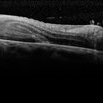
B-mode ultrasound of a patient with long-standing poorly controlled diabetes demonstrates characteristic findings of advanced proliferative diabetic retinopathy. The examination reveals moderate vitreous hemorrhage appearing as diffuse hyperechoic opacities throughout the vitreous cavity, along with a posterior hyaloid membrane densely infiltrated by hemorrhagic material, showing irregular thickening and increased reflectivity. A mild subhyaloid hemorrhage is visible as a subtle hyphema-like space anterior to the retinal surface. The study documents a total tractional retinal detachment, evidenced by rigid retinal folds with clear insertion points of vitreous strands, accompanied by a significant subretinal hemorrhage seen as a prominent hyperechoic collection beneath the elevated retina. These findings collectively illustrate the severe vitreoretinal interface pathology characteristic of end-stage diabetic eye disease, with predominant tractional components and distinct echographic stratification of hemorrhagic layers - from anterior vitreous involvement to deeper subretinal blood accumulation.
Photographer: Gustavo U. Fonseca Aguirre, Hospital Conde de Valenciana, Ciudad de México
Condition/keywords: diabetic retinopathy, tractional retinal detachment, Vitreous hemorrhage
-
 A Classic Case of Retinal Ora Serrata Imaging
Jan 16 2025 by yuan duo
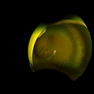
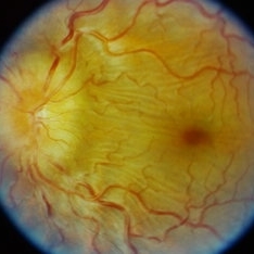
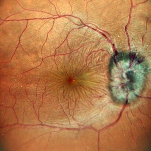
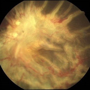
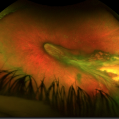
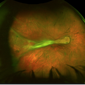
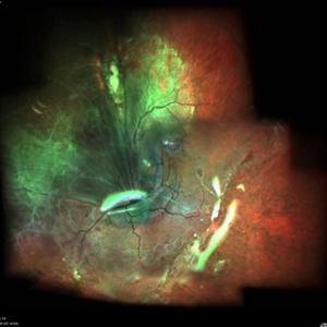
A 5-year-old girl, born full-term with no history of systemic disease, presented with poor vision since early childhood and underwent fundus examination. Anterior segments of both eyes showed no significant abnormalities. Fundus examination revealed retinal folds extending from the optic disc to the temporal peripheral retina, with blood vessels coursing through the folds (A, B). Avascular zones were observed in the peripheral retina, and the ora serrata’s boundaries were clearly visible, displaying dentate processes and bays (C, D). Retinal pigmentation was evident. Genetic testing confirmed the final diagnosis of bilateral Familial Exudative Vitreoretinopathy (FEVR).
Condition/keywords: Retinal Ora Serrata
-
 Familial Exudative Vitreoretinopathy
Jan 16 2025 by yuan duo
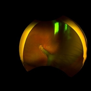
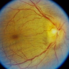
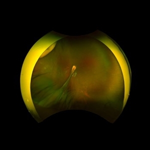
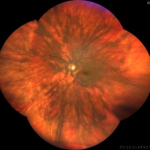
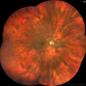
A 5-year-old girl, born full-term with no history of systemic disease, presented with poor vision since early childhood and underwent fundus examination. Anterior segments of both eyes showed no significant abnormalities. Fundus examination revealed retinal folds extending from the optic disc to the temporal peripheral retina, with blood vessels coursing through the folds (A, B). Avascular zones were observed in the peripheral retina, and the ora serrata’s boundaries were clearly visible, displaying dentate processes and bays (C, D). Retinal pigmentation was evident. Genetic testing confirmed the final diagnosis of bilateral Familial Exudative Vitreoretinopathy (FEVR).
Condition/keywords: Retinal Ora Serrata
-
 ERM
ERM
Jan 9 2025 by Richa Chaudhary, Mbbs,ms
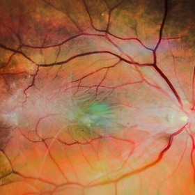
52 year old male presented with idipathic ERM, with pucker showing, retinal folds. Planned for surgical removal of the same.
Condition/keywords: ERM
-
 Venolymphatic Mass With Disc Edema
Dec 5 2024 by Tejaswita Verma
Fundus picture of a 26 year old male who presented with right eye abaxial proptosis, MRI confirmed venolymphatic mass inferomedial in location located near the optic disc with disc edema , nasal elevation ,retinal folds. Vision was 6/18 . He was planned for intralesional bleomycin injection.
Photographer: DR. TEJASWITA VERMA
Imaging device: MIRANTE
Condition/keywords: disc edema, intraorbital mass, proptosis
-
 Venolymphatic Mass with Retinal Folds
Nov 25 2024 by Tejaswita Verma
Fundus picture of a 26 year old male who presented with right eye abaxial proptosis, MRI confirmed venolymphatic mass inferomedial in location located near the optic disc with disc edema , nasal elevation ,retinal folds. Vision was 6/18 . He was planned for intralesional bleomycin injection.
Photographer: DR. TEJASWITA VERMA
Imaging device: MIRANTE
Condition/keywords: disc edema, intraorbital mass, proptosis, retinal folds
-
 Proliferative Vitreoretinopathy
Jun 9 2024 by Marcelo Zas, MD PhD
We present a case of a 20-year-old patient who underwent surgery for congenital cataract when he was born and 20 years after he developed a retinal detachment with proliferative vitreoretinopathy. Proliferative vitreoretinopathy (PVR), a major complication of rhegmatogenous retinal detachment (RRD), is an abnormal process whereby proliferative, contractile cellular membranes form in the vitreous and on both sides of the retina, resulting in tractional retinal detachment with fixed retinal folds. PVR arises in an estimated 5-10% of RRD cases, and therefore represents a major complication of retinal detachment. The best treatment of PVR is its prevention. Clinical factors associated with increased risk of PVR include: • Chronic RRD • 2 o more horseshoe retinal tears and RRD exposing three-disc diameters or more of RPE • RD associated with giant retinal • RD associated with choroidal detachment • Ocular Trauma • RRD associated with vitreous hemorrhage • Aphakia and RRD • Failure of previous surgery or multiple retinal surgeries • Aggressive retinitis, etc.
Photographer: Luciano Scorsetti MD
Condition/keywords: proliferative vitreoretinopathy (PVR)
-
 Posterior Scleritis
Sep 12 2023 by Ben Serar
Fundus photograph of LE showing Disc edema with Choroidal folds in a case of Posterior Scleritis
Condition/keywords: chorioretinal folds, disc edema, posterior scleritis
-
 Posterior Scleritis
Sep 12 2023 by Ben Serar
Fundus photograph of RE showing Disc edema with Choroidal folds in a case of Posterior Scleritis.
Condition/keywords: chorioretinal folds, disc edema, Posterior scleritis
-
 Birdshot Retinopathy
May 9 2023 by JEFFERSON R SOUSA, Tecg.º (Biomedical Systems Technology)
Female patient, 41 years old, with progressive low visual acuity, progressive history of autoimmune disease. In the multimodal retinal fundoscopic evaluation, important characteristics compatible with "Birdshot Retinopathy" were observed. Birdshot retinopathy, also known as birdshot chorioretinopathy or birdshot uveitis, is a rare, chronic inflammatory disorder that affects the retina and the choroid of the eye. It typically develops in adults between the ages of 30 and 60 years, and is more common in women than men. The name "birdshot" refers to the small, round, yellow-white spots that appear on the retina, which resemble the pattern of a shotgun blast. These spots are caused by inflammation in the eye, and can lead to vision loss if left untreated. Symptoms of birdshot retinopathy include blurred vision, floaters, loss of night vision, and difficulty adapting to changes in lighting. The condition can also cause inflammation in other parts of the eye, leading to redness, pain, and sensitivity to light. The exact cause of birdshot retinopathy is unknown, but it is believed to be an autoimmune disorder, in which the body's immune system mistakenly attacks the retina and choroid. Treatment typically involves the use of immunosuppressive medications, such as corticosteroids or biologic agents, to reduce inflammation and preserve vision. Close monitoring by an ophthalmologist is important, as the disease can progress even with.
Photographer: JEFFERSON ROCHA DE SOUSA - Retinal Department at Institute Dr. Suel Abujamra Sao Paulo-Brazil
Imaging device: Clarus 700 - Zeiss, composite of four 135 degree images.
Condition/keywords: bilateral chorioretinal folds, birdshot, birdshot chorioretinopathy, birdshot choroidopathy, birdshot retinochoroidopathy
-
 Birdshot Retinopathy
May 9 2023 by JEFFERSON R SOUSA, Tecg.º (Biomedical Systems Technology)
Female patient, 41 years old, with progressive low visual acuity, progressive history of autoimmune disease. In the multimodal retinal fundoscopic evaluation, important characteristics compatible with "Birdshot Retinopathy" were observed. Birdshot retinopathy, also known as birdshot chorioretinopathy or birdshot uveitis, is a rare, chronic inflammatory disorder that affects the retina and the choroid of the eye. It typically develops in adults between the ages of 30 and 60 years, and is more common in women than men. The name "birdshot" refers to the small, round, yellow-white spots that appear on the retina, which resemble the pattern of a shotgun blast. These spots are caused by inflammation in the eye, and can lead to vision loss if left untreated. Symptoms of birdshot retinopathy include blurred vision, floaters, loss of night vision, and difficulty adapting to changes in lighting. The condition can also cause inflammation in other parts of the eye, leading to redness, pain, and sensitivity to light. The exact cause of birdshot retinopathy is unknown, but it is believed to be an autoimmune disorder, in which the body's immune system mistakenly attacks the retina and choroid. Treatment typically involves the use of immunosuppressive medications, such as corticosteroids or biologic agents, to reduce inflammation and preserve vision. Close monitoring by an ophthalmologist is important, as the disease can progress even with.
Photographer: JEFFERSON ROCHA DE SOUSA - Retinal Department at Institute Dr. Suel Abujamra Sao Paulo-Brazil
Imaging device: Clarus 700 - Zeiss, composition of five 135 degree images.
Condition/keywords: bilateral chorioretinal folds, birdshot, birdshot chorioretinopathy, birdshot choroidopathy, birdshot retinochoroidopathy
-
 Hypotony Maculopathy
Jun 12 2022 by Pramod Kumar Suman, MBBS, MD
Fundus photograph of an 26-year-old male with retinal folds around the center of the fovea arranged in stellate pattern with optic disc edema.
Photographer: Pramod Kumar Suman, Retina Foundation, Ahmedabad
Condition/keywords: hypotony maculopathy
-
 Long-standing RD with PVR
Jan 28 2022 by Gayathri Mohan
Color fundus photograph showing a long standing RD with PVR with fixed retinal folds.
Photographer: Dr Gayathri Mohan
Imaging device: Canon
Condition/keywords: PVR, Retina Folds, Retinal Detachment
-
 Familial Exudative Vitreoretinopathy (FEVR)
Apr 24 2021 by Alexandre Grandinetti, MD, PhD
6-year-old girl with retinal folds on both eyes secondary to FEVR.
Photographer: Corina Szrek
Imaging device: Optos California
Condition/keywords: familial exudative vitreoretinopathy (FEVR)
-
 FEVR
FEVR
Apr 24 2021 by Alexandre Grandinetti, MD, PhD
6-year-old girl with retinal folds on both eyes secondary to FEVR.
Photographer: Corina Szrek
Imaging device: Optos California
Condition/keywords: familial exudative vitreoretinopathy (FEVR)
-
 Large Retinal Fold Masking the Optic Nerve
Sep 22 2020 by Sophia El Hamichi, MD
A 69-year-old female, with a history of choroidal melanoma in her left eye with exudative detachment, underwent tumor laser ablation. She then developed a complex combined tractional and rhegmatogenous retinal detachment with a giant retinal tear. The patient underwent surgical repair of her retinal detachment with pars plana vitrectomy and silicone oil. In the post-op, the patient developed large retinal folds masking the optic nerve depicted in the OCT photograph.
Photographer: Belinda Rodriguez, Murray Ocular Oncology and Retina, Miami
Condition/keywords: giant retinal tear, melanoma, pars plana vitrectomy (PPV), retinal fold, silicone oil
-
 Large Retinal Fold
Sep 22 2020 by Sophia El Hamichi, MD
A 69-year-old female, with a history of choroidal melanoma in her left eye with exudative detachment, underwent tumor laser ablation. She then developed a complex combined tractional and rhegmatogenous retinal detachment with a giant retinal tear. The patient underwent surgical repair of her retinal detachment with pars plana vitrectomy and silicone oil. In the post-op, the patient developed large retinal folds masking the optic nerve depicted in the fundus photograph.
Photographer: Belinda Rodriguez, Murray Ocular Oncology and Retina, Miami
Condition/keywords: melanoma, pars plana vitrectomy (PPV), retinal fold, silicone oil
-
 Hypotony Maculopathy
Apr 1 2019 by Anfisa Ayalon, MD
Fundus autofluorescence image of 81-year-old male with right eye ocular hypotony due to leaking bleb. Note severe hypotony maculopathy, peripheral choroidal detachments, multiple chorioretinal folds.
Photographer: Anfisa Ayalon, MD., Meir Medical Center, Kfar Saba, Israel.
Imaging device: California, Optos 200 DTX
Condition/keywords: choroidal detachment, choroidal folds, fundus autofluorescence (FAF), hypotonous retinopathy, hypotony maculopathy
-
 Ocular Hypotony Due to Leaking Bleb
Apr 1 2019 by Anfisa Ayalon, MD
81-year-old male who had trabeculectomy in his right eye 4 years ago, presented to the emergency room with complains of decreased vision in that eye for two months. Slit-lamp examination showed cystic bleb with leakage, intraocular pressure was 0 MMHg. Fundus examination showed hypotony maculopathy, peripheral choroidal detachments, multiple chorioretinal folds with subretinal fluid.
Photographer: Anfisa Ayalon, MD., Meir Medical Center, Kfar Saba, Israel.
Imaging device: California, Optos 200 DTX
Condition/keywords: choroidal detachment, hypotonous retinopathy, hypotony maculopathy
-
 C-R Folds
Mar 26 2019 by Gary R. Cook, MD, FACS
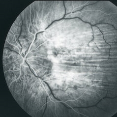
Early phase FA frame of the left eye of a WM with bilateral C-R folds showing alternating hyper- and hypofluorescent bands.
Imaging device: Topcon VT-50
Condition/keywords: bilateral chorioretinal folds, chorioretinal fold, FA early phase, fluorescein angiogram (FA)
-
 Bilateral C-R Folds
Mar 26 2019 by Gary R. Cook, MD, FACS
Fundus photo of the left eye of a white male with bilateral C-R folds.
Imaging device: Topcon VT-50
Condition/keywords: bilateral chorioretinal folds, chorioretinal fold
-
 C-R Folds
Mar 26 2019 by Gary R. Cook, MD, FACS
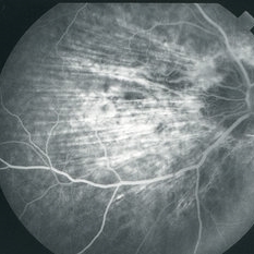
Mid-phase FA image of the right eye of a white male with bilateral C-R folds showing alternating hyper- and hypofluorescent bands.
Imaging device: Topcon VT-50
Condition/keywords: bilateral chorioretinal folds, chorioretinal fold, FA mid phase, fluorescein angiogram (FA)
-
 Bilateral C-R Folds
Mar 26 2019 by Gary R. Cook, MD, FACS
Fundus photo of the right eye of a white male with bilateral C-R folds.
Imaging device: Topcon VT-50
Condition/keywords: bilateral chorioretinal folds, chorioretinal fold
 Loading…