Search results (126 results)
-
 Gyrate Atrophy
Nov 22 2025 by Gaurav Kamble
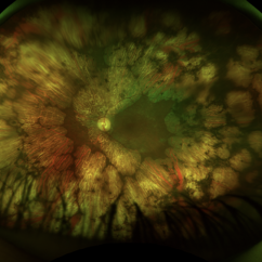
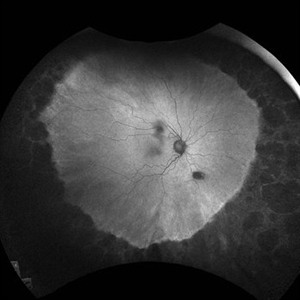
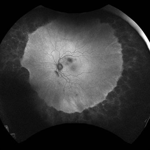
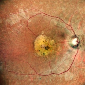
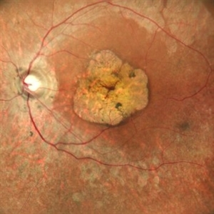
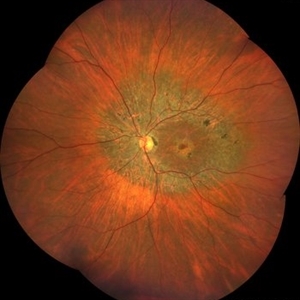
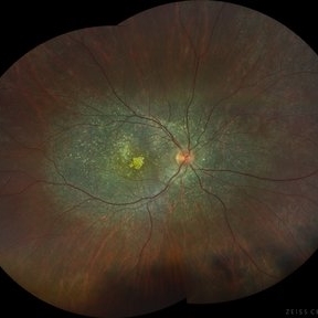
A 12-year-old female presented with progressive blurring of vision for distance and had a known history of convulsions. Ocular examination revealed bilateral proptosis and megalocornea. Fundus evaluation showed well-defined scalloped areas of peripheral chorioretinal degeneration characteristic of gyrate atrophy, along with cystoid macular edema involving the macular region. The overall clinical picture was consistent with gyrate atrophy.
Photographer: Ms. Vishaka Shah , Isha Eye Care Pvt Ltd ,Khadakpada, Kalyan
Imaging device: Optos Imaging Daytona
Condition/keywords: gyrate atrophy
-
 Cannula Tip Pressure
Mar 25 2025 by Robert Andrew Sisk, MD, FACS, FASRS
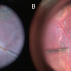
Color stills from surgical videos of subretinal delivery of gene augmentation therapy with A) voretigene neparvovec-ryzl and B) laru-zova. In the left panel, the cannula is slightly bent, and the retina and RPE are blanched white around the cannula tip engagement. The bleb was challenging to form in this patient with advanced retinal degeneration, and the bleb is shallow and mostly clear. In the right panel, the cannula tip is gently engaged, the cannula is straight, and it follows the retinotomy as the retina is elevated by the injection fluid.
Imaging device: Leica Proveo 8
Condition/keywords: gene therapy, genetic disorder, Leber's congenital amaurosis, retinitis pigmentosa, subretinal injection
-
 Progressive Chorioretinal Degeneration
Jun 27 2024 by Natalia Moraes
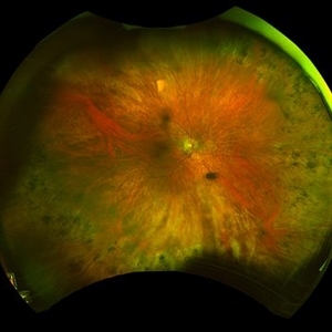
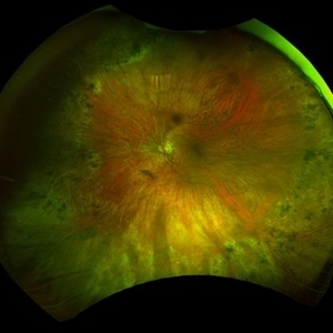
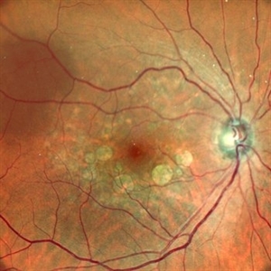
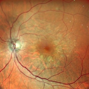
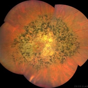
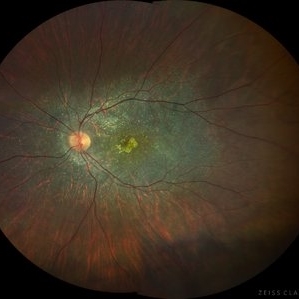
Funds photograph of a 63-year-old male with a progressive chorioretinal degeneration.
Photographer: Natália V. Moraes, Instituto Penido Burnier, Brazil
Imaging device: Daytona
Condition/keywords: gyrate atrophy
-
 Peripheral Retinal Degeneration (L-ORD)
Apr 17 2024 by Virginia Gebhart
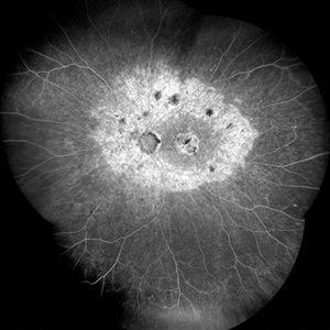
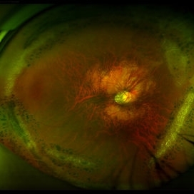
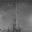
92 year old female with bilateral patchy, sharply demarcated circular areas of chorioretinal atrophy with hyperpigmented margins in the mid to far periphery. Labs showed normal plasma ornithine levels ruling out generalized gyrate atrophy. Also intermediate uveitis and CMD/CME. FTA-ABS, Quant gold, and HLA-A29 labs all negative.
Photographer: Virginia Gebhart
Imaging device: Optos California
Condition/keywords: cystoid macular degeneration, cystoid macular edema (CME), FA, Fluorescein angiography, peripheral retinal degeneration
-
 Peripheral retinal degenerations
Jan 29 2024 by Anupama Kiran Kumar
Fundus photo of a young man who underwent barrage laser after he presented to the clinic with floaters and was diagnosed to have lattices with horse shoe tears and retinal holes.
Photographer: Dr Anupama Kiran Kumar DNB FVR , Narayana Nethralaya Bangalore
Imaging device: Mirante SLO/OCT (Nidek Co., Gamagori, Japan)
Condition/keywords: lattice degeneration, peripheral retinal degeneration
-
 Alagille Syndrome UWF Color
Dec 4 2023 by Isaac Ezon, MD
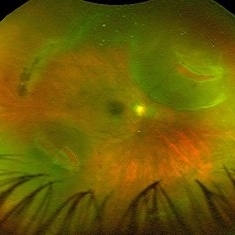
43 yo Female with known Alagille Syndrome, referred for peripheral retinal changes. Subjective nyctalopia but no other symtpoms. Alagille Syndrome UWF Color.
Photographer: Tara Murray
Imaging device: Optos
Condition/keywords: hereditary choroidal dystrophy, hereditary retinal degeneration
-
 Alagille Syndrome UWF Color
Dec 4 2023 by Isaac Ezon, MD
43 yo Female with known Alagille Syndrome, referred for peripheral retinal changes. Subjective nyctalopia but no other symtpoms. Alagille Syndrome UWF Color.
Photographer: Tara Murray
Imaging device: Optos
Condition/keywords: hereditary choroidal dystrophy, hereditary retinal degeneration
-
 Alagille Syndrome UWF Autofluorescence
Dec 4 2023 by Isaac Ezon, MD
43 yo Female with known Alagille Syndrome, referred for peripheral retinal changes. Subjective nyctalopia but no other symtpoms. Alagille Syndrome UWF Autofluorescence.
Photographer: Tara Murray
Imaging device: Optos
Condition/keywords: hereditary choroidal dystrophy, hereditary retinal degeneration
-
 White Without Pressure and Peripheral Retinoschisis
Dec 29 2022 by Gulnara Islamova
Fundus Photograph and OCT scan of an 18 year-old male with peripheral retinoschisis combined with WWOP lessions .Vitreoretinal traction is not visualized
Photographer: Gulnara Islamova, CENTER ZRENIYA Medical Clinic, LLC, Chelyabinsk, Russian Federation
Imaging device: Optovue XR Avanti
Condition/keywords: peripheral retinal degeneration
-
 Rod Cone dystrophy
Nov 29 2022 by Niloofar Piri, MD
Fundus autofluorescence of the left eye in a 58 yo male with rod cone dystrophy. He presented with night blindness and peripheral vision loss since youth and recent decrease in central vision for the past 10 years. Notice multiple coin shaped hypoautofluorescent pacthes within central 20 degrees which are coalescing centrally. (fundus photo uploaded separately) He has one pathogenic variants of both CEP290 and PRPH2 genes.
Photographer: Sean Kelso, Saint Louis University
Condition/keywords: hereditary retinal degeneration, hereditary retinal dystrophy, rod cone dystrophy
-
 HEREDITARY MACULAR DEGENERATION WITH ETHAMBUTOL TOXICITY
Nov 1 2022 by Akansha Sharma
COLOUR FUNDUS PHOTOGRAPH OF A 45 YEAR OLD MALE WITH HEREDITARY MACULAR DEGENERATION WITH ETHAMBUTOL TOXICITY
Photographer: Dr. Akansha Sharma-Retina Foundation, Ahmedabad
Condition/keywords: drug toxicity, hereditary retinal degeneration, toxic optic neuropathy
-
 HEREDITARY MACULAR DEGENERATION
Oct 15 2022 by Akansha Sharma
COLOUR FUNDUS PHOTOGRAPH OF A 43 YEAR OLD MALE PATIENT WITH HEREDITARY MACULAR DEGENERATION
Photographer: Dr. Akansha Sharma-Retina Foundation, Ahmedabad
Condition/keywords: hereditary retinal degeneration
-
 HEREDITARY MACULAR DEGENERATION
Oct 15 2022 by Akansha Sharma
COLOUR FUNDUS PHOTOGRAPH OF A 43 YEAR OLD MALE PATIENT WITH HEREDITARY MACULAR DEGENERATION
Photographer: Dr. Akansha Sharma-Retina Foundation, Ahmedabad
Condition/keywords: hereditary retinal degeneration
-
 Tapetoretinal Degeneration
Sep 7 2022 by JEFFERSON R SOUSA, Tecg.º (Biomedical Systems Technology)
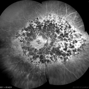
Patient 52 years old, Male, progressive loss of vision since the age of 20. Retinography showed mobilization of pigments in osteoblasts, extensive area of atrophy of the pigmentary epithelium and choroid. On fluorescein angiography, typical changes following the characteristic patterns of paracentra retinal retinitis pigmentosa. Autofluorescent fundus with a sectorial autohypofluorescence pattern in the regions of atrophies.
Photographer: JEFFERSON ROCHA DE SOUSA - Retinal Department at Instituto Dr. Suel Abujamra Sao Paulo-Brazil
Imaging device: Clarus 700 - Zeiss, composite of four 135 degree images.
Condition/keywords: pericentral retinitis pigmentosa, tapeoretinal degeneration
-
 Tapetoretinal Degeneration
Sep 7 2022 by JEFFERSON R SOUSA, Tecg.º (Biomedical Systems Technology)
Patient 52 years old, Male, progressive loss of vision since the age of 20. Retinography showed mobilization of pigments in osteoblasts, extensive area of atrophy of the pigmentary epithelium and choroid. On fluorescein angiography, typical changes following the characteristic patterns of paracentra retinal retinitis pigmentosa. Autofluorescent fundus with a sectorial autohypofluorescence pattern in the regions of atrophies.
Photographer: JEFFERSON ROCHA DE SOUSA - Retinal Department at Instituto Dr. Suel Abujamra Sao Paulo-Brazil
Imaging device: Clarus 700 - Zeiss, composite of four 135 degree images.
Condition/keywords: pericentral retinitis pigmentosa, tapeoretinal degeneration
-
 Snail Track Peripheral Retinal Degeneration
Apr 29 2022 by Otakar Dušek, M.D. Ph.D.
Colour fundus photograph of 22-year-old woman with incidentally found snail track retinal degeneration in the superior temporal periphery of the retina of the right eye.
Photographer: Otakar Dušek, Charles University, Prague
Imaging device: Zeiss Clarus
Condition/keywords: peripheral retinal degeneration
-
 Laser Treated Lattice Degeneration
Jul 12 2021 by Gabriel Costa Andrade, PhD
Fundus photograph of an 22-year-old man with peripheral lattice retinal degeneration treated with photocoagulation.
Photographer: Gabriel Andrade
Condition/keywords: lattice degeneration, retina
-
 Sea-Saw Vitreoretinal Dance
Mar 12 2021 by RUSHIK PATEL
Fundus photograph of 50-year-old female showing posterior vitreous detachment, 2 retinal tears with localized retinal detachment exactly 180 degrees apart with optic disc in between as a Falcrum giving an appearance of sea saw retinal tears. Macula was attached with lattice retinal degeneration superiorly.
Photographer: Rushik Patel, Netralaya Super Speciality Eye Hospital
Condition/keywords: peripheral lattice degeneration, retinal tear
-
 Bietti's Crystalline Dystrophy
Oct 3 2020 by SHISHIR VERGHESE, MS, FVRS, FAICO (Retina)
Fundus photo of the right eye of a 21-year-old gentleman with complaints of reduced vision, nyctalopia and visual field reduction. Fundus photograph showing numerous small glistening yellow-white retinal crystalline deposits in the retina.
Photographer: Shishir Verghese, Aravind Eye Hospital, Coimbatore
Condition/keywords: Bietti's crystalline dystrophy, hereditary retinal degeneration, heredomacular degeneration
-
 Bietti's Crystalline Dystrophy
Oct 3 2020 by SHISHIR VERGHESE, MS, FVRS, FAICO (Retina)
Fundus photo of the left eye of a 21-year-old gentleman with complaints of reduced vision, nyctalopia and visual field reduction. Fundus photograph showing numerous small glistening yellow-white retinal crystalline deposits in the retina.
Photographer: Shishir Verghese, Aravind Eye Hospital, Coimbatore
Condition/keywords: Bietti's crystalline dystrophy, heredomacular degeneration, retinal degeneration
-
 Fundus Autofluorescence in Central Areolar Choroidal Dystrophy
Dec 8 2019 by Anfisa Ayalon, MD
Fundus autofluorescence pictures of a 37-year-old male with CACD. The patient has visual acuity of 1/18 in the right eye and 6/30 in the left eye. Full-field ERG was normal under photopic and scotopic conditions.
Photographer: Anfisa Ayalon,MD., Meir Medical Center, Kfar Saba, Israel.
Condition/keywords: central areolar choroidal dystrophy (CACD), fundus autofluorescence (FAF), hereditary retinal degeneration
 Loading…