Search results (35 results)
-
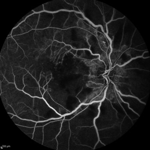
 Fluorescein and Indocyanine Green Angiography in Right Eye in Case of Choroidal Hemangioma
Nov 29 2024 by Anand Temkar
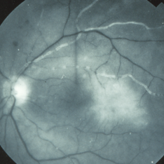
Right eye Fluorescein and Indocyanine green angiography of a 42 year old male in case of Choroidal hemangioma. Choroidal hemangioma have a unique pattern of circulation where the large blood vessels produce a “COARSE VASCULAR PATTERN.” Fluorescein angiography of circumscribed choroidal hemangiomas typically reveals very early hyperfluorescence of larger-caliber choroidal blood vessels either before or simultaneously with the initial filling of the retinal arterioles. Indocyanine green angiography typically shows filling of the intralesional vascular channels, intense hypercyanescence of the lesion by the intermediate frames (peaks around 3-4 minutes) and late washout of the central portion of the lesion.
Photographer: Dr.Anand Temkar- Retina Foundation, Ahmedabad
Imaging device: Mirante
Condition/keywords: Choroidal Hemangioma, FLUORESCEIN ANGIOGRAPHY, indocyanine green (ICG) angiography
-
 Retinal Arteriolar Variation
Oct 31 2024 by AVIK DEY SARKAR, MS, FVRS, FAICO(VR)
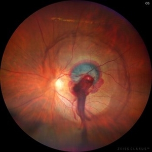
A 43-year-old hypertensive patient, diagnosed with Non-Ischemic Central retinal vein Occlusion in OS, presented with a striking anatomical variation in retinal vasculature. The inferior first-order retinal arteriole after initiating from the optic disc bifurcates, before reaching the fovea, and the superior branch after crossing the midline forms the superior arcade afterwards and produces dichotomous branching as usual. This defies basic anatomical considerations for retinal vasculature as they never cross the midline, also known as the watershed line for retinal vessels.1,2 References: 1. May CA, Rutkowski P. The Horizontal Raphe of the Human Retina and its Watershed Zones. Vision. 2019; 3(4):60. 2. May CA, Rutkowski P. Hypothesis: watershed zones in the human eye are a key for understanding glaucomatous retinal damage. Med Hypotheses. 2017;109:1-5.
Photographer: Dr. Avik Dey Sarkar, MBBS, MS, FVRS, FAICO, Consultant, Department of Vitreoretinal Services, Aravind Eye Hospital, Madurai, India
Imaging device: Wide angled Fundus imaging with Clarus 300
Condition/keywords: background diabetic retinopathy (BDR), Diabetic Retinopathy, retina, vascular anomaly
-
 Ruptured Retinal Arterial Macro-Aneurysm
Oct 27 2024 by César Adrián Gómez Valdivia, MD
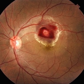
Ruptured retinal arterial macro-aneurysm found in a 56 YO female patient with history of untreated hypertension. Round or fusiform dilation of a retinal arteriole is usually seen within a third degree branch of one of the four main arcade arteries. Most common location for a symptomatic macroaneurysm is from a branch of the superotemporal arcade.
Photographer: @eyemissu2
Imaging device: TOPCON TRC-50DX
Condition/keywords: ruptured macroaneurysm
-
 Ruptured Retinal Artery Macroaneurysm
Jun 18 2024 by KANWALJEET HARJOT MADAN, M.S. (Ophthalmology), FAICO (Vitreous - Retina)
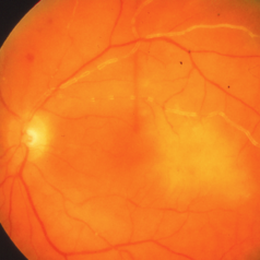
This is a fundus photo depicting ruptured Retinal Artery Macroaneurysm (RAM) in the left eye of a 63 years old female. RAM is an acquired saccular or fusiform dilatation of the retinal arterioles that usually occur within the first three orders of bifurcation. The Superotemporal artery is the most common location. RAM may be asymptomatic or cause a number of complications such as macular edema, serous macular detachment, and hemorrhages.
Photographer: Dr Kanwaljeet Harjot Madan
Condition/keywords: Haemorrhage, macroaneurysm, retinal arteriole
-
 Congenital Retinal Vessel Tortuosity
Apr 2 2024 by Pablo Angel Garcia Uribe
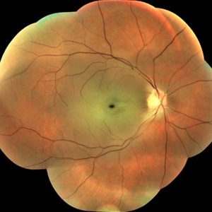
Fundus photograph of a 29-year-old man with bilateral congenital retinal vessel tortuosity. This image shows the sinuous course of retinal arterioles and a shiny internal limiting membrane.
Photographer: Pablo Ángel García-Uribe, Clínica Oftalmológica Salauno, Mexico City
Imaging device: NIDEK OCT RS-330 Duo 2
Condition/keywords: abnormal retinal vessel, anomalous vessels, Retina, tortuous vessels
-
 Idiopathic retinal vasculitis, aneurysms and neuroretinitis
Apr 24 2022 by Aniruddha K Agarwal, MD
Ultra-wide field fundus fluorescein angiography (FFA) of the left eye from an asymptomatic, healthy 33-year-old woman who was referred to the retina clinic from a refractive surgery unit due to the presence of vascular anomalies and hard exudates in both eyes. FFA revealed the characteristic sacular aneurysms at the bifurcation of retinal arterioles in the posterior pole, together with microvascular anomalies and capillary closure peripherally.
Photographer: Julio J GONZALEZ-LOPEZ, MD, PhD, FEBO and Teresa GONZALEZ-LOMAS, RN
Imaging device: Optos California
Condition/keywords: IRVAN Syndrome, IUSG, neuroretinitis, retinal vasculitis, uveitis
-
 Central Retinal Artery Occlusion
Mar 2 2021 by Renata Garcia Franco, Md
Retinal edema, cherry spot, retinal arteriolar attenuation and segmentation of blood in retinal arterioles.
Photographer: Guillermina Hernandez
Imaging device: Zeiss
Condition/keywords: central artery
-
 Central Retinal Artery Occlusion
Jan 22 2021 by Renata Garcia Franco, Md
65-year-old male, history of uncontrolled systemic arterial hypertension. Segmentation of blood in retinal arterioles, retinal whitening and cherry red spot.
Photographer: Fatima Hernandez, Instituto de la Retina del Bajio SC
Imaging device: Zeiss
Condition/keywords: central retinal artery occlusion (CRAO)
-
 Bilateral Calcific Retina Arteriolar Occlusions in a Patient with Metastatic Ovarian Carcinoma
Dec 10 2020 by McGill University Health Centre
47-year-old female with cough and fever. Imaging showed a right pulmonary infiltrate. Transbronchial needle biopsy revealed lymphangitic spread of papillary adenocarcinoma with psammoma bodies. MRI of thyroid, CT of abdomen and pelvis were negative. gynecologic evaluation negative at that time . The patient had bilateral floaters, VA: 20/40 OD and 20/20 OS. Fundus examination showed retinal arteriolar sheathing and a flat choroidal lesion OS and vitritis OD. Fluorescein angiogram showed staining of left superior temporal retinal arterioles and bilateral midperipheral patchy hyperfluorescence at RPE The patient vision in the OD deteriorated to 20/400, and in the OS 20/50. Four months later a new choroidal lesion was diagnosed OS. An abdominal mass consistent with a cystadenoma of the ovary was diagnosed. After a year patient developed systemic metastasis. Autopsy: Metastatic adenocarcinoma to the lung, both adrenals, para-aortic lymph nodes, left hip, right breast, occipital skin, serosal surface of liver, pituitary. In almost all metastatic lesions psammoma bodies were found. Presumptive diagnosis is a primary tumor of the ovary.
Condition/keywords: bilateral, calcification, metastatic adenocarcinoma, retinal arteriolar occlusion
-
 Bilateral Calcific Retina Arteriolar Occlusions in a Patient with Metastatic Ovarian Carcinoma
Dec 10 2020 by McGill University Health Centre
47-year-old female with cough and fever. Imaging showed a right pulmonary infiltrate. Transbronchial needle biopsy revealed lymphangitic spread of papillary adenocarcinoma with psammoma bodies (MRI of thyroid, CT of abdomen and pelvis were negative) gynecologic evaluation negative at that time . The patient had bilateral floaters, VA: 20/40 OD and 20/20 OS. Fundus examination showed retinal arteriolar sheathing and a flat choroidal lesion OS and vitritis OD. Fluorescein angiogram showed staining of left superior temporal retinal arterioles and bilateral midperipheral patchy hyperfluorescence at RPE. The patient vision in the OD deteriorated to 20/400, and in the OS 20/50. Four months later a new choroidal lesion was diagnosed OS. An abdominal mass consistent with a cystadenoma of the ovary was diagnosed. After a year patient developed systemic metastasis. Autopsy: Metastatic adenocarcinoma to the lung, both adrenals, para-aortic lymph nodes, left hip, right breast, occipital skin, serosal surface of liver, pituitary. In almost all metastatic lesions psammoma bodies were found. Presumptive diagnosis is a primary tumor of the ovary.
Imaging device: Fluoroscein angiogram
Condition/keywords: bilateral, calcification, metastatic adenocarcinoma, retinal arteriolar occlusion
-
 Bilateral Calcific Retina Arteriolar Occlusions in a Patient with Metastatic Ovarian Carcinoma
Dec 10 2020 by McGill University Health Centre
47-year-old female with cough and fever. Imaging showed a right pulmonary infiltrate. Transbronchial needle biopsy revealed lymphangitic spread of papillary adenocarcinoma with psammoma bodies (MRI of thyroid, CT of abdomen and pelvis were negative) gynecologic evaluation negative at that time . The patient had bilateral floaters, VA: 20/40 OD and 20/20 OS. Fundus examination showed retinal arteriolar sheathing and a flat choroidal lesion OS and vitritis OD. Fluorescein angiogram showed staining of left superior temporal retinal arterioles and bilateral midperipheral patchy hyperfluorescence at RPE The patient vision in the OD deteriorated to 20/400, and in the OS 20/50. Four months later a new choroidal lesion was diagnosed OS. An abdominal mass consistent with a cystadenoma of the ovary was diagnosed. After a year patient developed systemic metastasis. Autopsy: Metastatic adenocarcinoma to the lung, both adrenals, para-aortic lymph nodes, left hip, right breast, occipital skin, serosal surface of liver, pituitary. In almost all metastatic lesions psammoma bodies were found. Presumptive diagnosis is a primary tumor of the ovary. Histopathologic examination of both eyes disclosed : Bilateral metastatic adenocarcinoma to the vitreous with partially calcified proliferation along internal limiting membrane, OS. Metastatic adenocarcinoma to choroid, OS. Bilateral optic atrophy secondary to retinal arteriolar occlusion with calcification.
Condition/keywords: bilateral, calcification, histopathology, metastatic adenocarcinoma, pathology, retinal arteriolar occlusion
-
 Bilateral Calcific Retina Arteriolar Occlusions in a Patient with Metastatic Ovarian Carcinoma
Dec 10 2020 by McGill University Health Centre
47-year-old female with cough and fever. Imaging showed a right pulmonary infiltrate. Transbronchial needle biopsy revealed lymphangitic spread of papillary adenocarcinoma with psammoma bodies (MRI of thyroid, CT of abdomen and pelvis were negative) gynecologic evaluation negative at that time Patient had bilateral floaters, VA: 20/40 OD and 20/20 OS. Fundus examination showed retinal arteriolar sheathing and a flat choroidal lesion OS and vitritis OD. Fluorescein angiogram showed staining of left superior temporal retinal arterioles and bilateral midperipheral patchy hyperfluorescence at RPE The patient vision in the OD deteriorated to 20/400, and in the OS 20/50. Four months later a new choroidal lesion was diagnosed OS. An abdominal mass consistent with a cystadenoma of the ovary was diagnosed. After a year patient developed systemic metastasis. Autopsy: Metastatic adenocarcinoma to the lung, both adrenals, para-aortic lymph nodes, left hip, right breast, occipital skin, serosal surface of liver, pituitary. In almost all metastatic lesions psammoma bodies were found. Presumptive diagnosis is a primary tumor of the ovary. Histopathologic examination of both eyes disclosed : Bilateral metastatic adenocarcinoma to the vitreous with partially calcified proliferation along internal limiting membrane, OS. Metastatic adenocarcinoma to choroid, OS. Bilateral optic atrophy secondary to retinal arteriolar occlusion with calcification.
Condition/keywords: bilateral, calcification, histopathology, metastatic adenocarcinoma, pathology, retinal arteriolar occlusion
-
 Bilateral Calcific Retina Arteriolar Occlusions in a Patient with Metastatic Ovarian Carcinoma
Dec 10 2020 by McGill University Health Centre
47-year-old female with cough and fever. Imaging showed a right pulmonary infiltrate. Transbronchial needle biopsy revealed lymphangitic spread of papillary adenocarcinoma with psammoma bodies (MRI of thyroid, CT of abdomen and pelvis were negative) gynecologic evaluation negative at that time . The patient had bilateral floaters, VA: 20/40 OD and 20/20 OS. Fundus examination showed retinal arteriolar sheathing and a flat choroidal lesion OS and vitritis OD. Fluorescein angiogram showed staining of left superior temporal retinal arterioles and bilateral midperipheral patchy hyperfluorescence at RPE. The patient vision in the OD deteriorated to 20/400, and in the OS 20/50. Four months later a new choroidal lesion was diagnosed OS. An abdominal mass consistent with a cystadenoma of the ovary was diagnosed. After a year patient developed systemic metastasis. Autopsy: Metastatic adenocarcinoma to the lung, both adrenals, para-aortic lymph nodes, left hip, right breast, occipital skin, serosal surface of liver, pituitary. In almost all metastatic lesions psammoma bodies were found. Presumptive diagnosis is a primary tumor of the ovary. Histopathologic examination of both eyes disclosed : Bilateral metastatic adenocarcinoma to the vitreous with partially calcified proliferation along internal limiting membrane, OS. Metastatic adenocarcinoma to choroid, OS. Bilateral optic atrophy secondary to retinal arteriolar occlusion with calcification.
Condition/keywords: bilateral, calcification, histopathology, metastatic adenocarcinoma, pathology, retinal arteriolar occlusion
-
 Bilateral Calcific Retina Arteriolar Occlusions in a Patient with Metastatic Ovarian Carcinoma
Dec 10 2020 by McGill University Health Centre
47-year-old female with cough and fever. Imaging showed a right pulmonary infiltrate. Transbronchial needle biopsy revealed lymphangitic spread of papillary adenocarcinoma with psammoma bodies (MRI of thyroid, CT of abdomen and pelvis were negative) gynecologic evaluation negative at that time . The patient had bilateral floaters, VA: 20/40 OD and 20/20 OS. Fundus examination showed retinal arteriolar sheathing and a flat choroidal lesion OS and vitritis OD. Fluorescein angiogram showed staining of left superior temporal retinal arterioles and bilateral midperipheral patchy hyperfluorescence at RPE The patient vision in the OD deteriorated to 20/400, and in the OS 20/50. Four months later a new choroidal lesion was diagnosed OS. An abdominal mass consistent with a cystadenoma of the ovary was diagnosed. After a year patient developed systemic metastasis. Autopsy: Metastatic adenocarcinoma to the lung, both adrenals, para-aortic lymph nodes, left hip, right breast, occipital skin, serosal surface of liver, pituitary. In almost all metastatic lesions psammoma bodies were found. Presumptive diagnosis is a primary tumor of the ovary. Histopathologic examination of both eyes disclosed : Bilateral metastatic adenocarcinoma to the vitreous with partially calcified proliferation along internal limiting membrane, OS. Metastatic adenocarcinoma to choroid, OS. Bilateral optic atrophy secondary to retinal arteriolar occlusion with calcification.
Condition/keywords: bilateral, calcification, histopathology, metastatic adenocarcinoma, pathology, retinal arteriolar occlusion
-
 Bilateral Calcific Retina Arteriolar Occlusions in a Patient with Metastatic Ovarian Carcinoma
Dec 10 2020 by McGill University Health Centre
47-year-old female with cough and fever. Imaging showed a right pulmonary infiltrate. Transbronchial needle biopsy revealed lymphangitic spread of papillary adenocarcinoma with psammoma bodies (MRI of thyroid, CT of abdomen and pelvis were negative) gynecologic evaluation negative at that time . The patient had bilateral floaters, VA: 20/40 OD and 20/20 OS. Fundus examination showed retinal arteriolar sheathing and a flat choroidal lesion OS and vitritis OD. Fluorescein angiogram showed staining of left superior temporal retinal arterioles and bilateral midperipheral patchy hyperfluorescence at RPE. The patient vision in the OD deteriorated to 20/400, and in the OS 20/50. Four months later a new choroidal lesion was diagnosed OS. An abdominal mass consistent with a cystadenoma of the ovary was diagnosed. After a year patient developed systemic metastasis. Autopsy: Metastatic adenocarcinoma to the lung, both adrenals, para-aortic lymph nodes, left hip, right breast, occipital skin, serosal surface of liver, pituitary. In almost all metastatic lesions psammoma bodies were found. Presumptive diagnosis is a primary tumor of the ovary. Histopathologic examination of both eyes disclosed : Bilateral metastatic adenocarcinoma to the vitreous with partially calcified proliferation along internal limiting membrane, OS. Metastatic adenocarcinoma to choroid, OS. Bilateral optic atrophy secondary to retinal arteriolar occlusion with calcification.
Condition/keywords: bilateral, calcification, histopathology, metastatic adenocarcinoma, pathology, retinal arteriolar occlusion
-
 Central Retinal Artery Occlusion with Cilioretinal Sparing
Oct 28 2020 by Fang Helen Mi
Fundus photograph of an 61-year-old Chinese male showing central retinal artery occlusion with cilioretinal sparing. Photo shows diffuse ischemic retinal whitening and box-carring of the retinal arterioles.
Condition/keywords: central retinal artery occlusion (CRAO), cilioretinal sparing
-
 Embolic Central Retinal Artery Occlusion
Mar 26 2019 by Gary R. Cook, MD, FACS
58-year-old WM with embolic CRAO demonstrating a a cherry-red spot in macula, retinal whitening around the fovea, and the embolus in a inferotemporal branch retinal arteriole; VA= HM 6''
Imaging device: Topcon VT-50
Condition/keywords: central retinal artery occlusion (CRAO), cherry red spot, embolus, retinal whitening
-
 Slide 9-84
Feb 26 2019 by Lancaster Course in Ophthalmology
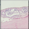
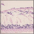
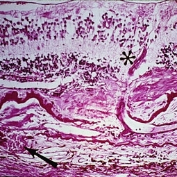
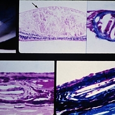
Senile macular degeneration with disciform scar. A retinal arteriole (asterisk) extends into the subretinal component of the scar, through a break in the thickened and detached inner layer of Bruch's membrane, and then into the vascularized intra-Bruch's-membrane component of the scar. Study of serial sections disclosed this retinal vessel to anastomose with the choroidal vessel (arrow) which extends through a branch in Bruch's membrane.
Condition/keywords: Bruch's membrane, disciform scar, macular degeneration, retinal arteriole
-
 Slide 9-20
Feb 26 2019 by Lancaster Course in Ophthalmology
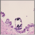
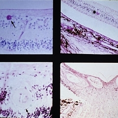
Cholesterol emboli to retina and choroid. There is a large microinfarction of the nerve fiber layer (arrows). A cholesterol embolus is lodged in a retinal arteriole (upper right) proximal to the microinfarction. Cholesterol emboli were also found in the choroid, and in one (lower right) erythrocytes (arrow) could be seen in the periphery as they were presumably going around the embolus.
Condition/keywords: choroid, emboli, embolus, erythrocytes
-
 Slide 9-19
Feb 26 2019 by Lancaster Course in Ophthalmology
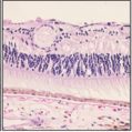
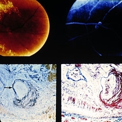
Retinal arterial macroaneurysm. A ring of retinal exudate partially surrounds the macroaneurysm (upper left), which is more clearly delineated by fluorescein (upper right). The retinal arteriole is greatly dilated, and the stain for elastic tissue shows a localized area of disruption and loss of the internal elastic membrane (arrow). The surrounding retina is thickened by edema and some hemorrhage. The ectatic area of the vessel wall is greatly thickened by the accumulation of a laminated fibrinous material. (Courtesy of Alan Friedman, M.D.)
Condition/keywords: retinal arterial macroaneurysm, retinal exudates
-
 Slide 9-18
Feb 26 2019 by Lancaster Course in Ophthalmology
Malignant hypertension with retinal arterioles that are thickened and have fibrinoid necrosis (arrows). Retinal exudates (asterisk) and papilledema are also present. Papilledema is evidenced by fullness of the optic nerve head and peripapillary crowding of the retina (lower right).
Condition/keywords: fibrinoid, malignant hypertension, papilledema, retinal arteriole, retinal exudates
-
 Slide 4-6
Feb 20 2019 by Lancaster Course in Ophthalmology
Optic atrophy. Note the marked narrowing of the retinal arterioles.
Condition/keywords: optic atrophy, retinal arteriole
-
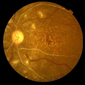
 Primary Hyperoxaluria and Oxalosis
Oct 10 2015 by Hamid Ahmadieh, MD
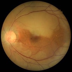
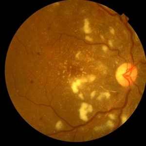
Color fundus photograph of the right eye of a 55-year-old woman with primary hyperoxaluria and oxalosis leading to intraretinal and subretinal deposition of calcium oxalate crystals . In addition, deposition of these crystals in the retinal vessels has led to the occlusion of retinal arterioles and venules leading to multiple cotton wools and dot and blot retinal hemorrhages.
Photographer: shabnam Pooreh, Negah Eye Center, Tehran, Iran
Condition/keywords: color fundus photograph, oxalosis, primary hyperoxaluria
-
 Primary Hyperoxaluria and Oxalosis
Oct 10 2015 by Hamid Ahmadieh, MD
Late venous phase FA image of the right of a 55-year-old woman with primary hyperoxaluria and oxalosis . Notice macular infarction and areas of capillary non -perfusion in retinal mid periphery due to the occlusion of retinal arterioles.
Photographer: Shabnam Pooreh, Negah Eye Center, Tehran, Iran
Condition/keywords: oxalosis, primary hyperoxaluria
-
 Primary Hyperoxaluria and Oxalosis
Oct 10 2015 by Hamid Ahmadieh, MD
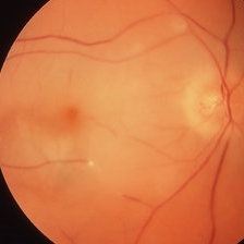
Color fundus photograph of the left eye of a 55-year-old woman with primary hyperoxaluria and oxalosis leading to intraretinal and subretinal deposition of calcium oxalate crystals . In addition, deposition of these crystals in the retinal vessels has led to the occlusion of retinal arterioles and venules leading to multiple cotton wools and dot and blot retinal hemorrhages.
Photographer: Shabnam Pooreh, Negah Eye Center, Tehran, Iran
Condition/keywords: color fundus photograph, oxalosis, primary hyperoxaluria
 Loading…