Search results (38 results)
-
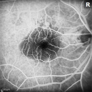
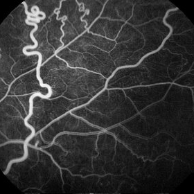
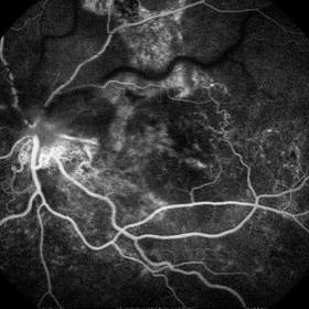
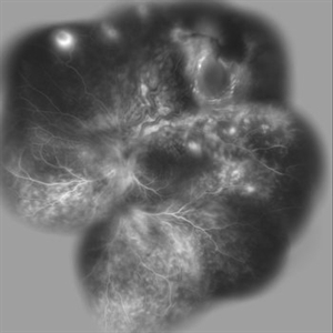
 Retinal angiomatous proliferation (RAP)
Jun 15 2022 by Priyanka Raj, MBBS, MS
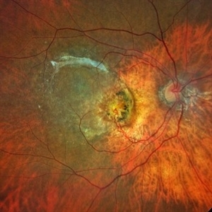
Retinochoroidal anastomosis seen in stage III Retinal angiomatous proliferation (RAP).
Photographer: Sushil Mishra
Imaging device: Zeiss Clarus 500
Condition/keywords: age-related macular degeneration (AMD), retinal angiomatous proliferation (RAP), retinochoroidal anastomosis
-
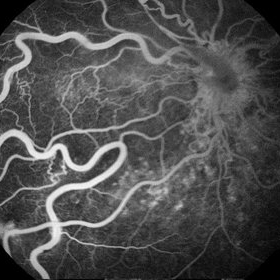
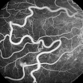
 Retinal Angiomatous Proliferation
May 7 2021 by Dhaivat Shah
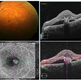
Retinal angiomatous proliferation (RAP) is a distinct variant of neovascular age-related degeneration (AMD) that usually initiates at the retina and progresses posteriorly into sub retinal space. In most recent study, it was suggested that angiogenesis may begin in the retina, choroid, or both, and introduced a new name for the process: Type 3 neovascularization. The frequency of RAP has been studied in many studies, with figures ranging from 10% to 21% of exudative AMD. Clinically, three stages were originally described as intraretinal neovascularization (IRN), subretinal neovascularization (SRN), choroidal neovascularization (CNV). RAP predominantly intraretinal hard exudates, and intra/pre retinal hemorrhages along with intraretinal edema, associated pigment epithelial detachment beneath it, at times retinochoroidal, retino-retinal anastomosis. Apart from conventional OCT, FFA and ICG, OCT-A has now been used primarily as a tool in the diagnosis RAP. Here we present imaging of a 30-year-old young male diagnosed as RAP stage 3 (Type 3 CNVM). Patient was started on intravitreal anti-VEGF monotherapy therapy.
Photographer: Choithram Netralaya Indore
Condition/keywords: retinal angiomatous proliferation (RAP)
-
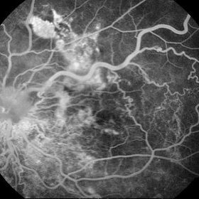
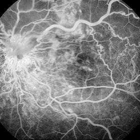
 Retinal Angiomatous Proliferation RAP
Mar 11 2020 by RAFAEL REIS PEREIRA, MD
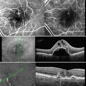
Retinal angiomatous proliferation (RAP) is a unique variant of neovascular age-related macular degeneration. Published studies have estimated that up to 15% of patients with neovascular age-related macular degeneration have RAP. Clinical features frequently associated with RAP include bilateral disease, presence of pigment epithelial detachments, and reticular pseudodrusen. RAP is more frequently associated with the development of retinal pigment epithelial tears and geographic atrophy that can lead to severe vision loss. We present a stereo fluorescein angiography and ICG (upper right and left image respectively) and OCT of left and right eye (middle and inferior image) of a RAP choroidal neovascularization in an 89-year-old patient.
Photographer: Rafael Reis Pereira
Imaging device: HRA Heildelberg Spectralis
Condition/keywords: retinal angiomatous proliferation (RAP)
-
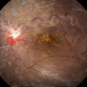
 Retinal Angiomatous Proliferation
Sep 10 2018 by Gabriela Lopezcarasa Hernandez, MD
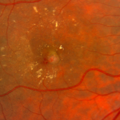
75-year-old patient with decrease in visual acuity right eye with metamorphopsia, in the FA and ICG we can see a RAP lesion.
Photographer: Azucena Rios
Imaging device: Heidelberg Spectralis
Condition/keywords: FA mid phase, indocyanine green (ICG) angiography, RAP lesion, retinal angiomatous proliferation (RAP)
-
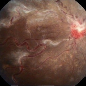
 Retinal Angiomatous Proliferation
Jan 29 2017 by ADRIANO FERREIRA
Fundus photograph of an 56-year-old man with a retinal angiomatous proliferation (RAP). RAP has been known as a variant of exudative age-related macular degeneration (AMD) with a unfavorable prognosis.
Photographer: Laercio
Condition/keywords: vascular anomaly
-
 Stereo View of Retinal Angiomatous Proliferation in Age-Related Macular Degeneration
Jan 21 2016 by James B. Soque, CRA, OCT-C, COA, FOPS
Stereo pair of 75-year-old white male with classic SRN with RAP lesion of right eye, actively receiving anti-VEGF treatment. 50 Degrees, no mag, L and R stereo pair. Single View of OD also visible in this case.
Condition/keywords: age-related macular degeneration (AMD), anti-VEGF, retinal angiomatous proliferation (RAP), stereo pair, subretinal neovascularization (SRNV)
-
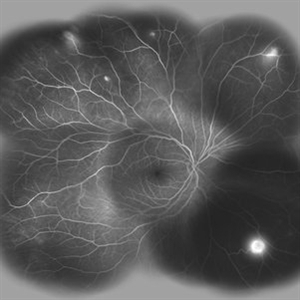
 RAP lesions
Sep 29 2014 by Thomas A. Ciulla, MD, MBA, FASRS
Fluorescein angiogram of an 81-year-old man revealing several RAP lesions superior to fovea.
Photographer: Stuart Alfred CRA
Condition/keywords: choroidal neovascular membrane (CNVM), neovascular age-related macular degeneration (AMD), retinal angiomatous proliferation (RAP), wet age-related macular degeneration (wet AMD)
-
 RAP Lesions
Sep 29 2014 by Thomas A. Ciulla, MD, MBA, FASRS
Fluorescein angiogram of an 81-year-old man revealing several RAP lesions superior to fovea.
Photographer: Stuart Alfred CRA
Condition/keywords: choroidal neovascular membrane (CNVM), neovascular age-related macular degeneration (AMD), retinal angiomatous proliferation (RAP), wet age-related macular degeneration (wet AMD)
-
 RAP lesions
Sep 29 2014 by Thomas A. Ciulla, MD, MBA, FASRS
Fluorescein angiogram of an 81-year-old man revealing several RAP lesions superior to fovea.
Photographer: Stuart Alfred CRA
Condition/keywords: choroidal neovascular membrane (CNVM), neovascular age-related macular degeneration (AMD), retinal angiomatous proliferation (RAP), wet age-related macular degeneration (wet AMD)
-
 RAP lesions
Sep 29 2014 by Thomas A. Ciulla, MD, MBA, FASRS
Late frame fluorescein angiogram of an 81-year-old man revealing several RAP lesions superior to fovea.
Photographer: Stuart Alfred CRA
Condition/keywords: choroidal neovascular membrane (CNVM), neovascular age-related macular degeneration (AMD), retinal angiomatous proliferation (RAP), wet age-related macular degeneration (wet AMD)
-
 RAP lesions
Sep 29 2014 by Thomas A. Ciulla, MD, MBA, FASRS
OCT of an 81-year-old man revealing several RAP lesions superior to fovea.
Photographer: Stuart Alfred CRA
Condition/keywords: choroidal neovascular membrane (CNVM), neovascular age-related macular degeneration (AMD), retinal angiomatous proliferation (RAP), wet age-related macular degeneration (wet AMD)
-
 Racemose Hemangioma and Retinal Vein Occlusion
Mar 5 2013 by Eduardo Torres-Porras, MD
A 13-year-old woman had a history of decreased vision for 2 years. Visual acuity was 20/400.
Photographer: Camelia Rosales Lara
Condition/keywords: retinal angiomatous proliferation (RAP)
-
 Racemose Hemangioma and Retinal Vein Occlusion
Mar 5 2013 by Eduardo Torres-Porras, MD
A 13-year-old woman had a history of decreased vision for 2 years. Visual acuity was 20/400.
Photographer: Camelia Rosales Lara
Condition/keywords: retinal angiomatous proliferation (RAP)
-
 Racemose Hemangioma and Retinal Vein Occlusion
Mar 5 2013 by Eduardo Torres-Porras, MD
A 13-year-old woman had a history of decreased vision for 2 years. Visual acuity was 20/400.
Photographer: Camelia Rosales Lara
Condition/keywords: retinal angiomatous proliferation (RAP)
-
 Racemose Hemangioma and Retinal Vein Occlusion
Mar 5 2013 by Eduardo Torres-Porras, MD
A 13-year-old woman had a history of decreased vision for 2 years. Visual acuity was 20/400.
Photographer: Camelia Rosales Lara
Condition/keywords: retinal angiomatous proliferation (RAP)
-
 Racemose Hemangioma and Retinal Vein Occlusion
Mar 5 2013 by Eduardo Torres-Porras, MD
A 13-year-old woman had a history of decreased vision for 2 years. Visual acuity was 20/400.
Photographer: Camelia Rosales Lara
Condition/keywords: retinal angiomatous proliferation (RAP)
-
 Racemose Hemangioma and Retinal Vein Occlusion
Mar 5 2013 by Eduardo Torres-Porras, MD
A 13-year-old woman had a history of decreased vision for 2 years. Visual acuity was 20/400.
Photographer: Camelia Rosales Lara
Condition/keywords: retinal angiomatous proliferation (RAP)
-
 Racemose Hemangioma and Retinal Vein Occlusion
Mar 5 2013 by Eduardo Torres-Porras, MD
A 13-year-old woman had a history of decreased vision for 2 years. Visual acuity was 20/400.
Photographer: Camelia Rosales Lara
Condition/keywords: retinal angiomatous proliferation (RAP)
-
 Racemose Hemangioma and Retinal Vein Occlusion
Mar 5 2013 by Eduardo Torres-Porras, MD
A 13-year-old woman had a history of decreased vision for 2 years. Visual acuity was 20/400.
Photographer: Camelia Rosales Lara
Condition/keywords: retinal angiomatous proliferation (RAP)
-
 Racemose Hemangioma and Retinal Vein Occlusion
Mar 5 2013 by Eduardo Torres-Porras, MD
A 13-year-old woman had a history of decreased vision for 2 years. Visual acuity was 20/400.
Photographer: Camelia Rosales Lara
Condition/keywords: retinal angiomatous proliferation (RAP)
-
 Racemose Hemangioma and Retinal Vein Occlusion
Mar 5 2013 by Eduardo Torres-Porras, MD
A 13-year-old woman had a history of decreased vision for 2 years. Visual acuity was 20/400.
Photographer: Camelia Rosales Lara
Condition/keywords: retinal angiomatous proliferation (RAP)
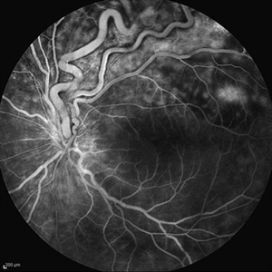
-
 Angiomatous Retinae
Mar 4 2013 by Judy E. Kim, MD, FARVO, FASRS
Fluorescein angiogram of the lesion in inferior retina.
Condition/keywords: retinal angiomatous proliferation (RAP)
-
 Von Hippel-Lindau
Oct 13 2012 by Hamid Ahmadieh, MD
Wide field FA image of the right eye of a 25-year-old woman with retinal angiomatosis (Von Hippel-Lindau). Fundus of the right eye seemed to be normal in ophthalmoscopy.
Photographer: Soodabeh Fooladin, Negah Eye Center, Tehran
Imaging device: Heidelberg Spectralis
Condition/keywords: exudative retinal detachment, retinal angiomatous proliferation (RAP), Von Hippel-Lindau
-
 Von Hippel-Lindau
Oct 13 2012 by Hamid Ahmadieh, MD
Wide field FA image of the left eye of a 25-year-old woman with exudative retinal detachment secondary to retinal angiomatosis (Von Hippel-Lindau).
Photographer: Soodabeh Fooladin, Negah Eye Center, Tehran
Imaging device: Heidelberg Spectralis
Condition/keywords: exudative retinal detachment, retinal angiomatous proliferation (RAP), Von Hippel-Lindau
-
 Von Hippel-Lindau
Oct 13 2012 by Hamid Ahmadieh, MD
Late FA image of the left eye of a 25-year-old woman with exudative retinal detachment secondary to retinal angiomatosis (Von Hippel-Lindau).
Photographer: Soodabeh Fooladin, Negah Eye Center, Tehran
Imaging device: Heidelberg Spectralis
Condition/keywords: exudative retinal detachment, retinal angiomatous proliferation (RAP)
 Loading…