Search results (65 results)
-
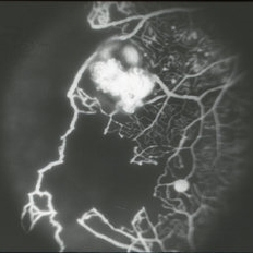
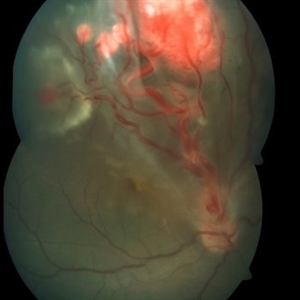
 Imaging Feeder Vessels on OCT-A in a case of Retinal Angiomatous Proliferation
Feb 24 2023 by Dhaivat Shah
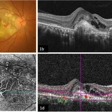
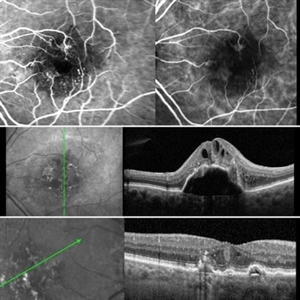
A 58-year-old male patient, a chronic smoker, came to our OPD with complaints of a diminution of vision in the right eye (BCVA: 2/60). On examination, the following findings were observed in the patient. On fundus examination (Image 1a) - Large areas of exudation with multiple superficial and deep hemorrhages at the macula were noted. On SD-OCT imaging (Image 1b) - Multiple intraretinal spaces were seen along with shallow subretinal fluid and hyperreflective dots (indicative of phagocytosed photoreceptors). On the foveal area- a hyperreflective membrane was noted which seemed to dip down and establish a retino-choroidal anastomosis. On OCT-A imaging (Image 1c) - In the ORCC complex, the neovascularization frown, correlating with the membrane complex on the fundus and structural OCT, was visible which was noted to be supplied by small feeder vessels coming from the superior aspect of the fovea. On OCT-A blood flow analysis imaging (Image 1d) - The blood flow analysis showed increased blood flow signals at the level of the membrane (indicative of increased color signal). Based on the findings of the above investigations and clinical examination, the patient was diagnosed with a Case of CNVM Type 3, also described as RAP, and was managed by Anti VEGF injections. This condition usually requires more injections as compared to Type 1 and 2 CNVMs, and the visual prognosis is guarded. Hence, it's very important to counsel the patient before initiating the treatment that the treatment would be long-term and the aim would be preservation of existing vision.
Photographer: Choithram Netralaya, Indore
Condition/keywords: feeder vessel, OCTA, RAP lesion
-
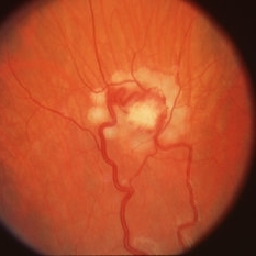
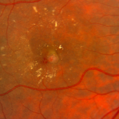
 Retinal angiomatous proliferation (RAP)
Jun 15 2022 by Priyanka Raj, MBBS, MS
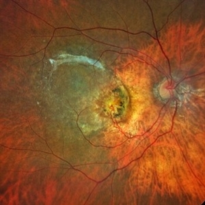
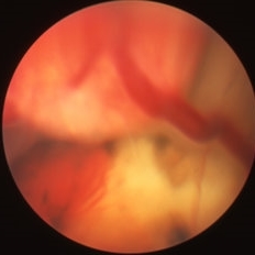
Retinochoroidal anastomosis seen in stage III Retinal angiomatous proliferation (RAP).
Photographer: Sushil Mishra
Imaging device: Zeiss Clarus 500
Condition/keywords: age-related macular degeneration (AMD), retinal angiomatous proliferation (RAP), retinochoroidal anastomosis
-
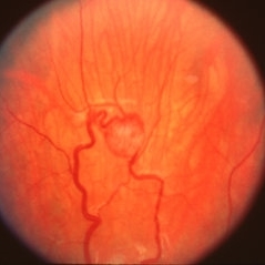
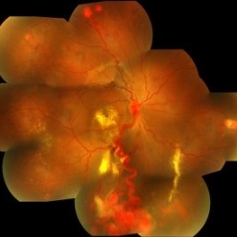
 Retinal Angiomatous Proliferation
May 7 2021 by Dhaivat Shah
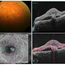
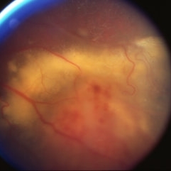
Retinal angiomatous proliferation (RAP) is a distinct variant of neovascular age-related degeneration (AMD) that usually initiates at the retina and progresses posteriorly into sub retinal space. In most recent study, it was suggested that angiogenesis may begin in the retina, choroid, or both, and introduced a new name for the process: Type 3 neovascularization. The frequency of RAP has been studied in many studies, with figures ranging from 10% to 21% of exudative AMD. Clinically, three stages were originally described as intraretinal neovascularization (IRN), subretinal neovascularization (SRN), choroidal neovascularization (CNV). RAP predominantly intraretinal hard exudates, and intra/pre retinal hemorrhages along with intraretinal edema, associated pigment epithelial detachment beneath it, at times retinochoroidal, retino-retinal anastomosis. Apart from conventional OCT, FFA and ICG, OCT-A has now been used primarily as a tool in the diagnosis RAP. Here we present imaging of a 30-year-old young male diagnosed as RAP stage 3 (Type 3 CNVM). Patient was started on intravitreal anti-VEGF monotherapy therapy.
Photographer: Choithram Netralaya Indore
Condition/keywords: retinal angiomatous proliferation (RAP)
-
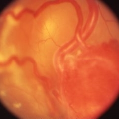
 Angiomatosis Retinae
Jul 22 2020 by MOHIT GUPTA
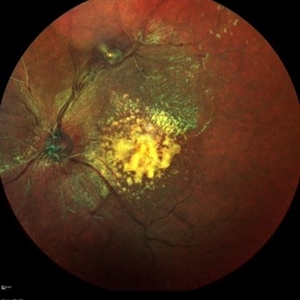
39-year-old female presented with retinal angiomas along superior arcade with macular exudation.
Photographer: Dr Mohit Gupta, Prakash Netra Kendr, Lucknow, India
Imaging device: Heidelberg spectralis Fundus camera
Condition/keywords: angiomatosis retinae
-
 Retinal Angiomatos Proliferans
Jun 10 2020 by Manish Nagpal, MD, FRCS (UK), FASRS
RAP lesion
Photographer: Gayathri Mohan, Retina Foundation
Imaging device: NIDEK SLO MIRANTE
Condition/keywords: RAP lesion
-
 Retinal Angiomatous Proliferation RAP
Mar 11 2020 by RAFAEL REIS PEREIRA, MD
Retinal angiomatous proliferation (RAP) is a unique variant of neovascular age-related macular degeneration. Published studies have estimated that up to 15% of patients with neovascular age-related macular degeneration have RAP. Clinical features frequently associated with RAP include bilateral disease, presence of pigment epithelial detachments, and reticular pseudodrusen. RAP is more frequently associated with the development of retinal pigment epithelial tears and geographic atrophy that can lead to severe vision loss. We present a stereo fluorescein angiography and ICG (upper right and left image respectively) and OCT of left and right eye (middle and inferior image) of a RAP choroidal neovascularization in an 89-year-old patient.
Photographer: Rafael Reis Pereira
Imaging device: HRA Heildelberg Spectralis
Condition/keywords: retinal angiomatous proliferation (RAP)
-
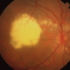
 Coats' Disease - Stage 3A
Aug 21 2019 by Victor M Villegas, MD
Coats' Disease - stage 3A.
Condition/keywords: abnormal retina, Coats' disease, diffuse lipid exudate, edema, foveal hard exudates, pediatic retina, retcam, retinal angioma
-
 Von Hippel-Lindau
Apr 8 2019 by Gary R. Cook, MD, FACS
20-year-old patient with a peripheral retinal angioma OS and dilated feeder vessel secondary to Von Hippel-Lindau.
Condition/keywords: retinal angioma, Von Hippel-Lindau
-
 Retinal Angioma
Apr 8 2019 by Gary R. Cook, MD, FACS
21 year-old male with retinal angioma with exudation OS; VA = 20/40.
Condition/keywords: angioma, exudate, lipid exudation, telangiectatic vessels
-
 Peripapillary Angioma
Apr 8 2019 by Gary R. Cook, MD, FACS
57-year-old white female with a peripapillary angioma OS secondary to Von Hippel-Lindau.
Condition/keywords: retinal angioma, Von Hippel-Lindau
-
 Von Hippel-Lindau
Apr 8 2019 by Gary R. Cook, MD, FACS
Large retinal angioma OS secondary to Von Hippel-Lindau.
Condition/keywords: retinal angioma, Von Hippel-Lindau
-
 Von Hippel-Lindau
Apr 8 2019 by Gary R. Cook, MD, FACS
Fundus photo immediately after laser photocoagulation treatment of the retinal angioma OD in Von Hippel-Lindau.
Condition/keywords: laser photocoagulation, retinal angioma, Von Hippel-Lindau
-
 Von Hippel-Lindau
Apr 8 2019 by Gary R. Cook, MD, FACS
Retinal angioma OD in Von Hippel-Lindau.
Condition/keywords: retinal angioma, Von Hippel-Lindau
-
 Von Hippel-Lindau
Apr 8 2019 by Gary R. Cook, MD, FACS
FA image of capillary non-perfusion and retinal angiomas of Von Hippel-Lindau OS.
Condition/keywords: fluorescein angiogram (FA), Von Hippel-Lindau
-
 Retinal Angioma
Apr 8 2019 by Gary R. Cook, MD, FACS
34-year-old white female with a retinal angioma and heavy lipid exudate secondary to it in the macula of her right eye; V.A. = counting fingers at 8 feet.
Imaging device: Topcon VT-50
Condition/keywords: lipid exudation, retinal angioma
-
 Retinal Cavernous Hemangioma
Nov 30 2018 by Brenda Fallas
2-year-old female with retinal cavernous hemangioma.
Photographer: Brenda Fallas
Imaging device: Retcam 130 lens
Condition/keywords: cluster of grapes, retinal angioma
-
 Retinal Angiomatous Proliferation
Sep 10 2018 by Gabriela Lopezcarasa Hernandez, MD
75-year-old patient with decrease in visual acuity right eye with metamorphopsia, in the FA and ICG we can see a RAP lesion.
Photographer: Azucena Rios
Imaging device: Heidelberg Spectralis
Condition/keywords: FA mid phase, indocyanine green (ICG) angiography, RAP lesion, retinal angiomatous proliferation (RAP)
-
 Von-Hippel Lindau Syndrome
Nov 20 2017 by Nichole Lewis
44-year-old female with Von-Hippel Lindau Syndrome and worsening retinal angioma.
Photographer: Nichole
Condition/keywords: retinal angioma, Von Hippel-Lindau
-
 Von Hippel-Lindau Syndrome
Nov 20 2017 by Nichole Lewis
44-year-old female with Von-Hippel Lindau Syndrome and worsening retinal angioma.
Photographer: Nichole Lewis
Condition/keywords: retinal angioma, Von Hippel-Lindau
-
 Retinal Angiomatous Proliferation
Jan 29 2017 by ADRIANO FERREIRA
Fundus photograph of an 56-year-old man with a retinal angiomatous proliferation (RAP). RAP has been known as a variant of exudative age-related macular degeneration (AMD) with a unfavorable prognosis.
Photographer: Laercio
Condition/keywords: vascular anomaly
-
 Von Hippel-Lindau Syndrome
Mar 12 2016 by Sjakon G Tahija, MD
Fundus photograph of a patient with Von Hipple Lindau Disease and retinal angiomas.
Photographer: Avris Siahaan, Klinik Mata Nusantara
Condition/keywords: Von Hippel-Lindau
-
 Stereo View of Retinal Angiomatous Proliferation in Age-Related Macular Degeneration
Jan 21 2016 by James B. Soque, CRA, OCT-C, COA, FOPS
Stereo pair of 75-year-old white male with classic SRN with RAP lesion of right eye, actively receiving anti-VEGF treatment. 50 Degrees, no mag, L and R stereo pair. Single View of OD also visible in this case.
Condition/keywords: age-related macular degeneration (AMD), anti-VEGF, retinal angiomatous proliferation (RAP), stereo pair, subretinal neovascularization (SRNV)
-
 Retinal Capillary Hemangioblastoma
Oct 6 2015 by Pukhraj P Rishi, MBBS, MS, DO, FRCS, FRCSEd, FASRS, FACS
Fundus photograph of an 18-year-old Asian Indian male with multiple retinal capillary hemangiomas with sub retinal fluid.
Photographer: M S KRISHNA
Imaging device: Zeiss FF4
Condition/keywords: retinal angioma, tumor, Von Hippel-Lindau
-
 Retinal Angiomatosis in a 21-Year-Old Male - 1
Aug 11 2015 by Roy Schwartz, MD
Fundus photograph of a 21-year-old man, who on routine examination was found to have two capillary hemangioblastomas in his left eye. He was diagnosed with retinal angiomatosis.
Photographer: Galit Yair Pur
Condition/keywords: retinal angioma
-
 Retinal Angiomatosis in a 21-Year-Old Male - 2
Aug 11 2015 by Roy Schwartz, MD
Fundus photograph of a 21-year-old man, who on routine examination was found to have two capillary hemangioblastomas in his left eye. He was diagnosed with retinal angiomatosis.
Photographer: Galit Yair Pur
Condition/keywords: retinal angioma
 Loading…