Search results (56 results)
-
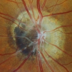
 Collar Button Melanoma
Mar 27 2025 by Virginia Gebhart
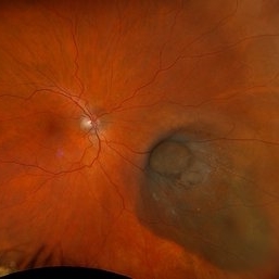
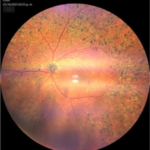
62 year old male with large pigmented lesion with collar button. Pt states he was never aware of any lesion/nevus in the past. Fluid and orange pigment present, appears to be chronic. Pt will be scheduled for brachytherapy pending CT scan results.
Photographer: Virginia Gebhart, Retina Consultants of Carolina
Imaging device: Optos California
Condition/keywords: choroidal melanoma, collar button
-
 Suspicious Lesion 18 Years s/p Iris Resection
Oct 15 2024 by Virginia Gebhart
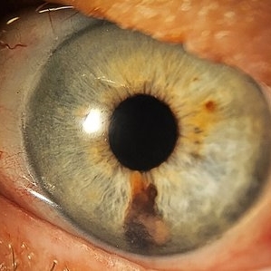
85 year old female with small pigmented lesion present s/p sectoral iridectomy in 2006. Lesion is suspicious for recurrence of melanoma after 18 years. Stable compared to previous exam in March 2024, unclear if this is a new lesion or has been present for an extended time. Will monitor closely.
Photographer: Virginia Gebhart, Retina Consultants of Carolina
Imaging device: Samsung Galaxy
Condition/keywords: iris melanoma, melanoma
-
 Torpedo Retinopathy
Sep 16 2024 by Sriharanathan Poopalaratnam, MD,FRCS
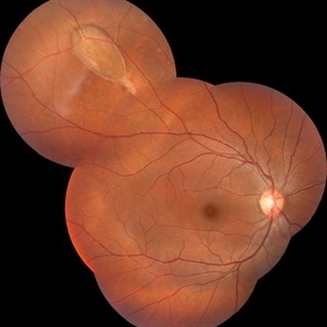
Fundus photograph of a 13-year-old boy asymptomatic accidental finding of unilateral, extramacular oval hypopigmented lesion, with its tip directed toward the central macula, suggestive of a "torpedo" lesion in the nonclassical location.
Photographer: Ms.Samitha
Condition/keywords: torpedo Retinopathy
-
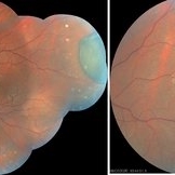
 Multifocal Choroiditis
Jul 13 2024 by Tejaswita Verma
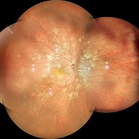
RE fundus montage of a 34 y/o male showing old and active hypopigmented lesions with macular involvement .He presented with DOV since a month,treated with oral steroids for 15 days elsewhere,with BCVA of CF2mt and positive Mantoux test.
Photographer: DR. TEJASWITA VERMA
Imaging device: MIRANTE
Condition/keywords: multifocal choroiditis
-
 Melanocytoma of the Optic Nerve
Apr 6 2024 by Hector Gabriel Moreno Solano, MD, MHA
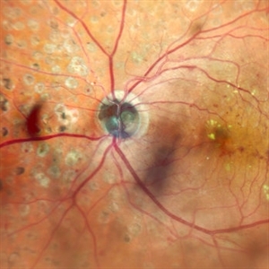
Optic Nerve laser scan image reconstruction of a 57-year-old male presented for an ophthalmological evaluation with a chief complaint of progressive visual loss. Indirect ophthalmoscopy revealed proliferative diabetic retinopathy, without macular edema, and a hyperpigmented lesion at the optic disc which corresponds to a melanocytoma.
Photographer: Héctor Gabriel Moreno-Solano, MD, MHA
Imaging device: Mirante
Condition/keywords: intraocular tumor, macular edema, melanocytoma, optic nerve
-
 Melanocytoma of the Optic Nerve
Apr 6 2024 by Hector Gabriel Moreno Solano, MD, MHA
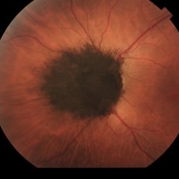
Fundus photograph of a 57-year-old male presented for an ophthalmological evaluation with a chief complaint of progressive visual loss. Indirect ophthalmoscopy revealed proliferative diabetic retinopathy, without macular edema, and a hyperpigmented lesion at the optic disc which corresponds to a melanocytoma.
Photographer: Héctor Gabriel Moreno-Solano
Imaging device: Clarus 700
Condition/keywords: diabetic retinopathy, intraocular tumor, melanocytoma, optic nerve
-
 Iris Melanoma
Feb 1 2024 by Virginia Gebhart
90 year old female with elevated pigmented lesion, amelanotic portion extending toward the angle, questionable vascularity on UBM.
Photographer: Virginia Gebhart
Imaging device: Samsung Galaxy Z Flip
Condition/keywords: iris lesion, iris melanoma
-
 Melanocytoma of Optic Disc
Nov 3 2023 by Virginia Gebhart
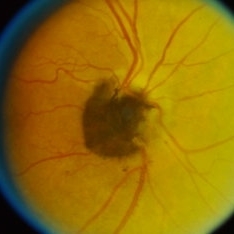
69 year-old female with pigmented lesion that covers the optic nerve. Patient has been aware for over 30 years. Remains stable and unchanged
Photographer: Virginia Gebhart
Imaging device: Topcon
Condition/keywords: benign melanocytoma, Melanocytoma, optic disc melanocytoma
-
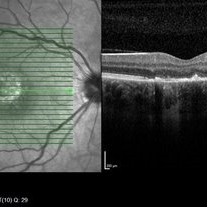
 Melanocytoma of the optic disc
Oct 10 2023 by Navneet Mehrotra, DNB
melanocytoma of the optic nerve head in a 48 year old female diagnosed on routine examination
Photographer: Dharti, Retina Care , Ahmedabad
Imaging device: Nidek RS 330
Condition/keywords: benign pigmented lesion, melanocytoma
-
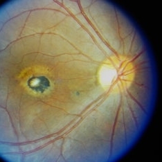
 Optic Disc Melanocytoma
Sep 14 2023 by Ben Serar
Fundus photograph showing hyper pigmented lesion at the optic disc in a case of Optic disc melanocytoma.
Condition/keywords: optic disc melanocytoma
-
 Choroidal Melanocytosis
Sep 14 2023 by Ben Serar
Fundus photograph showing hyper pigmented lesion near the disc, at the level of the choroid, in a case of choroidal melanocytosis.
Condition/keywords: choroidal melanocytosis
-
 Optic Disc Melanocytoma
Sep 14 2023 by Ben Serar
Fundus photograph showing hyper pigmented lesion at the optic disc in a case of Optic disc melanocytoma.
Condition/keywords: optic disc melanocytoma
-
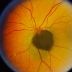
 Macular scar
Sep 14 2023 by Ben Serar
Fundus photograph of RE showing hyper pigmented lesion at the posterior pole indicative of scarring at the macula.
Condition/keywords: macular scar
-
 Optic Disc Melanocytoma
Sep 14 2023 by Ben Serar
Fundus photograph of the LE showing hyper pigmented lesion at the optic disc in a case of Optic disc melanocytoma.
Condition/keywords: optic disc melanocytoma
-
 Sunset Glow Fundus
May 15 2022 by Manuel Ángel Alcántara Delgado, MD
Optomap ultra-widefield retinal imaging of an 35-year-old woman showed sunset glow fundus, multiple nummular chorioretinal atrophic lesions, macular subretinal fibrosis and pigment clumping in chronic recurrent stage of Vogt-Koyanagi-Harada disease.
Photographer: Manuel Ángel Alcántara Delgado. Conde de Valenciana.
Condition/keywords: abnormal retina, benign pigmented lesions, pigment clumps, retinal fibrosis, uveitis, Vogt-Koyanagi-Harada
-
 CHRPE
CHRPE
Jan 15 2021 by Priya Rasipuram Chandrasekaran, MBBS, DO, DNB, FRCS
This is the fundus photo and fundus photo montage of the left eye of a 25-year-old male showing flat, solitary, round, greyish pigmented lesion situated AT THE equator with a scalloped margin. Vessels overlying the lesion are normal and there is a clear demarcation line between this and normal retina. The margins are hypopigmented with few hypopigmented lacunae inside.
Condition/keywords: congenital hypertrophy of the retinal pigment epithelium (CHRPE)
-
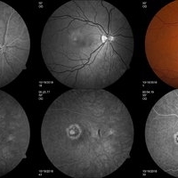
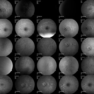 Unilateral Acute Idiopathic Maculopathy FA
May 7 2019 by William Ensor
A 37-year-old female presented with a two-week history of vision loss in the right eye. She experienced a flu-like illness including rash on the hands, feet, and mouth 2 days prior to her vision change. Her 3-year-old son had a similar illness diagnosed as hand, foot, and mouth disease by his pediatrician one week prior. Her visual acuity was 20/150 of the right eye, and 20/20 of the left eye. On dilated fundus examination, the left eye was unremarkable; the right eye revealed a circular, variably pigmented lesion of the macula. OCT imaging showed areas of RPE loss and clumping, with overlying loss of the photoreceptor layer. Fluorescein angiography showed central and peripheral hyperfluorescence consistent with window defect, and blockage in area of RPE loss. No treatment was initiated at this time. The patient returned 10 days later; her visual acuity improved to 20/50 in the right eye. Dilated fundus exam showed increased pigmentation of the macular lesion. OCT of the right eye showed further RPE clumping without recovery of the photoreceptor layer, despite her improved visual acuity.
Condition/keywords: unilateral acute idiopathic maculopathy
-
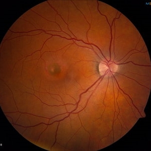 Unilateral Acute Idiopathic Maculopathy Fundus
May 7 2019 by William Ensor
A 37-year-old female presented with a two-week history of vision loss in the right eye. She experienced a flu-like illness including rash on the hands, feet, and mouth 2 days prior to her vision change. Her 3-year-old son had a similar illness diagnosed as hand, foot, and mouth disease by his pediatrician one week prior. Her visual acuity was 20/150 of the right eye, and 20/20 of the left eye. On dilated fundus examination, the left eye was unremarkable; the right eye revealed a circular, variably pigmented lesion of the macula. OCT imaging showed areas of RPE loss and clumping, with overlying loss of the photoreceptor layer. Fluorescein angiography showed central and peripheral hyperfluorescence consistent with window defect, and blockage in area of RPE loss. No treatment was initiated at this time. The patient returned 10 days later; her visual acuity improved to 20/50 in the right eye. Dilated fundus exam showed increased pigmentation of the macular lesion. OCT of the right eye showed further RPE clumping without recovery of the photoreceptor layer, despite her improved visual acuity.
Condition/keywords: unilateral acute idiopathic maculopathy
-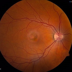
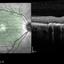 Unilateral Acute Idiopathic Maculopathy OCT Macula
May 7 2019 by William Ensor
A 37-year-old female presented with a two-week history of vision loss in the right eye. She experienced a flu-like illness including rash on the hands, feet, and mouth 2 days prior to her vision change. Her 3-year-old son had a similar illness diagnosed as hand, foot, and mouth disease by his pediatrician one week prior. Her visual acuity was 20/150 of the right eye, and 20/20 of the left eye. On dilated fundus examination, the left eye was unremarkable; the right eye revealed a circular, variably pigmented lesion of the macula. OCT imaging showed areas of RPE loss and clumping, with overlying loss of the photoreceptor layer. Fluorescein angiography showed central and peripheral hyperfluorescence consistent with window defect, and blockage in area of RPE loss. No treatment was initiated at this time. The patient returned 10 days later; her visual acuity improved to 20/50 in the right eye. Dilated fundus exam showed increased pigmentation of the macular lesion. OCT of the right eye showed further RPE clumping without recovery of the photoreceptor layer, despite her improved visual acuity.
Condition/keywords: unilateral acute idiopathic maculopathy
-
 Iris Pigmented Lesion
Apr 27 2018 by Mark Lazcano
Gonio photograph of 20-year-old male with pigmented iris lesion consistent with melanocytoma
Photographer: mark Lazcano,University of Miami , Bascom Palmer Eye Institute
Imaging device: gonio Prism
Condition/keywords: pigmented lesion
-
 Retinal hyperplasia
Feb 19 2018 by JEFFERSON R SOUSA, Tecg.º (Biomedical Systems Technology)
Female patient, 28 years in monitoring to control a hyperpigmented lesion in the temporal retina of the right eye.
Photographer: Photographer JEFFERSON ROCHA DE SOUSA, Clinic Dr. Marco Antonio Albhy Oftalmology, Institute Dr. Suel Abujamra São Paulo-Brazil
Imaging device: Retinografo Topcin TRC-NW6S. Mosaic, Flash 25.
Condition/keywords: hyperplasia, hyperplastic retinal pigment epithelium (RPE)
 Loading…