Search results (135 results)
-
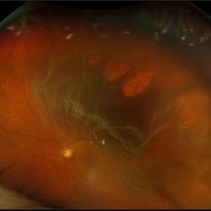
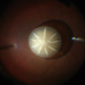 Dislocated Cataractous Lens
Jun 19 2025 by Mrinali Gupta, MD, FASRS
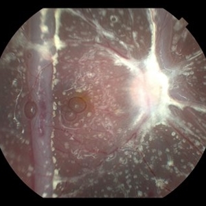
Intraoperative image of a chronically dislocated cataractous lens. The patient underwent pars plana vitrectomy, lensectomy, and placement of an anterior chamber intraocular lens, with improvement in vision from Count Fingers to 20/20 without correction.
Photographer: Mrinali Gupta, MD
Imaging device: Intraoperative surgical video (Zeiss Lumera scope, Resight lens)
Condition/keywords: dislocated crystalline lens
-
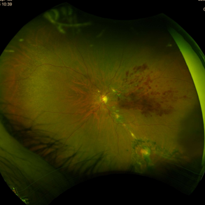
 Inadvert Globe Perfuration After Peribulbar Block
Mar 13 2025 by Bruno B Ribeiro
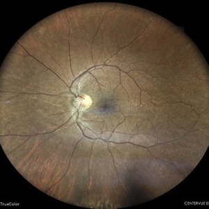
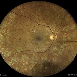
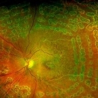
Fundus photograph of a 74-year-old woman who underwent pars plana vitrectomy OS due to rhegmatogenous retinal detachment. A horseshoe retinal tear can be seen at 5h. Intraoperative evaluation revealed a chorioretinal scar with the shape of the needle track at the same location. Despite rare, globe perfuration after peri or retrobulbar block may happen, even by the most experienced anesthesiologist.
Photographer: Bruno Barbosa Ribeiro, Angelina Meireles
Imaging device: Optos California
Condition/keywords: retinal detachment
-
 Inadvert Globe Perfuration After Peribulbar Block
Mar 13 2025 by Bruno B Ribeiro
Fundus photograph of a 74-year-old woman who underwent pars plana vitrectomy OS due to rhegmatogenous retinal detachment. A horseshoe retinal tear can be seen at 5h. Intraoperative evaluation revealed a chorioretinal scar with the shape of the needle track at the same location. Despite rare, globe perfuration after peri or retrobulbar block may happen, even by the most experienced anesthesiologist.
Photographer: Bruno Barbosa Ribeiro, Angelina Meireles
Imaging device: Optos California
Condition/keywords: hemmorhage
-
 Persistent Fetal Vasculature
Sep 23 2024 by Carlos Augusto Moreira, MD, PhD
Persistent Fetal Vasculature - an intraocular cotton swab appearance.
Photographer: Carlos Augusto Moreira-Neto, Hospital de Olhos do Paraná
Imaging device: NGENUITY Visualization System
Condition/keywords: pars plana vitrectomy (PPV), persistent fetal vasculature (PFV)
-
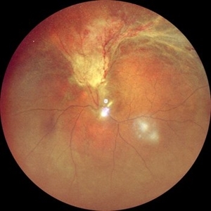 RD with PVR in CMV Retinitis in an HIV Positive Patient
Jul 31 2024 by Tejaswita Verma
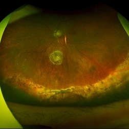
Fundus photograph of a 48 year old male with CF 1.5 mt vision having history of CMV retinitis, on HAART with CD4 count 81, showing retinal detachment with proliferative vitreoretinopathy changes. He was advised pars plana vitrectomy with silicon oil infusion.
Photographer: DR. TEJASWITA VERMA
Imaging device: MIRANTE
Condition/keywords: CMV retinitis with retinal detachment, HIV
-
 RPE-Transplantation
Jul 25 2024 by Gabriel Costa Andrade, PhD
Postoperative period of RPE-transplantation in a patient with neovascular AMD after RPE tear.
Photographer: Gabriel Andrade
Condition/keywords: neovascular age-related macular degeneration (AMD), pars plana vitrectomy (PPV), wet age-related macular degeneration (wet AMD)
-
 Status Post Pars Plana Vitrectomy With Silicone Oil Infusion Followed by Silicone Oil Removal
Jun 17 2024 by Akansha Sharma
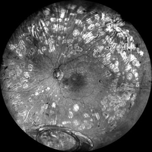
Color fundus photograph of a 47 year old male with retina on status post pars plana vitrectomy with silicone oil infusion followed by silicone oil removal.
Photographer: Dr. Akansha Sharma, Bharati Eye Hospital
Condition/keywords: RD SURGERY, silicone oil
-
 What Lies Beneath
Jun 14 2024 by SHISHIR VERGHESE, MS, FVRS, FAICO (Retina)
Grey color fundus photograph of the left eye of a 78 year old gentleman who has undergone pars plana vitrectomy for proliferative diabetic retinopathy, shows dislocated intraocular lens bag complex lying on the inferior retina
Photographer: SHISHIR VERGHESE
Condition/keywords: dislocated intraocular lens (IOL), dislocated IOL, proliferative diabetic retinopathy (PDR)
-
 Fish Hook Eye Trauma
Jun 12 2024 by Miguel Brito, MD, FASRS
Fundus photograph of a 15-year-old boy post cataract aspiration, pars plana vitrectomy, suprachoroidal drainage, and retinal reattachment surgery secondary to traumatic endophthalmitis.
Photographer: Miguel Brito
Condition/keywords: endophthalmitis, PFCL, Retinal detachment under Silicon Oil, retinal fold
-
 Rhegmatogenous Macula Off Retinal Detachment with Multiple Breaks
May 29 2024 by Alexis Singstock
Ultra widefield fundus photograph of a 66 year old male with rhegmatogenous macula off retinal detachment with multiple breaks. Patient presented emergently for a curtain/veil in inferonasal visual field. Patient reports the curtain/veil in left eye started about 1 week prior, yet denied seeing flashes and floaters. Patient's vision was hand motion. Dr. Edward Korot examined the patient and scheduled him for a scleral buckle along with pars plana vitrectomy surgery.
Photographer: Alexis Singstock, Retina Specialists of Michigan
Imaging device: Optos California
Condition/keywords: fundus photography, left eye, macula off retinal detachment, OPTOS CALIFORNIA, pars plana vitrectomy (PPV), rhegmatogenous retinal detachment, scleral buckle, ULTRA WIDE FIELD
-
 Status Post Pars Plana Vitrectomy in a Case of Vitreous Hemorrhage in Proliferative Diabetic Retinopathy
May 7 2024 by Akansha Sharma
Color fundus photograph of a 39 year old female with stable retina status post pars plana vitrectomy in a case of vitreous hemorrhage in proliferative diabetic retinopathy.
Photographer: Dr. Akansha Sharma, Bharati Eye Hospital
Condition/keywords: PDR, POST SURGERY, proliferative diabetic retinopathy (PDR)
-
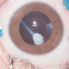
 Silicone Oil in the Anterior Chamber
Mar 27 2024 by Emma Paulina Carrillo Haro
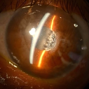
Anterior chamber photograph of a 67-year-old male with silicone oil in the anterior chamber after pars plana vitrectomy (PPV) and silicone oil injection into the vitreous cavity.
Photographer: Emma Paulina Carrillo Haro
Imaging device: Iphone
Condition/keywords: anterior chamber, silicone oil
-
 Subretinal Gas After Pneumatic Retinopexy
Mar 6 2024 by James P Dossett, MD
Pseudocolor fundus photograph of a 68-year-old man who presented with a macula-on rhegmatogenous retinal detachment with a single horseshoe tear at 12 o'clock. Pneumatic retinopexy was performed with cryopexy to the retinal break. He returned to clinic three days later and the entire SF6 gas bubble was noted to have migrated to the subretinal space through the retinal break. Pars plana vitrectomy was performed that day with retinal reattachment and improvement in vision to 20/40 now 6 months postoperatively.
Imaging device: Optos
Condition/keywords: pneumatic retinopexy, subretinal gas bubble
-
 Posteriorly Dislocated Intraocular Lens
Feb 22 2024 by Nikhil K Bommakanti, MD
A woman in her seventies with a history of retinal detachment repair presented with poor vision and was found to have a posteriorly dislocated intraocular lens. She subsequently underwent pars plana vitrectomy, intraocular lens extraction, and scleral-fixated intraocular lens implantation.
Condition/keywords: dislocated intraocular lens (IOL), dislocated posterior chamber intraocular lens (PCIOL)
-
 Serous Retinal Detachment in Advanced Proliferative Diabetic Retinopathy
Feb 15 2024 by Annaka Gooding
Ultra-Wide fundus photograph of a 29 year old female with a Serous Retinal Detachment in Advanced PDR. Patient present to clinic with LP vision following PPV and fill in PRP. Physician recommended oral prednisone treatment and to reassess at their following visit.
Photographer: Annaka Gooding, CPO
Imaging device: Optos California RGB
Condition/keywords: Diabetes, diabetic macular edema, fundus photography, OPTOS CALIFORNIA, pan-retinal photocoagulation (PRP), pars plana vitrectomy (PPV), proliferative diabetic retinopathy (PDR), serous retinal detachment, ultra-wide field imaging
-
 Dislocated Lens, Posterior OD
Jan 26 2024 by Corey Grant
OPTOS California photo presents a 71 year old male patient with a dislocated lens, posterior in the right eye. Presented on 1/26/24 with posteriorly dislocated SN60WF with a Soemmerring ring. Associated retinal hemorrhage within retinoschisis as well. This will result in a PPV/IOL exchange/SFIOL/STK for the right eye.
Photographer: Corey Grant, Ophthalmic Imager, Retina Specialist of Michigan
Imaging device: OPTOS California
Condition/keywords: color photo, IOL, OD, Optos, OPTOS CALIFORNIA, pars plana vitrectomy (PPV), retina
-
 Macrocysts in Kickboxer
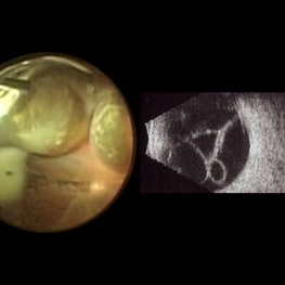
Nov 17 2023 by Bradley T. Smith, MD, FASRS
Intraoperative photo and preoperative b scan of chronic retinal detachment with macrocysts in a kickboxer
Condition/keywords: B scan ultrasound, chronic retinal detachment, ocular trauma, pars plana vitrectomy (PPV), retinal macrocyst
-
 Before and After Vitrectomy
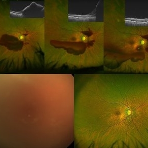
Nov 17 2023 by Bradley T. Smith, MD, FASRS
Middle age male diabetic retinopathy and resolving exudate following repair of tractional detachment with membrane peeling.
Condition/keywords: coats-like response, Diabetes, fibrotic neovascularization, fibrovascular proliferation, pars plana vitrectomy (PPV), proliferative diabetic retinopathy (PDR), tractional retinal detachment
-
 Chronic Full Thickness Macular Hole
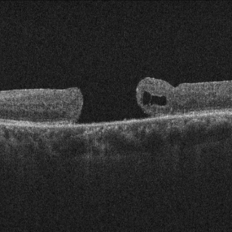
Oct 25 2023 by Jessica Hampton, BS
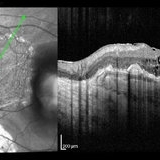
Optical-coherence tomography image of a 65-year old woman with a chronic full-thickness macular hole in the left eye, recurred following three attempts at repair with pars plana vitrectomy, membrane peel, and gas tamponade.
Photographer: Dr. Diana Do, Stanford Medicine, Byers Eye Institute
Condition/keywords: full thickness macular hole, optical coherence tomography (OCT)
-
 Ectopia Lentis
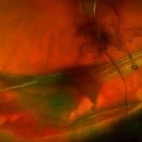
Oct 8 2023 by Nassim Alejandro Abreu Arbaje, MD
Frame capture of a 3 year old boy during pars plana vitrectomy and lensectomy due to congenital ectopia lentis
Photographer: Nassim Abreu-Arbaje
Imaging device: Alcon NGenuity systems
Condition/keywords: ectopia lentis, lensectomy, pars plana vitrectomy (PPV)
-
 Vitreoretinal Traction with Adjacent Tear and Vitreous Hemorrhage
Oct 3 2023 by Alexis Singstock
Ultra-widefield fundus photograph of a 76 year old woman with vitreoretinal traction, an adjacent retinal tear and vitreous hemorrhage affecting the left eye. Patient was referred for retinal detachment and vitreous hemorrhage. Patient reports waking up the day prior to their appointment with "a lot of lines coming down the front, like swirling dirt in the left eye". Patient's vision was counting fingers at 1 ft. Dr. Joseph Boss noticed a horseshoe tear inferior to traction on exam and with the help of ultra-widefield imaging. Dr. Boss performed laser retinopexy to tear and impending tear at site of traction. Patient is scheduled for pars plana vitrectomy for dense vitreous hemorrhage.
Photographer: Alexis Singstock
Imaging device: Optos California
Condition/keywords: acute posterior vitreous detachment, fundus photography, left eye, Optos, OPTOS CALIFORNIA, pseudocolor, ULTRA WIDE FIELD, vitreoretinal traction, vitreous hemorrhage
-
 Ruptured retinal artery macroaneurysm
Sep 6 2023 by PRATIK SHENOY, MBBS, DNB, FVRS
A 66-year-old female presented with a ruptured retinal artery macroaneurysm and a visual acuity of finger counting close to face. The multi layered hemorrhage receded on its own inferiorly with an improvement of visual acuity. However, the patient developed a breakthrough bleed with vitreous hemorrhage three weeks later with a drop in visual acuity to hand movements. She underwent pars plana vitrectomy for the same with an improvement in visual acuity to 6/9.
Photographer: Gaurav Kamble, Isha Netralaya
Imaging device: Optos
Condition/keywords: OCT, Optos, pars plana vitrectomy (PPV), retinal arterial macroaneurysm, vitreous hemorrhage
-
 Endolaser in Status-Post Vitrectomy
Aug 28 2023 by Aditya S Kelkar, MS, FRCS, FASRS,FRCOphth
Endolaser in Status-Post Vitrectomy.
Photographer: Optom Komal Jangam, National Institute of Ophthalmology, Pune, India.
Imaging device: OPTOS DAYTONA
Condition/keywords: endolaser, pars plana vitrectomy (PPV), vitrectomy
-
 Intra-op picture of SFIOL explantation with pars plana bleed
Aug 23 2023 by Harsh Vardhan Singh, MS
45-old-male with history of trauma & subluxated SFIOl - undergone IOL explantation & repeat SFIOL. Image showing pars plana bleed during IOL explantation
Photographer: Harsh Vardhan Singh
Condition/keywords: intraoperative, IOL explantation
-
 Microspherophakia
Jun 29 2023 by Ethan K Sobol, MD
A 23-year-old male with no past medical history presented with elevated intraocular pressure in the right eye secondary to pupillary block, unresponsive to maximum medical therapy. In the right eye, there was zonular laxity, as well as a spherical appearance of the crystalline lens with a small horizontal diameter and an elongated anteroposterior diameter in both eyes. A diagnosis of microspherophakia was made, and the patient was scheduled for pars plana vitrectomy and lensectomy of the right eye. The image shows a small spherical lens with a clearly visible lens equator, immediately prior to surgical removal.
Condition/keywords: lensectomy, microspherophakia, pars plana vitrectomy (PPV), pupillary block, secondary glaucoma
 Loading…