Search results (48 results)
-
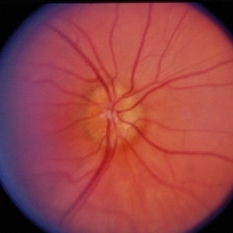
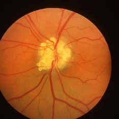
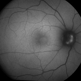
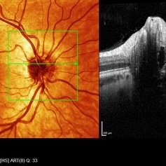 Right eye SD- OCT-RNFL of optic nerve head drusen showing hypo reflective centre with hyper reflective margins.
Aug 5 2022 by Kavitha Duraipandi, MD DNB FICO FRCS
A 20 year old patient referred to the clinic with blurred disc margins to rule out papilledema.
Photographer: Natalie Fox- Bussell
Condition/keywords: optic nerve drusen, optical coherence tomography (OCT)
-
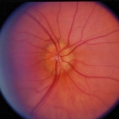
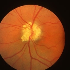
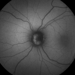
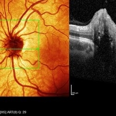 Left eye SD- OCT-RNFL of optic nerve head drusen showing hypo reflective centre with hyper reflective margins.
Aug 5 2022 by Kavitha Duraipandi, MD DNB FICO FRCS
A 20 year old patient referred to the clinic with blurred disc margins to rule out papilledema.
Photographer: Natalie Fox- Bussell
Condition/keywords: optic nerve drusen, optical coherence tomography (OCT)
-
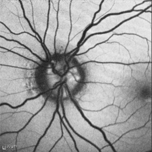
 Optic Nerve Head Drusen
Feb 9 2018 by Olivia Rainey
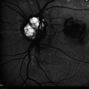
Fundus autofluorescence of a 49-year-old female with optic nerve head drusen affecting her left eye. The patient has pseudoxanthoma elasticum with choroidal neovascularization and has been receiving anti-VEGF treatment for many years.
Photographer: Olivia Rainey
Imaging device: Heidelberg Spectralis
Condition/keywords: 30 degrees, anti-VEGF, choroidal neovascularization (CNV), fundus autofluorescence (FAF), Heidelburg Spectralis, left eye, optic disc, optic nerve drusen, pseudoxanthoma elasticum (PXE)
-
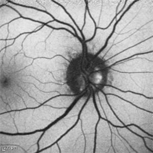
 Optic Nerve Head Drusen With Idiopathic CNV
Feb 17 2017 by Kristen Wagner
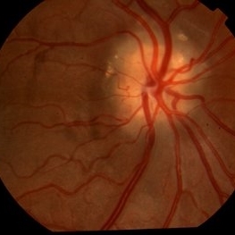
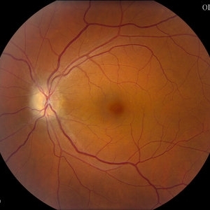
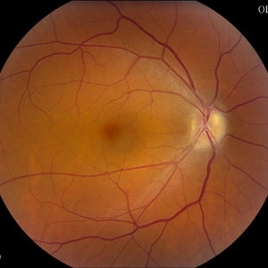
22-year-old female fundus photograph of a right eye with Optic Nerve Drusen with Idiopathic CNV.
Photographer: Kristen Wagner, COT, OSC Ophthalmic Photographer, Tennessee Retina, Nashville TN
Condition/keywords: choroidal neovascularization (CNV), drusen of optic disc, optic disc drusen
-
 Optic Disc Drusen
Jul 31 2016 by Mitzy E Torres Soriano, MD
Optic Disc Drusen (Right eye)
Photographer: Mitzy E. Torres Soriano. Retina Department. Hospital Provincial del Centenario. Rosario, Argentina
Imaging device: TOPCON
Condition/keywords: optic disc drusen, optic nerve drusen
-
 ON Drusen
Dec 11 2014 by H. Michael Lambert, MD
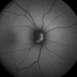
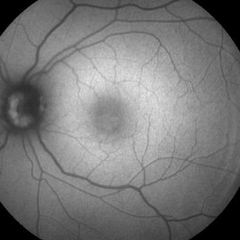
Optic Nerve Drusen - right eye
Condition/keywords: optic nerve drusen
-
 ON Drusen
Dec 11 2014 by H. Michael Lambert, MD
Optic Nerve Drusen - right eye
Condition/keywords: optic nerve drusen
-
 ON Drusen
Dec 11 2014 by H. Michael Lambert, MD
Optic Nerve Drusen - right eye
Condition/keywords: optic nerve drusen
-
 Optic Nerve Drusen Autofluorescence
Nov 28 2014 by Thomas A. Ciulla, MD, MBA, FASRS
This 55-year-old man was noted to have optic nerve drusen in each eye, which autofluoresce prominently.
Photographer: Thomas Steele
Condition/keywords: optic nerve drusen
-
 Optic Nerve Drusen Autofluorescence
Nov 28 2014 by Thomas A. Ciulla, MD, MBA, FASRS
This 55-year-old man was noted to have optic nerve drusen in each eye, which autofluoresce prominently.
Photographer: Thomas Steele
Condition/keywords: optic nerve drusen
-
---thumb.jpg/image-square;max$300,300.ImageHandler) Extruded ONH Drusen
Extruded ONH Drusen
Apr 4 2014 by H. Michael Lambert, MD
Extruded ONH Druse
Photographer: Donald Lowd
Condition/keywords: optic nerve drusen
-
 Optic Nerve Head Drusen - Red Free
Oct 5 2013 by Roy Schwartz, MD
Optice nerve head drusen in a right eye, red-free image.
Photographer: Galit Yair-Pur
Condition/keywords: disc drusen, drusen of optic disc, optic nerve drusen, red-free
-
 Optic Nerve Head Drusen - Fundus Image
Oct 5 2013 by Roy Schwartz, MD
Optice nerve head drusen in a right eye, fundus image.
Photographer: Galit Yair-Pur
Condition/keywords: disc drusen, drusen of optic disc, optic nerve drusen, red-free
-
 Optic Nerve Head Drusen
Oct 2 2013 by Jerald A. Bovino, MD
There are optic nerve head drusen. These are also visualized on fundus autofluorescence and B-scan ultrasonography.
Condition/keywords: optic nerve drusen
-
 Optic Nerve Head Drusen
Oct 2 2013 by Jerald A. Bovino, MD
There are optic nerve head drusen. These are also visualized on fundus autofluorescence and B-scan ultrasonography.
Condition/keywords: optic nerve drusen
-
 Optic Nerve Drusen
Jul 18 2013 by Jason S. Calhoun
Asymptomatic patient with 20/20 vision in both eyes. Fundus photo shows optic nerve drusen verified by FAF photos.
Photographer: Jason S. Calhoun, Department of Ophthalmology, Mayo Clinic Jacksonville, Florida
Imaging device: TOPCON TRC 50-EX
Condition/keywords: optic nerve drusen
-
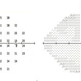 Optic Disc Drusen
Mar 27 2013 by Henry J. Kaplan, MD
Perimetry demonstrates slightly enlarged blind spot in the same patient #5.
Condition/keywords: drusen of optic disc, optic disc drusen, optic nerve drusen
-
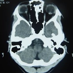 Orbital CT Scan in Optic Nerve Drusen
Mar 27 2013 by Henry J. Kaplan, MD
Axial CT scan of orbit demonstrates high density spot on optic nerve head on both sides #4.
Condition/keywords: drusen of optic disc, optic disc drusen, optic nerve drusen
 Loading…