Search results (11 results)
-
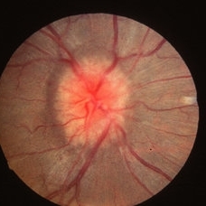
 A Vein in Vain: Ischemic CRVO
Dec 6 2024 by Jasmeet Kaur Chandi
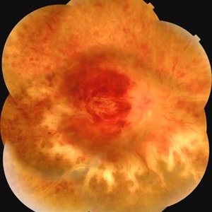
Fundus photo of a 55 year-old female with extensive flame-shaped and dot-blot hemorrhages in all four quadrants. Tortuous and dilated veins with cotton-wool spots. Optic disc swelling with hyperemia and macular edema.
Photographer: Dr. Jasmeet Kaur Chandi
Condition/keywords: Ischemic Central Retinal Vein Occlusion
-
 Multimodal Imaging for Differentiating Unilateral Pseudo Optic Disc Swelling(Buried Drusen) From True Optic Disc Swelling
Feb 7 2024 by Fawwaz F Al Mamoori, MD, Medical Retina Consultant
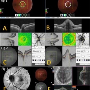
27-year-old male, medically free, presented with left unilateral optic disc swelling. BCVA=1.0(OU), color vision, and contrast sensitivity were normal (OU)with no RAPD in the left eye. Swept Source OCT: showed elevated left optic disc with hyporeflective mass (Fig-1 B). Enface OCT: Showed left peripapillary multiple ovoid mass lesions(drusen) (Fig-2 d, Fig3 F). FAF: of the left eye showed superonasal hyper autofluorescent drusenoid lesions)(Fig3 E). Orbital MRI with contrast was requested to exclude any compressive lesions like tumors(menigioma)or inflammatory lesions like granuloma(sarcoid granuloma). orbital MRI result was normal.
Photographer: Hana.S.Owais
Imaging device: TRITON(TOPCON,Swept Source OCT)
Condition/keywords: fundus autofluorescence (FAF), multimodal imaging, OCT EN FACE, optic disc drusen, optic disc edema, swept source
-
 Multimodal Imaging for Differentiating Unilateral Pseudo Optic Disc Swelling(Buried Drusen) From True Optic Disc Swelling
Feb 7 2024 by Fawwaz F Al Mamoori, MD, Medical Retina Consultant
A 27-year-old male patient, medically free, presented with unilateral left optic disc swelling. BCVA=1.0(OU), color vision, and contrast sensitivity were normal (OU) with no RAPD in the left eye. SS-OCT: showed left optic disc elevation with hyporeflective mass lesion (Fig-1 B). Enface OCT: showed left peripapillary hyperreflective ovoid mass lesions(Fig-2 D, Fig-3 F), FAF: showed left superonasal hyperautofluorescent drusenoid lesions. Orbital MRI with contrast was requested to exclude any optic nerve compressive lesions like (tumors: like mengioma or inflammatory lesions like granuloma (sarcoidosis). the result of orbital MRI was normal.
Photographer: Hana.S.Owais
Imaging device: TRITON(TOPCON,Swept Source OCT)
Condition/keywords: fundus autofluorescence (FAF), multimodal imaging, OCT EN FACE, optic disc drusen, optic disc edema
-
 Non invasive multimodal imaging for differentiating unilateral pseudo swelling buried optic disc drusen from true optic disc swelling
Feb 7 2024 by Fawwaz F Al Mamoori, MD, Medical Retina Consultant
27-year-old male, medically free, routine fundus examination showed left optic dic swelling, BCVA =1.0(OU), color vision, and contrast sensitivity were normal with no RAPD (OU). SS-OCT of the left optic disc showed a hyporeflective mass. Enface OCT shadogram showed peripapillary ovoid structures (drusen).FAF: showed drusenoid autofluorescence in the superonasal part only. Orbital MRI with contrast was requested to exclude any optic nerve tumor and it was normal.
Photographer: Hana.S.Owais
Imaging device: TRITON(OCT) Topcon
Condition/keywords: multimodal imaging, optic disc drusen, optic disc swelling
-
 Central Retinal Vein Occlusion
Dec 22 2018 by FELIPE PEREIRA
25-year-old male patient with acute and painless vision loss of left eye. The fundus examination demonstrate optic disc swelling, venous tortuosity, diffuse intraretinal hemorrhage and severe macular edema. There is also extensive exudative retinal detachment with lipid deposits in the posterior pole, mainly around the vessels. The systemic work up was negative, including serologies, rheumatologic and hematological markers and cholesterol and triglycerides within normal limits.
Photographer: Felipe Pereira
Imaging device: Vizucan, Zeiss
Condition/keywords: central vein occlusion, ischemic CRVO
-
 Diabetic Retinopathy Optic Nerve Edema, Fluorescein Angiogram, Stereo
Apr 11 2015 by James B. Soque, CRA, OCT-C, COA, FOPS
Optic Nerve Edema and Leakage on fluorescein angiography in this 48-year-old patient with a 10 year history of diabetes. 50 degree stereo photo fluorescein angiogram.
Photographer: James B. Soque, CRA, COA
Imaging device: Topcon TRC 50 DX, OIS 5 MP Digital Camera, MERGE Software
Condition/keywords: background diabetic retinopathy (BDR), diabetes, disc leakage, fluorescein leakage, optic disc swelling, optic nerve edema, stereo pair
-
 Papillitis
May 2 2013 by Henry J. Kaplan, MD
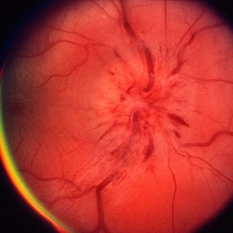
Anterior optic neuropathy or papillitis in the right eye; notice the blurred optic disc margin, engorged capillaries and flame shaped hemorrhages at the margin.
Condition/keywords: optic disc edema, optic disc swelling, papillitis
-
 Papilledema
May 2 2013 by Henry J. Kaplan, MD
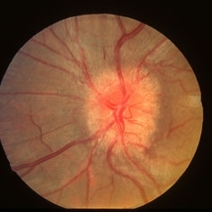
Optic disc swelling due to RICP . Left eye; #2.
Condition/keywords: disc swelling, papilledema, raised intracranial pressure (RICP)
-
 Papilledema
May 2 2013 by Henry J. Kaplan, MD
Optic disc swelling due to RICP. Right Eye; #1.
Condition/keywords: optic disc edema, raised intracranial pressure (RICP)
-
---thumb.jpg/image-square;max$300,300.ImageHandler) Anterior Ischemic Optic Neuropathy
Anterior Ischemic Optic Neuropathy
Mar 29 2013 by Henry J. Kaplan, MD
Anterior Ischemic Optic Neuropathy; notice the typical pale optic disc swelling and faint splinter hemorrhages.
Condition/keywords: anterior ischemic optic neuropathy
-
---thumb.jpg/image-square;max$300,300.ImageHandler) Optic Disc Drusen
Optic Disc Drusen
Mar 27 2013 by Henry J. Kaplan, MD
An 11-year-old boy presented with transient blurry vision, VA:20/20 bilaterally. He has pseudo optic disc swelling only in the right eye ; margin is blurred but the pattern of vessels are normal and there are some yellowish deposits on the superior of ON #1. AF, B-scan, CT scan, and VF are uploaded in the following slides.
Condition/keywords: drusen of optic disc, optic disc drusen, optic nerve drusen
 Loading…