Search results (70 results)
-
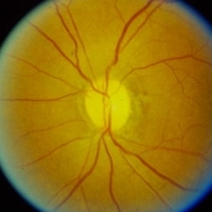
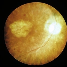
 Central Serous Chorioretinopathy (CSR)
Sep 21 2023 by Ben Serar
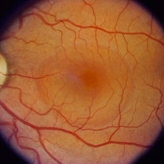
Fundus photograph showing increased cup-disc ratio with nasalisation of vessels , with thinning of Neuroretinal rim and bayonetting of vessels in a case of Glaucomatous Optic Atrophy (GOA) Fundus photograph of LE showing serous macular detachment in a case of Central Serous Chorioretinopathy (CSR).
Condition/keywords: Central Serous Chorioretinopathy (CSR)
-
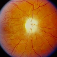
 Glaucomatous Optic Atrophy (GOA)
Sep 21 2023 by Ben Serar
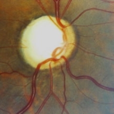
Fundus photograph showing increased cup-disc ratio with nasalisation of vessels , with thinning of Neuroretinal rim and bayonetting of vessels in a case of Glaucomatous Optic Atrophy (GOA).
Condition/keywords: Glaucomatous Optic Atrophy (GOA)
-
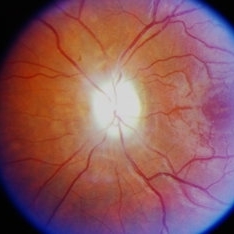
 Optic Atrophy
Sep 14 2023 by Ben Serar
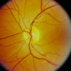
Fundus photograph of the LE showing disc pallor with well-defined disc margins in a case of optic atrophy.
Condition/keywords: optic atrophy
-
 Optic atrophy
Sep 14 2023 by Ben Serar
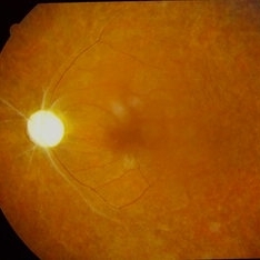
Fundus photograph of LE showing Pale disc in a case of optic atrophy, with arteriolar attenuation with vascular sheathing and granular fundus.
Condition/keywords: optic atrophy
-
 Optic Atrophy
Sep 12 2023 by Ben Serar
Fundus photograph of the LE showing disc pallor with well-defined disc margins in a case of primary optic atrophy
Condition/keywords: optic atrophy
-
 Glaucomatous Optic Atrophy (GOA)
Sep 12 2023 by Ben Serar
Fundus photograph of the LE showing enlarged optic cup with disc pallor in a case of Glaucomatous Optic Atrophy (GOA)
Condition/keywords: Glaucomatous Optic Atrophy, GOA
-
 Optic Atrophy
Sep 12 2023 by Ben Serar
Fundus photograph of the RE showing disc pallor with hazy disc margins in a case of secondary optic atrophy
Condition/keywords: optic atrophy
-
 Optic Atrophy
Sep 12 2023 by Ben Serar
Fundus photograph of the LE showing disc pallor with hazy disc margins in a case of secondary optic atrophy
Condition/keywords: optic atrophy
-
 Pseudo Foster Kennedy Syndrome
Oct 13 2022 by Aditya S Kelkar, MS, FRCS, FASRS,FRCOphth
Colour fundus photograph of a 44-year-old man showing bilateral small discs with optic atrophy on the right eye and disc edema on the left eye resulting from consecutive NAAION in both eyes.
Photographer: Dr Sukanya Mondal, National Institute of Ophthalmology, Pune. India
Imaging device: Zeiss Clarus 500
Condition/keywords: ischemic optic neuropathy, optic atrophy, optic disc edema
-
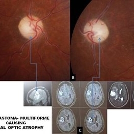 Glioblastoma Multiform Causing Bilateral Optic Atrophy
Mar 8 2021 by Deepak Bhojwani, MS
A 27-year-old male presented with complaints of severe headache associated with progressive loss of vision in both eyes since last 2- 3 months. On ocular examination his visual acuity was no perception of light in both eyes with bilateral dilated pupils not reacting to light. Fundus examination revealed bilateral optic atrophy (Figures A &B). He was advised an MRI scan for neurological evaluation. The patient was advised to review with Neurologist after getting MRI scans and review again with us. To our surprise this young patient was harboring a massive brain tumor reported as glioblastoma multiforme by the radiologist (Figure C). He has been advised tumor resection and chemoreduction by the neuro-oncologist. This case highlights neurological evaluation of all patients with bilateral optic atrophy. The presenting complaint of headache also prompted us for getting neurological examination. So its rightly said : eyes are the window to the body.
Photographer: DEEPAK BHOJWANI
Imaging device: ZEISS VISUCAM 524
Condition/keywords: optic atrophy
-
 Bilateral Calcific Retina Arteriolar Occlusions in a Patient with Metastatic Ovarian Carcinoma
Dec 10 2020 by McGill University Health Centre
47-year-old female with cough and fever. Imaging showed a right pulmonary infiltrate. Transbronchial needle biopsy revealed lymphangitic spread of papillary adenocarcinoma with psammoma bodies (MRI of thyroid, CT of abdomen and pelvis were negative) gynecologic evaluation negative at that time . The patient had bilateral floaters, VA: 20/40 OD and 20/20 OS. Fundus examination showed retinal arteriolar sheathing and a flat choroidal lesion OS and vitritis OD. Fluorescein angiogram showed staining of left superior temporal retinal arterioles and bilateral midperipheral patchy hyperfluorescence at RPE The patient vision in the OD deteriorated to 20/400, and in the OS 20/50. Four months later a new choroidal lesion was diagnosed OS. An abdominal mass consistent with a cystadenoma of the ovary was diagnosed. After a year patient developed systemic metastasis. Autopsy: Metastatic adenocarcinoma to the lung, both adrenals, para-aortic lymph nodes, left hip, right breast, occipital skin, serosal surface of liver, pituitary. In almost all metastatic lesions psammoma bodies were found. Presumptive diagnosis is a primary tumor of the ovary. Histopathologic examination of both eyes disclosed : Bilateral metastatic adenocarcinoma to the vitreous with partially calcified proliferation along internal limiting membrane, OS. Metastatic adenocarcinoma to choroid, OS. Bilateral optic atrophy secondary to retinal arteriolar occlusion with calcification.
Condition/keywords: bilateral, calcification, histopathology, metastatic adenocarcinoma, pathology, retinal arteriolar occlusion
-
 Bilateral Calcific Retina Arteriolar Occlusions in a Patient with Metastatic Ovarian Carcinoma
Dec 10 2020 by McGill University Health Centre
47-year-old female with cough and fever. Imaging showed a right pulmonary infiltrate. Transbronchial needle biopsy revealed lymphangitic spread of papillary adenocarcinoma with psammoma bodies (MRI of thyroid, CT of abdomen and pelvis were negative) gynecologic evaluation negative at that time Patient had bilateral floaters, VA: 20/40 OD and 20/20 OS. Fundus examination showed retinal arteriolar sheathing and a flat choroidal lesion OS and vitritis OD. Fluorescein angiogram showed staining of left superior temporal retinal arterioles and bilateral midperipheral patchy hyperfluorescence at RPE The patient vision in the OD deteriorated to 20/400, and in the OS 20/50. Four months later a new choroidal lesion was diagnosed OS. An abdominal mass consistent with a cystadenoma of the ovary was diagnosed. After a year patient developed systemic metastasis. Autopsy: Metastatic adenocarcinoma to the lung, both adrenals, para-aortic lymph nodes, left hip, right breast, occipital skin, serosal surface of liver, pituitary. In almost all metastatic lesions psammoma bodies were found. Presumptive diagnosis is a primary tumor of the ovary. Histopathologic examination of both eyes disclosed : Bilateral metastatic adenocarcinoma to the vitreous with partially calcified proliferation along internal limiting membrane, OS. Metastatic adenocarcinoma to choroid, OS. Bilateral optic atrophy secondary to retinal arteriolar occlusion with calcification.
Condition/keywords: bilateral, calcification, histopathology, metastatic adenocarcinoma, pathology, retinal arteriolar occlusion
-
 Bilateral Calcific Retina Arteriolar Occlusions in a Patient with Metastatic Ovarian Carcinoma
Dec 10 2020 by McGill University Health Centre
47-year-old female with cough and fever. Imaging showed a right pulmonary infiltrate. Transbronchial needle biopsy revealed lymphangitic spread of papillary adenocarcinoma with psammoma bodies (MRI of thyroid, CT of abdomen and pelvis were negative) gynecologic evaluation negative at that time . The patient had bilateral floaters, VA: 20/40 OD and 20/20 OS. Fundus examination showed retinal arteriolar sheathing and a flat choroidal lesion OS and vitritis OD. Fluorescein angiogram showed staining of left superior temporal retinal arterioles and bilateral midperipheral patchy hyperfluorescence at RPE. The patient vision in the OD deteriorated to 20/400, and in the OS 20/50. Four months later a new choroidal lesion was diagnosed OS. An abdominal mass consistent with a cystadenoma of the ovary was diagnosed. After a year patient developed systemic metastasis. Autopsy: Metastatic adenocarcinoma to the lung, both adrenals, para-aortic lymph nodes, left hip, right breast, occipital skin, serosal surface of liver, pituitary. In almost all metastatic lesions psammoma bodies were found. Presumptive diagnosis is a primary tumor of the ovary. Histopathologic examination of both eyes disclosed : Bilateral metastatic adenocarcinoma to the vitreous with partially calcified proliferation along internal limiting membrane, OS. Metastatic adenocarcinoma to choroid, OS. Bilateral optic atrophy secondary to retinal arteriolar occlusion with calcification.
Condition/keywords: bilateral, calcification, histopathology, metastatic adenocarcinoma, pathology, retinal arteriolar occlusion
-
 Bilateral Calcific Retina Arteriolar Occlusions in a Patient with Metastatic Ovarian Carcinoma
Dec 10 2020 by McGill University Health Centre
47-year-old female with cough and fever. Imaging showed a right pulmonary infiltrate. Transbronchial needle biopsy revealed lymphangitic spread of papillary adenocarcinoma with psammoma bodies (MRI of thyroid, CT of abdomen and pelvis were negative) gynecologic evaluation negative at that time . The patient had bilateral floaters, VA: 20/40 OD and 20/20 OS. Fundus examination showed retinal arteriolar sheathing and a flat choroidal lesion OS and vitritis OD. Fluorescein angiogram showed staining of left superior temporal retinal arterioles and bilateral midperipheral patchy hyperfluorescence at RPE The patient vision in the OD deteriorated to 20/400, and in the OS 20/50. Four months later a new choroidal lesion was diagnosed OS. An abdominal mass consistent with a cystadenoma of the ovary was diagnosed. After a year patient developed systemic metastasis. Autopsy: Metastatic adenocarcinoma to the lung, both adrenals, para-aortic lymph nodes, left hip, right breast, occipital skin, serosal surface of liver, pituitary. In almost all metastatic lesions psammoma bodies were found. Presumptive diagnosis is a primary tumor of the ovary. Histopathologic examination of both eyes disclosed : Bilateral metastatic adenocarcinoma to the vitreous with partially calcified proliferation along internal limiting membrane, OS. Metastatic adenocarcinoma to choroid, OS. Bilateral optic atrophy secondary to retinal arteriolar occlusion with calcification.
Condition/keywords: bilateral, calcification, histopathology, metastatic adenocarcinoma, pathology, retinal arteriolar occlusion
-
 Bilateral Calcific Retina Arteriolar Occlusions in a Patient with Metastatic Ovarian Carcinoma
Dec 10 2020 by McGill University Health Centre
47-year-old female with cough and fever. Imaging showed a right pulmonary infiltrate. Transbronchial needle biopsy revealed lymphangitic spread of papillary adenocarcinoma with psammoma bodies (MRI of thyroid, CT of abdomen and pelvis were negative) gynecologic evaluation negative at that time . The patient had bilateral floaters, VA: 20/40 OD and 20/20 OS. Fundus examination showed retinal arteriolar sheathing and a flat choroidal lesion OS and vitritis OD. Fluorescein angiogram showed staining of left superior temporal retinal arterioles and bilateral midperipheral patchy hyperfluorescence at RPE. The patient vision in the OD deteriorated to 20/400, and in the OS 20/50. Four months later a new choroidal lesion was diagnosed OS. An abdominal mass consistent with a cystadenoma of the ovary was diagnosed. After a year patient developed systemic metastasis. Autopsy: Metastatic adenocarcinoma to the lung, both adrenals, para-aortic lymph nodes, left hip, right breast, occipital skin, serosal surface of liver, pituitary. In almost all metastatic lesions psammoma bodies were found. Presumptive diagnosis is a primary tumor of the ovary. Histopathologic examination of both eyes disclosed : Bilateral metastatic adenocarcinoma to the vitreous with partially calcified proliferation along internal limiting membrane, OS. Metastatic adenocarcinoma to choroid, OS. Bilateral optic atrophy secondary to retinal arteriolar occlusion with calcification.
Condition/keywords: bilateral, calcification, histopathology, metastatic adenocarcinoma, pathology, retinal arteriolar occlusion
-
 BRVO With Optic Atrophy
Nov 27 2020 by Sham Talati, DOMS
A 55-year-old male patient presented with glaucomatous optic atrophy with ST BRVO in the right eye.
Photographer: Dr. Sham Talati,Retina Foundation,Ahmedabad
Imaging device: Nidek Mirante
Condition/keywords: branch retinal vein occlusion (BRVO), branch vein occlusion (BVO), glaucomatous atrophy of optic disc, optic atrophy
-
 Radiation Retinopathy
Apr 2 2019 by Gary R. Cook, MD, FACS
White male with radiation retinopathy OS; he also had glaucoma and radiation-induced optic atrophy present in this eye
Condition/keywords: radiation optic neuropathy, radiation retinopathy, retinal hemorrhage
-
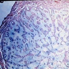
 Slide 12-29
Feb 27 2019 by Lancaster Course in Ophthalmology
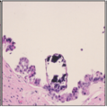
Sequelae. Cavernous degeneration of the optic nerve (Schnabel's cavernous atrophy) shows cystic spaces which appear clear in hematoxylin and eosin stained sections (H&E x21).
Condition/keywords: optic atrophy, sequelae
-
 Slide 11-36
Feb 26 2019 by Lancaster Course in Ophthalmology
Schnabel's cavernous optic atrophy. After treatment with hyaluronidase, the mucopolysaccharide stain is negative.
Condition/keywords: optic atrophy
-
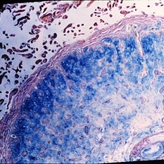
 Slide 11-35
Feb 26 2019 by Lancaster Course in Ophthalmology
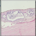
Schnabel's cavernous optic atrophy. Large spaces within the nerve are filled with acid mucopolysaccharide (Aician blue stain x40).
Condition/keywords: acid mucopolysaccharide, optic atrophy
-
 Slide 11-34
Feb 26 2019 by Lancaster Course in Ophthalmology
Glaucoma cup ( x16). Histopathology shows a posterior bowing of the lamina cribrosa, an excavation of disk margins, a loss of nerve fiber layers, and optic atrophy. (Scheie Eye Institute, No. 6596.)
Condition/keywords: glaucoma, lamina cribrosa
-
 Slide 11-32
Feb 26 2019 by Lancaster Course in Ophthalmology
Ascending optic atrophy. Clinical appearance of optic atrophy associated with retinitis pigmentosa. The arterioles are nearly obliterated due to profound retinal atrophy.
Condition/keywords: optic atrophy, retinitis pigmentosa
-
 Slide 11-31
Feb 26 2019 by Lancaster Course in Ophthalmology
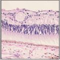
Primary or descending optic atrophy. Higher-power longitudinal section of the optic disk showing loss of nerve fibers (Bodian stain x40). (Scheie Eye Institute, No. 3580.)
Condition/keywords: optic atrophy
-
 Slide 11-30
Feb 26 2019 by Lancaster Course in Ophthalmology
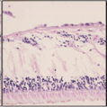
Primary or descending optic atrophy. Horizontal section of the optic disk ( x 16). Note the increase in thickness of the pial septa in the nerve, the loss of ganglion cell and nerve fiber layers in the adjacent retina, and the loss of the optic cup.
Condition/keywords: optic atrophy, pial septa
-
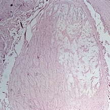
 Slide 11-29
Feb 26 2019 by Lancaster Course in Ophthalmology
Optic atrophy. Early atrophy is accompanied by thickening of the pial septa ( x 110). (Scheie Eye Institute, No. 5838.)
Condition/keywords: optic atrophy
 Loading…