Search results (50 results)
-
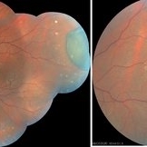
 Macular Coloboma
Jun 5 2025 by César Adrián Gómez Valdivia, MD
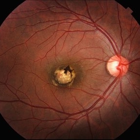
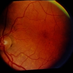
Macular Coloboma found in a 28 year-old male patient, visual acuity was 20/60. Resulting due to fusion failure of the optic fissure, colobomas are commonly found in the infero-nasal quadrant. If the retina is involved, it is reduced to glial tissue with no underlying RPE or choroid. This appears as an area of whitening often with pigment deposition at the junction of the coloboma and normal retina. Findings were bilateral.
Photographer: @eyemissu2
Imaging device: TOPCON TRC-50DX
Condition/keywords: coloboma
-
 Representative Electrooculogram Responses
May 13 2024 by Gabrielle Hallai
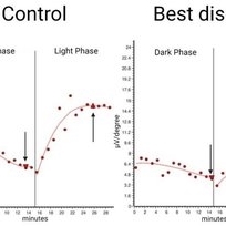
Electrooculogram responses on the left from a control individual with no known retinal pathology. There is a clear dark trough around 13 minutes (arrow down) and a light peak around 25 minutes (arrow up). The Arden ratio, or the light peak to dark trough ratio, is 2.54, indicative of normal retinal pigment epithelium function (normal > 1.80, abnormal < 1.65). On the right-hand side, there is a representative image from an individual with Best macular dystrophy. Note the reduced responses for both the dark and light phase. There is a reduced Arden ratio of 1.23, suggestive of abnormal retinal pigment epithelium function. An abnormal Arden ratio is universal in Best vitelliform macular dystrophy and is the most common electroretinographic change in this disease. Other bestrophinopathies such as autosomal recessive bestrophinopathy may have normal EOG. EOG testing was completed on the Diagnosys ColorDome.
Photographer: Gabrielle Hallai, PhD, Cleveland Clinic Cole Eye Institute
Imaging device: Diagnosys ColorDome
Condition/keywords: Best disease, electrooculogram, electroretinography, EOG
-
 Congenital Retinal Vessel Tortuosity
Apr 2 2024 by Pablo Angel Garcia Uribe
Fundus photograph of a 29-year-old man with bilateral congenital retinal vessel tortuosity. This image shows the sinuous course of retinal arterioles and a shiny internal limiting membrane.
Photographer: Pablo Ángel García-Uribe, Clínica Oftalmológica Salauno, Mexico City
Imaging device: NIDEK OCT RS-330 Duo 2
Condition/keywords: abnormal retinal vessel, anomalous vessels, Retina, tortuous vessels
-
 Von Hippel Lindau with retinal capillary hemangioma
Nov 2 2023 by Marcelo Zas, MD PhD
30-year-old female patient diagnosed with Syndrome VHL (Von Hippel Lindau). Stage II. In the first wide-field retinography of the right eye we can observe the exophytic retinal hemangiomas, rounded, slightly delimited, located in the peripheral retina in the upper and lower temporal quadrants and due to the exudation produced by them, hard exudates are observed in the star hemisphere, affecting the macula.
Photographer: Mariano Cotic MD
Imaging device: Silverstone SS OCT Optos
Condition/keywords: abnormal retinal vessel
-
 Sunset Glow Fundus
May 15 2022 by Manuel Ángel Alcántara Delgado, MD
Optomap ultra-widefield retinal imaging of an 35-year-old woman showed sunset glow fundus, multiple nummular chorioretinal atrophic lesions, macular subretinal fibrosis and pigment clumping in chronic recurrent stage of Vogt-Koyanagi-Harada disease.
Photographer: Manuel Ángel Alcántara Delgado. Conde de Valenciana.
Condition/keywords: abnormal retina, benign pigmented lesions, pigment clumps, retinal fibrosis, uveitis, Vogt-Koyanagi-Harada
-
 Spontaneously dropped Nucleus In a Congenital Rubella Retina disease
Apr 29 2022 by NEIFFER RABELO
Intraoperative Fundus Photograph of an 68 years old with past medical history of
Photographer: Rodrigo Dos Anjos Vesiani, Retina Institute - Belo Horizonte - Brazil
Imaging device: OPMI LUMERA® 700
Condition/keywords: abnormal retina, dropped nucleus, retina surgery, rubella
-
 Retinal Capillary Hemangioma
Sep 9 2021 by Jesus Lozano, MD
60 year-old woman with a Peripheral RCH treated with laser photocoagulation.
Photographer: Yair Bet Yosef, Hadassah Medical Center. Israel
Imaging device: Optos Silverstone
Condition/keywords: abnormal retinal vessel, anomalous vessels, dilated tortuous vessels, hemangioma, retina
-
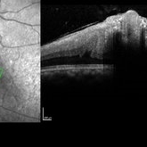
 CSNB-OCT-OD
Aug 23 2021 by Jennifer Carstens
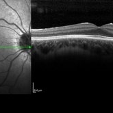
OCT/infrared image showing myopic fundus with normal retinal structure in patient with CACNA1F associated X-linked CSNB (OD).
Photographer: Jing Zhang, Ophthalmic Photographer
Condition/keywords: congenital stationary night blindness (CSNB), infrared image, optical coherence tomography (OCT)
-
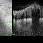
 CSNB-OCT-OS
Aug 23 2021 by Jennifer Carstens
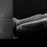
OCT/infrared image showing myopic fundus with normal retinal structure in patient with CACNA1F associated X-linked CSNB (OS).
Photographer: Jing Zhang, Ophthalmic Photographer
Condition/keywords: congenital stationary night blindness (CSNB), infrared image, optical coherence tomography (OCT)
-
 CHRPE
CHRPE
Jan 15 2021 by Priya Rasipuram Chandrasekaran, MBBS, DO, DNB, FRCS
This is the fundus photo and fundus photo montage of the left eye of a 25-year-old male showing flat, solitary, round, greyish pigmented lesion situated AT THE equator with a scalloped margin. Vessels overlying the lesion are normal and there is a clear demarcation line between this and normal retina. The margins are hypopigmented with few hypopigmented lacunae inside.
Condition/keywords: congenital hypertrophy of the retinal pigment epithelium (CHRPE)
-
 Presumed Combined Hamartoma of the Retina and the Retinal Pigment Epithelium
Dec 7 2020 by Martin J Siemerink, MD, PhD
7-year-old girl, right eye VA 20/200, left eye unremarkable.
Photographer: Faroch Payman, Bergman Clinics Doetinchem
Condition/keywords: abnormal retinal vessel, combined hamartoma
-
 Central Retinal Artery Occlusion with Cilioretinal Sparing - Optical Coherence Tomography
Oct 28 2020 by Fang Helen Mi
Optical coherence tomography showed hyper-reflective inner retinal layers, indicating intracellular oedema of the affected retina, with normal retinal layers in the area perfused by the cilioretinal artery.
Condition/keywords: central retinal artery occlusion (CRAO), cilioretinal sparing
-
 Left Eye- Posterior Segment
Aug 10 2020 by RITESH VERMA
Normal posterior segment of the left eye.
Photographer: Dr. Ritesh Verma, Regional institute of Ophthalmology, Rohtak, Haryana, India
Imaging device: CR-2AF CANON
Condition/keywords: left eye, normal retina
-
 Combined Hamartoma of Retina and RPE
Jul 15 2020 by Itzel Ocampo
Fundus photograph of a 53-year-old man with a combined hamartoma of retina and RPE; visual capacity of hand movement. No systemic associations.
Photographer: Itzel Ocampo, Universidad Autonoma de Mexico, Hospital General de Mexico "Eduardo Liceaga"
Condition/keywords: abnormal retina, combined hamartoma
-
 Combined Hamartoma of the Retina and Retinal Pigment Epithelium (CHRRPE)
Jan 21 2020 by Pierre-Henry Gabrielle, MD
Coupled OCT B-scan and IR imaging of a 17-year-old man with combined hamartomas of the retina and retinal pigment epithelium (CHRRPE) at the posterior pole of the left eye. One can see a highly reflective elevated macular lesion with hyporeflective shadowing of the underlying tissue and obscuration of the normal retinal layers.
Photographer: Pierre-Henry Gabrielle, Ophthalmology department, Dijon University Hospital, France
Imaging device: Heidelberg Spectralis
Condition/keywords: combined hamartoma, optical coherence tomography (OCT)
-
 Combined Hamartoma of the Retina and Retinal Pigment Epithelium (CHRRPE)
Jan 21 2020 by Pierre-Henry Gabrielle, MD
Coupled OCT B-scan and IR imaging of a 17-year-old man with Combined hamartomas of the retina and retinal pigment epithelium (CHRRPE) at the posterior pole of the left eye. One can see a highly reflective elevated macular lesion with hyporeflective shadowing of the underlying tissue and obscuration of the normal retinal layers.
Photographer: Pierre-Henry Gabrielle, Ophthalmology department, Dijon University Hospital, France
Imaging device: Heidelberg Spectralis
Condition/keywords: combined hamartoma, optical coherence tomography (OCT)
-
 Coats' Disease - Stage 3A
Aug 21 2019 by Victor M Villegas, MD
Coats' Disease - stage 3A.
Condition/keywords: abnormal retina, Coats' disease, diffuse lipid exudate, edema, foveal hard exudates, pediatic retina, retcam, retinal angioma
-
 Advanced Coats' Disease with Neovascular Glaucoma
Aug 21 2019 by Victor M Villegas, MD
Advanced Coats' Disease with neovascular glaucoma.
Photographer: Giselle Deoliveira, Bascom Palmer Eye Institute, University of Miami
Imaging device: RetCam III
Condition/keywords: abnormal retinal vessel, bullous retinal detachment, Coats' disease, diffuse lipid exudate, foveal hard exudates, neovascular glaucoma, pediatric retina
-
 Slide 13-28
Mar 4 2019 by Lancaster Course in Ophthalmology
Pseudoglioma: abnormal retina behind the lens with tunica vasculosa lentis in a maldeveloped eye associated with chromosomal defect ( x25).
Condition/keywords: abnormal retina, chromosomal defect, pseudoglioma, tunica vasculosa lentis
-
 Normal Fundus image
Aug 30 2018 by Timothy Adeyemo
Fundus photograph of a healthy 31-year-old lady being examined for glaucoma.
Photographer: Adeyemo Timothy, National Eye Centre, Kaduna, Nigeria.
Imaging device: Topcon's TRC- NW8 plus
Condition/keywords: normal retina
-
 IJFT
IJFT
Mar 26 2018 by Purva Patwari
Defective vision.
Photographer: Dr Purva Patwari, Patwari Retina Center, Ahmedabad, Gujarat , India
Condition/keywords: abnormal retinal vessel, IJFT, IJT
-
 Vascular Anormalities
Jan 6 2016 by Andrea Arriola-Lopez, MD MSc
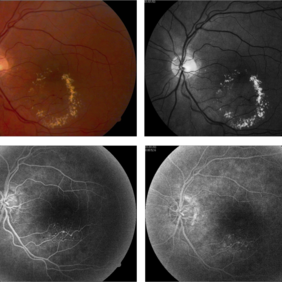
77-year-old man. Decrease of visual acuity OS. VA 20/30 IOP 14mmHg. Fundus examination findings: Hard exudates, microaneurysms near to fovea. OCT shows IRF. Late leakage on FA.
Photographer: Andrea Elizabeth Arriola-Lopez, MSc MD
Condition/keywords: abnormal retinal vessel, aneurysm, hard exudates, vascular anomaly
-
 Normal
Jan 7 2015 by H. Michael Lambert, MD
Color photograph of normal fundus.
Condition/keywords: normal eye, normal retina
-
---thumb.jpg/image-square;max$300,300.ImageHandler) Normal Retina
Normal Retina
-
---thumb.jpg/image-square;max$300,300.ImageHandler) Normal Retina
Normal Retina
 Loading…