Search results (323 results)
-
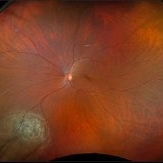
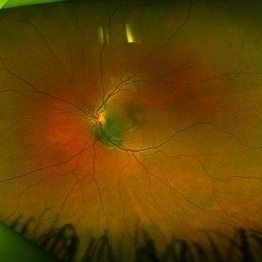
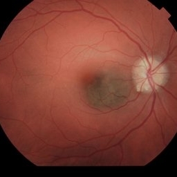
 Nevus to Melanoma in 1 Year
Dec 12 2025 by Virginia Gebhart
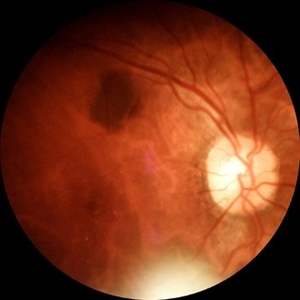
83 year old male presents with partially amelanotic choroidal nevus, which has been followed for the last 10 years. On exam there is a new area of orange pigment, and change in diameter compared to photos from 2024. Ultrasound shows small increase in height, medium to low internal reflectivity, and vascularity. Pt is asymptomatic. Due to personal matters pt wishes to reassess in few months and schedule treatment.
Photographer: Virginia Gebhart, Retina Consultants of Carolina
Imaging device: Optos California
Condition/keywords: choroidal melanoma, choroidal nevus
-
 Giant Nevus of the Macula
Nov 5 2025 by Virginia Gebhart
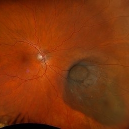
68 year old female with stable large pigmented lesion throughout the whole posterior pole, extending beyond the superior and inferior arcades. Pt has been aware of nevus for 45 years. Lesion has been stable since initial photos in 2013. Will continue to observe. Vision CF
Photographer: Virginia Gebhart, Retina Consultants of Carolina
Imaging device: Optos California
Condition/keywords: Choroidal nevus, pigmented lesion, pigmented nevus
-
 Optic Disc Melanocytoma Hyperpigmented Magnocellular Nevus of the Optic Disk (HMNOD)
Aug 5 2025 by SHRADDHA ASHOK CHANDORKAR, DNB DO FVRS
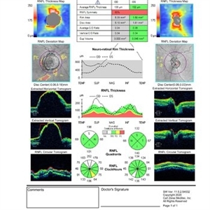
OCT DISC image of a case of optic nerve melanocytoma.
Photographer: Dr.Shraddha A Chandorkar
Imaging device: zeiss
Condition/keywords: optic disc melanocytoma
-
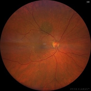
 Choroidal Nevus
Jun 4 2025 by Paulina Araujo
The 55-degree central fundus photograph of the right eye reveals a choroidal nevus measuring 0.5 disc diameters along the superior temporal arcade.
Photographer: Paulina D.Araujo Martínez, Asociación para Evitar la Ceguera en México I.A.P., Hospital Dr Luis Sánchez Bulnes.
Condition/keywords: Choroidal nevus
-
 Collar Button Melanoma
Mar 27 2025 by Virginia Gebhart
62 year old male with large pigmented lesion with collar button. Pt states he was never aware of any lesion/nevus in the past. Fluid and orange pigment present, appears to be chronic. Pt will be scheduled for brachytherapy pending CT scan results.
Photographer: Virginia Gebhart, Retina Consultants of Carolina
Imaging device: Optos California
Condition/keywords: choroidal melanoma, collar button
-
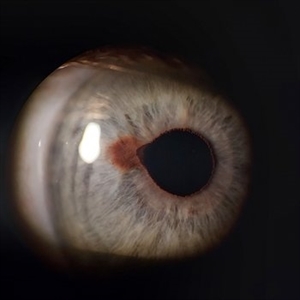
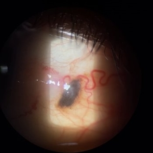
 Iris Nevus
Jan 28 2025 by Korey Starkey
Slit-lamp image of an 89-year-old patient with an iris nevus. Nevus appeared stable on exam, will continue to monitor.
Photographer: Korey Starkey
Imaging device: Slit lamp camera
Condition/keywords: ectropion uveae, iris nevus, slit lamp photo
-
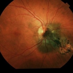
 Suspicious Nevus
Jan 15 2025 by Virginia Gebhart
14 year female with suspicious nevus located adjacent to the optic nerve. Questionable orange pigment present and worsening SRF compared to previous photos/OCT. RPE atrophy also present from previous fluid. No elevation. Will continue observation. BCVA 20/25
Photographer: Virginia Gebhart, Retina Consultants of Carolina
Imaging device: Topcon 50DX
Condition/keywords: choroidal nevus, nevus
-
 Choroidal Nevus
Nov 7 2024 by Ashley Romero-Martinez
57 year old female presents with choroidal nevus in left eye. Has been present for 20 years, remains stable and will continue to observe.
Photographer: Ashley Romero-Martinez
Imaging device: Optos
Condition/keywords: choroidal nevus
-
 Choroidal Nevus
Nov 7 2024 by Ashley Romero-Martinez
57 year old female presents with choroidal nevus in left eye. Has been present for 20 years, remains stable and will continue to observe.
Photographer: Ashley Romero-Martinez
Imaging device: Optos
Condition/keywords: choroidal nevus
-
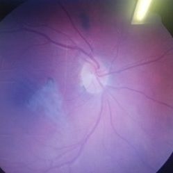
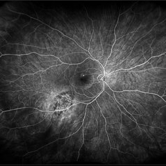 Suspicious Nevus / CSR
Aug 8 2024 by Virginia Gebhart
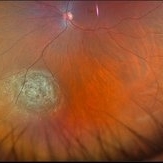
Fluorescein angiogram of 54 year old male with a suspicious appearing choroidal nevus and central serous retinopathy. Will monitor closely and follow up with serial exams.
Photographer: Virginia Gebhart
Imaging device: Optos California
Condition/keywords: central serous retinopathy (CSR), choroidal nevus, nevus
-
 Iris Nevus
Jul 3 2024 by Zach Seim
Slit Lamp Photograph of an 88 year old man with an Iris Nevus. Patient presented with DCC 20/60+1. Plan to monitor.
Photographer: Zach Seim
Imaging device: Slit Lamp photography with Samsung Galaxy 7
Condition/keywords: iris, iris nevus, nevus, right eye, slit lamp photo, slit lamp photography
-
 Choroidal Nevus
May 25 2024 by Gustavo Del Castillo-Marquez, MD
Fundus photograph of a 65 year old woman with a choroidal nevus.
Photographer: Gustavo Del Castillo-Márquez, Asociación Para Evitar la Ceguera en México, CDMX
Imaging device: Zeiss Clarus
Condition/keywords: choroidal nevus
-
 Suspicious Choroidal Nevus
May 8 2024 by Virginia Gebhart
13 year old female with suspicious appearing choroidal nevus. High risk features present, adjacent to optic nerve, questionable orange pigment, SRF. No significant elevation on ultrasound. Will follow up with serial exams.
Photographer: Virginia Gebhart
Imaging device: Optos California
Condition/keywords: choroidal nevus, nevus
-
 Macula-threatening PEHCR causing a nasal visual-field defect in a patient with AMD
Apr 15 2024 by David A Reichstein, MD
(A) Ultra-widefield color fundus photograph demonstrates a PEHCR encroaching upon the temporal macula. Lipid exudation is apparent at the lesion’s anterior and inferior border. Subretinal hemorrhage is apparent at the lesion’s inferior border. Drusen are apparent in the macula. An unrelated, small choroidal nevus is apparent in the inferior fundus. (B) Ultra-widefield FA taken in early stage demonstrates hypofluorescence within the lesion consistent with blockage by possible sub-RPE or subretinal heme. (C) Ultra-widefield fundus photograph taken 6 months following the initiation of monthly anti-VEGF therapy demonstrates considerable reduction in the size of the lesion and resolution of the subretinal hemorrhage and lipid exudation. (D) Ultra-widefield fundus photograph taken 1 year after presentation where a treat-and-extend approach was performed for the most recent 6 months. The lesion had almost completely resolved.
Condition/keywords: peripheral exudative hemorrhagic chorioretinopathy (PEHCR)
-
 Blue Nevus
Apr 9 2024 by Hector Gabriel Moreno Solano, MD, MHA
48-year-old Hispanic male patient who comes to the clinic due to the presence of pterygium. Upon examination, a 2 mm blue nevus is found, which the patient reports having had since he was 15 years old.
Photographer: Héctor Gabriel Moreno-Solano.
Condition/keywords: conjunctiva, nevus
-
 Suspicious Nevus
Feb 14 2024 by Virginia Gebhart
61 year old female with a suspicious choroidal nevus involving the optic nerve head. Patient asymptomatic, will continue to observe.
Photographer: Virginia Gebhart
Imaging device: Topcon TRC 50DX
Condition/keywords: choroidal nevus, nevus
-
 Suspicious Choroidal Nevus
Dec 6 2023 by Virginia Gebhart
55 year old female with suspicious pigmented choroidal nevus. 3 high risk features present. Ultrasound shows SRF and high internal reflectivity. Will observe closely
Photographer: Virginia Gebhart
Imaging device: Topcon
Condition/keywords: choroidal nevus, orange pigment
-
 Benign Nevus
Dec 6 2023 by Virginia Gebhart
71 year old female with benign choroidal nevus with fibrotic PED
Photographer: Virginia Gebhart
Imaging device: Topcon
Condition/keywords: choroidal nevus, fibrotic neovascularization, nevus
-
 Suspicious Choroidal Nevus
Dec 6 2023 by Virginia Gebhart
82 year old male with suspicious choroidal nevus and ERM. Remains unchanged since first visit 6 mos ago.
Photographer: Virginia Gebhart
Imaging device: Topcon
Condition/keywords: choroidal nevus
-
 Suspicious Choroidal Nevus / Optic Disc Drusen
Nov 1 2023 by Virginia Gebhart
29 year-old female with suspicious choroidal nevus adjacent to optic nerve and extending into fovea. Optic disc drusen OU. Pt is asymptomatic
Photographer: Virginia Gebhart
Imaging device: Topcon
Condition/keywords: choroidal nevus, disc drusen, drusen of optic disc, nevus
-
 Choroidal-Nevus
Feb 25 2023 by Aditya S Kelkar, MS, FRCS, FASRS,FRCOphth
Color fundus photograph of the left eye of a 55 year old male showing large choroidal nevus.
Photographer: Dr. Apoorva Jadhav, National Institute of Ophthalmology, Pune. India.
Imaging device: Zeiss Clarus 500
Condition/keywords: Choroidal nevus
-
 Choroidal nevus with halo
Aug 21 2022 by Niloofar Piri, MD
Fundus photograph of the right eye in a 43 yo patient demonstrating a flat choroidal nevsu with halo nasal to the optic nerve
Photographer: Andrew Polk, MD
Condition/keywords: Choroidal nevus, nevus with halo
-
 Nevus
Nevus
Jan 21 2021 by AGNES KIM
Fundus photograph of 30-year-old female of choroidal nevus. Another nevus was found in the same eye in the periphery. Macula has a lens artifact.
Condition/keywords: choroidal nevus
-
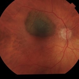
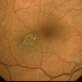
 Macular Nevus
Jan 20 2021 by Jamin S. Brown, MD
Macular Nevus as well as CSR.
Photographer: Stefanie Palmer CRA, Retina Vitreous Surgeons of CNY
Condition/keywords: choroidal nevus, macula lesion
-
 Choroidal Nevus Associated with Drusen
Jan 11 2021 by Gabriel Costa Andrade, PhD
Fundus photograph of an 53-year-old man with a macular melanotic nevus.
Photographer: Gabriel Andrade
Condition/keywords: choroidal nevus
 Loading…