Search results (61 results)
-
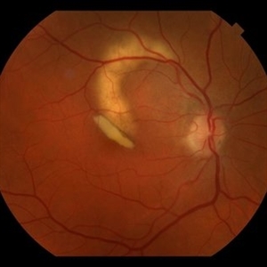
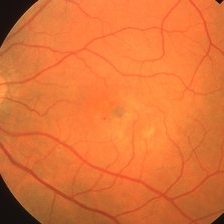
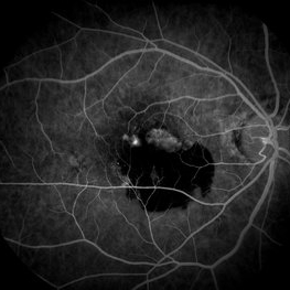
 Neovascular AMD with Active CNV
May 22 2025 by Kimberly Wakester
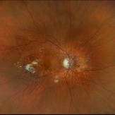
Optomap RGB of an 82-year-old man with Neovascular AMD with Active CNV and Dry AMD in the right eye. There is advanced atrophic changes without subfoveal involvement located temporally to the fovea. Patient is to continue follow up care with dilated exam, repeat OCT, and treatment of intravitreal injection of Vabysmo every 5 weeks at this time.
Photographer: Kimberly Wakester, COA, OCT-C
Imaging device: Optos California
Condition/keywords: advanced geographic atrophy, dry age-related macular degeneration (dry AMD), neovascular age-related macular degeneration (AMD)
-
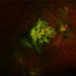
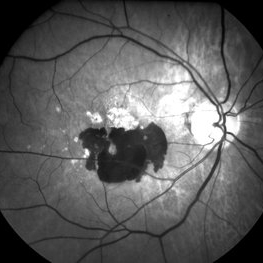
 Peripheral Exudative Hemorrhagic Chorioretinopathy
Nov 19 2024 by Toolie Winters
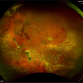
Ultra-wide field fundus photograph of an 85-year-old woman with Peripheral Exudative Hemorrhagic Chorioretinopathy (PECHR) affecting the right eye. Patient presented with a blind spot centrally in the right eye which she first noticed 4 months prior to this image being taken. The patient states that in the month prior to this image, she began noticing bright lights flash across her vision 4-5x/day which last about 15 seconds. The flashes are either black with a blue ring around them or yellow, and their frequency has increased over time. The patient's vision at the time of this appointment was Dcc20/100+1 PHNI. This photo also shows diffuse hemorrhage, lipid, and an eccentric disciform lesion.
Photographer: Toolie Winters
Imaging device: Optos California
Condition/keywords: fundus photography, neovascular age-related macular degeneration (AMD), Optos, OPTOS CALIFORNIA, peripheral exudative hemorrhagic chorioretinopathy (PEHCR), pseudocolor, ultra-wide field imaging, wet age-related macular degeneration (wet AMD)
-
 RPE-Transplantation
Jul 25 2024 by Gabriel Costa Andrade, PhD
Postoperative period of RPE-transplantation in a patient with neovascular AMD after RPE tear.
Photographer: Gabriel Andrade
Condition/keywords: neovascular age-related macular degeneration (AMD), pars plana vitrectomy (PPV), wet age-related macular degeneration (wet AMD)
-
 RPE Rip
Jan 25 2024 by Virginia Gebhart
69 year old female with Neovascular AMD. New RPE rip and increased IRF on OCT 10 weeks s/p Eylea injection. Switched to Vabysmo to extend intervals
Photographer: Virginia Gebhart
Imaging device: Topcon
Condition/keywords: neovascular age-related macular degeneration (AMD)
-
 Hemorrhagic Pigment Epithelial Detachment
Jan 25 2024 by Virginia Gebhart
64 year old male with persistent hemorrhagic PED with oxidized SRH involving the central macula. Continued improvement with 12 week intervals of Eylea. BCVA 20/80
Photographer: Virginia Gebhart
Imaging device: Topcon
Condition/keywords: neovascular age-related macular degeneration (AMD), pigment epithelial detachment (PED)
-
 The Phoenix (Mythological)
Aug 23 2023 by Angela Rico
93 year-old female with Neovascular AMD with Active CNV OD
Photographer: Angela Rico M.D.
Imaging device: Optos
Condition/keywords: neovascular age-related macular degeneration (AMD)
-
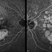 Macular Degeneration with Extensive Geographic Atrophy
Jan 26 2022 by Olivia Rainey
Heidelberg Spectralis fluorescein angiography of a 94-year-old woman with Macular Degeneration affecting both eyes. The FA reveals transmission defects consistent with RPE changes and geographic atrophy of RPE of both eyes, as well as window defects consistent with peripheral scarring in the right eye. The patient's vision was Dcc20/70 in both eyes at the visit the images were taken.
Photographer: Olivia Rainey, OCT-C, COA
Imaging device: Heidelberg Spectralis
Condition/keywords: 30-degrees, choroidal neovascularization (CNV), dry age-related macular degeneration (dry AMD), early phase, fluorescein angiogram (FA), geographic atrophy, heidelberg spectralis, macular degeneration, neovascular age-related macular degeneration (AMD)
-
 Neovascular Age-Related Macular Degeneration (2)
Apr 28 2021 by Ambar Faridi, MD
80-year-old woman with neovascular age-related macular degeneration with interval resolution of large subretinal hemorrhage and vascular exudation followed over time.
Photographer: Jennifer Tu-Bui, VA Portland Health Care System
Condition/keywords: neovascular age-related macular degeneration (AMD), reabsorbing subretinal hemorrhage
-
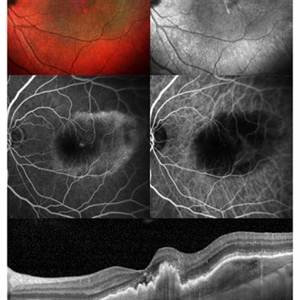 Neovascular AMD
Jun 27 2020 by Aayesha Khanum
Multicolor image and infra-red image showing fibrovascular pigment epithelial detachment (PED),FFA and ICG confirms the diagnosis., SD-OCT shows notched fibrovascular PED
Photographer: Puttaswamy,Ravikrishna
Imaging device: Heidelberg Spectralis
Condition/keywords: neovascular age-related macular degeneration (AMD)
-
 Neovascular AMD with Active CNV
May 21 2019 by Carolyn Daley
Fluorescein angiogram image of an 86-year-old woman with neovascular AMD. This image was taken 1 minute and 22 seconds into the FA testing, after dye was injected.
Photographer: Carolyn Daley, COA, Retina Specialists of Michigan
Imaging device: Heidelberg Spectralis
Condition/keywords: choroidal neovascularization (CNV), fluorescein angiogram (FA), neovascular age-related macular degeneration (AMD)
-
 Subfoveal Neovascular Membrane
Mar 26 2019 by Gary R. Cook, MD, FACS
63-year-old white male with subfoveal CNVM secondary to AMD OS; VA= 20/60.
Imaging device: Topcon VT-50
Condition/keywords: neovascular age-related macular degeneration (AMD), subfoveal neovascular membrane
-
 Recurrent Subfoveal CNVM
Mar 26 2019 by Gary R. Cook, MD, FACS
63-year-old white male with exudative AMD and a subfoveal CNV 6 weeks after Argon laser photocoagulation OS per MPS Protocol for subfoveal treatment; VA decreased to 20/100.
Imaging device: Topcon VT-50
Condition/keywords: neovascular age-related macular degeneration (AMD), subfoveal choroidal neovascularization, subretinal neovascularization (SRNV), treated recurrence
-
 Submacular Hemorrhage
Apr 24 2018 by Pauline T Merrill, MD, FASRS
Fundus photo of left eye of a 65-year-old AMD patient one week following vitrectomy with subretinal tPA and air-fluid exchange. There was significant displacement of the submacular hemorrhage, and vision was beginning to improve (20/350).
Photographer: Karen Parque, Illinois Retina Associates, Chicago, Illinois
Imaging device: Topcon 50DX
Condition/keywords: neovascular age-related macular degeneration (AMD), submacular hemorrhage
-
 Submacular Hemorrhage
Apr 24 2018 by Pauline T Merrill, MD, FASRS
Fundus photo of left eye of a 65-year-old AMD patient, 3 days following injection of C3F8 and face-down positioning; there was little change in the large submacular hemorrhage. There was no further change one week later, at which time the patient elected surgery.
Photographer: Karen Parque, Illinois Retina Associates, Chicago, Illinois
Imaging device: Topcon 50DX
Condition/keywords: neovascular age-related macular degeneration (AMD), submacular hemorrhage
-
 Submacular Hemorrhage
Apr 24 2018 by Pauline T Merrill, MD, FASRS
Fundus photo of left eye of a 65-year-old AMD patient who presented with sudden drop of vision from 20/30 to CF due to a large submacular hemorrhage, 7 months following her last Eylea injection. She underwent immediate injection of C3F8 in the office, with little effect. 10 days later vitrectomy with subretinal tPA and air-fluid exchange was performed, with successful displacement of the hemorrhage.
Photographer: Ermelinda Diaz, Illinois Retina Associates, Chicago, Illinois
Imaging device: Topcon 50DX
Condition/keywords: neovascular age-related macular degeneration (AMD), submacular hemorrhage
-
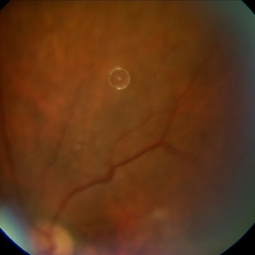
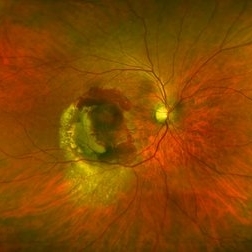 Polypoidal Choroidal Vasculopathy
Mar 13 2018 by Gabriel Costa Andrade, PhD
Fundus image of the right eye of a 76-year-old man with Polypoidal Choroidal Vasculopathy in the right eye.
Photographer: Gabriel Andrade, MD
Imaging device: Optos® California
Condition/keywords: neovascular age-related macular degeneration (AMD)
-
 Neovascular AMD with Active CNV
Jan 2 2018 by Carolyn Daley
30 degree fluorescein angiogram of an 80-year-old woman with neovascular AMD with active CNV in the left eye. Patient is being treated with Avastin.
Photographer: Carolyn Daley, Retina Specialists of Michigan
Imaging device: Heidelberg Spectralis
Condition/keywords: 30 degrees, choroidal neovascularization (CNV), Heidelburg Spectralis, left eye, neovascular age-related macular degeneration (AMD)
-
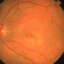
 Neovascular AMD
Jan 3 2017 by Jason Griffith
75-year-old male with neovascular AMD with disciform scar OS
Photographer: Jason Griffith
Imaging device: Topcon TRC-50EX
Condition/keywords: disciform scar, neovascular age-related macular degeneration (AMD)
-
 Neovascular AMD
Jan 3 2017 by Jason Griffith
65-year-old female dx neovascular AMD with active CNV.
Photographer: Jason Griffith, Tennessee Retina
Imaging device: Topcon TRC-50EX
Condition/keywords: choroidal neovascularization (CNV), neovascular age-related macular degeneration (AMD)
-
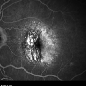
 Subretinal Hemorrhage Due to SRNVM, Fluorescein Angiogram Photograph
Dec 1 2016 by James B. Soque, CRA, OCT-C, COA, FOPS
89-year-old white male with NVAMD and new subretinal hemorrhage, fluorescein angiogram, early phase, of the right eye. Currently receiving anti VEGF treatment.
Photographer: James Soque, CRA, OCT-C, COA, Island Retina, Shirley, NY
Imaging device: Topcon TRC 50 DX, with MERGE software
Condition/keywords: hemorrhage, Hot spot, neovascular age-related macular degeneration (AMD), subretinal hemorrhage, subretinal blood, wet age-related macular degeneration (wet AMD)
-
 Subretinal Hemorrhage Due to SRNVM, Red Free Photograph
Dec 1 2016 by James B. Soque, CRA, OCT-C, COA, FOPS
89-year-old white male with NVAMD and new subretinal hemorrhage, red free photograph of the right eye. Currently receiving anti VEGF treatment.
Photographer: James Soque, CRA- OCT-C, COA, Island Retina, Shirley, NY
Imaging device: Topcon TRC 50 DX, with MERGE software
Condition/keywords: hemorrhage, neovascular age-related macular degeneration (AMD), red-free, subretinal hemorrhage, wet age-related macular degeneration (wet AMD)
-
 Silicone Oil Droplet After Avastin Injection
Aug 23 2016 by Roger A. Goldberg, MD, MBA
Silicone oil droplet in the eye of a patient being treated with Avastin for neovascular AMD (August 2016).
Photographer: Alexandra Estrella
Imaging device: Zeiss FF450
Condition/keywords: neovascular age-related macular degeneration (AMD), silicone oil
-
 Retinal Pigment Epithelium Detachment
Jan 26 2016 by Andrea Arriola-Lopez, MD MSc
89-year-old woman, VA 20/800, IOP 13 mmHg. OCT showed subretinal fluid and PED. Anti-VEGF was administrated.
Photographer: Andrea Elizabeth Arriola MD, MSc
Imaging device: Cirrus
Condition/keywords: neovascular age-related macular degeneration (AMD), neovascularization (NV), sub-retinal pigment epithelium (RPE)
-
 Active Neovascular AMD With Disciform Scar
Apr 30 2015 by Mitzy E Torres Soriano, MD
Active neovascular AMD with disciform scar.
Photographer: Mitzy E. Torres Soriano, MD; Centro medico Cagua-Estado Aragua. Venezuela
Imaging device: TOPCON
Condition/keywords: disciform scar, disciform with hemorrhage, neovascular age-related macular degeneration (AMD), wet age-related macular degeneration (wet AMD)
-
 Chronical Submacular Hemorrhage in the Setting of Neovascular AMD
Mar 23 2015 by Rita Couceiro, MD, MS
An 80-year-old male, with a history of hypertension and high cholesterol, complained of acute and painless vision loss in his left eye (OS) in the previous 5 months. On observation best corrected visual acuity in OS was hand motion. A dense vitreous opacity in OS precluded fundus examination. Ocular ultrasound revealed vitreous hemorrhage and thickening of the macular area. The patient was submitted to pars plana vitrectomy, which disclosed a large submacular hemorrhage with chronical features and disciform scarring in the setting of neovascular AMD.
Imaging device: Intraoperative fundus photograph
Condition/keywords: neovascular age-related macular degeneration (AMD), submacular hemorrhage, wet age-related macular degeneration (wet AMD)
 Loading…