Search results (18 results)
-
 Myopic Traction Maculopathy
Mar 17 2025 by Drew Mitchell
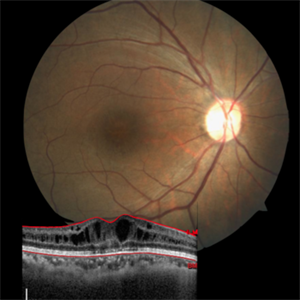
HD 1 line 100x 9 mm scan of a right eye with MTM at stage 3c. Macular Schisis Detachment.
Photographer: Drew Mitchell OCT-C
Imaging device: Zeiss Cirrus 5000
Condition/keywords: full thickness macular hole, Macular hole, myopic foveoschisis, myopic macular schisis, myopic traction maculopathy, PVD
-
 Foveoschisis
Jul 31 2018 by Pushkar Dhir
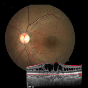
This 37-year-old male presented to us with BCVA of 6/18 . Fundus showed typical cartwheel appearance and was diagnosed with B/L foveoschisis. HRA-OCT was done for documentation which showed typical schitic pattern of fovea . Currently started on dorzolamide eye drops.
Photographer: Pushkar Dhir , Sri Sankaradeva Nethralaya, Guwahati,India
Imaging device: Zeiss
Condition/keywords: cartwheel appearance, myopic foveoschisis
-
 Foveoschisis
Jul 31 2018 by Pushkar Dhir
This 37-year-old male presented to us with BCVA of 6/18 . Fundus showed typical cartwheel appearance and was diagnosed with B/L foveoschisis. HRA-OCT was done for documentation which showed typical schitic pattern of fovea . Currently started on dorzolamide eye drops.
Photographer: Pushkar Dhir
Condition/keywords: cartwheel appearance, myopic foveoschisis
-
 Bilateral Myopic Foveoschisis
Feb 11 2016 by Mallika Goyal, MD
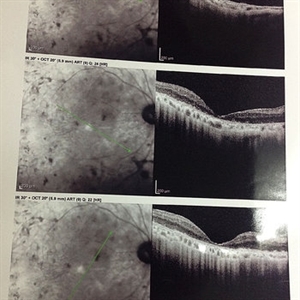
Left eye OCT of a 22-year-old lady with myopic foveoschisis & an outer lamellar macular hole who presented with complaint of Left Eye vision drop 4 months prior to presentation. BCVA was 20/400. The lamellar macular hole accounts for the vision loss.
Condition/keywords: myopic foveoschisis
-
 Bilateral Myopic Foveoschisis
Feb 10 2016 by Mallika Goyal, MD
Left eye OCT of a 22-year-old lady with bilateral myopic foveoschisis who presented with complaint of left eye vision drop 4 months prior to presentation. BCVA was 20/400. This OCT scan is taken inferior to foveal centre. The scan through foveal centre reveals an outer lamellar macular hole accounting for vision loss.
Photographer: Mallika Goyal, MD, Apollo Health City, Hyderabad, India
Condition/keywords: myopic foveoschisis
-
 Bilateral Myopic Foveoschisis
Feb 10 2016 by Mallika Goyal, MD
Left eye OCT of a 22-year-old lady with bilateral myopic foveoschisis who presented with complaint of left eye vision drop 4 months prior to presentation. BCVA was 20/400. This OCT scan is taken superior to foveal centre. The scan through foveal centre reveals an outer lamellar macular hole accounting for vision loss.
Photographer: Mallika Goyal, MD, Apollo Health City, Hyderabad, India
Condition/keywords: myopic foveoschisis
-
 Bilateral Myopic Foveoschisis
Feb 10 2016 by Mallika Goyal, MD
Left eye OCT of a 22-year-old lady with myopic foveoschisis & an outer lamellar macular hole who presented with complaint of left eye vision drop 4 months prior to presentation. BCVA was 20/400. The lamellar macular hole accounts for the vision loss.
Photographer: Mallika Goyal, MD, Apollo Health City, Hyderabad, India
Condition/keywords: myopic foveoschisis
-
 Bilateral Myopic Foveoschisis
Feb 10 2016 by Mallika Goyal, MD
Right eye OCT of a 22-year-old lady with bilateral myopic foveoschisis. BCVA is 20/25, and she is visually asymptomatic in this eye.
Photographer: Mallika Goyal, MD, Apollo Health City, Hyderabad, India
Condition/keywords: myopic foveoschisis
-
 Bilateral Myopic Foveoschisis
Feb 10 2016 by Mallika Goyal, MD
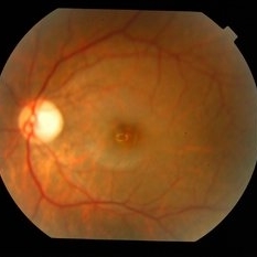
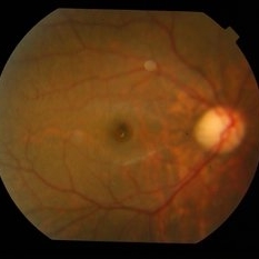
Left eye fundus of a 22-year-old lady with bilateral myopic foveoschisis who presented with complaint of left eye vision drop 4 months prior to presentation. BCVA was 20/400. OCT scan through foveal centre revealed an outer lamellar macular hole accounting for vision loss.
Photographer: Mallika Goyal, MD, Apollo Health City, Hyderabad, India
Condition/keywords: myopic foveoschisis
-
 Bilateral Myopic Foveoschisis
Feb 10 2016 by Mallika Goyal, MD
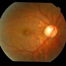
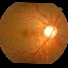
Right eye fundus of a 22-year-old lady with bilateral myopic foveoschisis. BCVA is 20/25, and she is visually asymptomatic in this eye.
Photographer: Mallika Goyal, MD, Apollo Health City, Hyderabad, India
Condition/keywords: myopic foveoschisis
-
 Bilateral Myopic Foveoschisis
Feb 10 2016 by Mallika Goyal, MD
Right eye fundus of a 22-year-old lady with bilateral myopic foveoschisis. BCVA is 20/25, and she is visually asymptomatic in this eye.
Photographer: Mallika Goyal, MD, Apollo Health City, Hyderabad, India
Condition/keywords: myopic foveoschisis
-
 Myopic Foveoschisis
May 19 2014 by Ahmad Gawady
OCT Lt of a 56-year-old female with drop of vision OS bilateral high myopia -6.0 D. Normal I.O.P., BCVA 6/18 OD , 2/60 OS., clear media, myopic fundus OD. No abnormalities OD, myopic fundus Ill defined fovea abnormality OS. OCT macular area OS : inferior juxtafoveal splitting of inner retinal layer (myopic foveoschisis) No evidence of PVD, CNV or leakage.
Condition/keywords: high myopia, optical coherence tomography (OCT)
-
 Myopic Foveoschisis
May 19 2014 by Ahmad Gawady
OCT Of 56-year-old highly myopic female with drop of vision BCVA 2/60. Clear media, myopic fundus , Ill defined macular abnormality. OCT shows inferior juxtafoveal splitting of Inner retinal layer. No evidence of PVD, CNV or leakage .
Condition/keywords: myopic foveoschisis, optical coherence tomography (OCT)
-
---thumb.jpg/image-square;max$300,300.ImageHandler) Myopic Traction Maculopathy
Myopic Traction Maculopathy
Mar 23 2014 by Hamid Ahmadieh, MD
OCT image of the left eye of a 55-year-old woman with decreased vision secondary to myopic traction maculopathy.
Photographer: Solmaz Shahmohammad , Negah Eye Center , Tehran
Imaging device: Specteralis
Condition/keywords: myopic foveoschisis, myopic traction maculopathy, optical coherence tomography (OCT)
-
---thumb.jpg/image-square;max$300,300.ImageHandler) Myopic Traction Maculopathy
Myopic Traction Maculopathy
Mar 23 2014 by Hamid Ahmadieh, MD
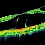
OCT image of the right eye of a 55-year-old woman with marked visual reduction due to myopic traction maculopathy manifesting as foveoschisis and neurosensory retinal detachment.
Photographer: Solmaz Shahmohammad , Negah Eye Center , Tehran
Imaging device: Specteralis
Condition/keywords: myopic foveoschisis, myopic traction maculopathy, neurosensory detachment of retina, optical coherence tomography (OCT)
 Loading…