Search results (49 results)
-
 Myelinated Nerve Fiber
Dec 5 2025 by Vishal Agrawal, MD, FRCS,FACS,FASRS
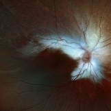
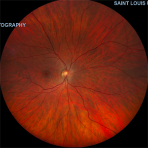
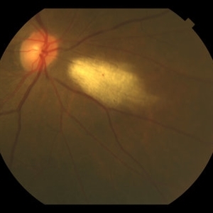
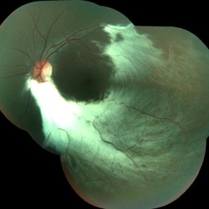
Fundus picture of left eye shows dense area of myelinated retinal nerve fibers involving the peripapillary region and extending along the superior and inferior temporal arcades.
Photographer: Dr Ayushi Gupta, Agrawal Hospital, Jaipur
Imaging device: Clarus 700
Condition/keywords: amblyopia, myelinated nerve fiber layer
-
 Myelinated Nerve Fiber Layer
Aug 13 2025 by Kimberly Wakester
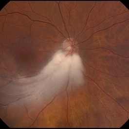
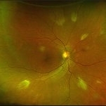
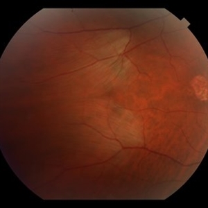
Optomap RGB of a 3-year-old girl that presents with extensive myelinated never fiber in the right eye sparing the fovea. Patient is to return in 6 months for follow up visit with repeat Optos imaging.
Photographer: Kimberly Wakester, COA, OCT-C, Retina Consultants of Carolina
Imaging device: Optos California
Condition/keywords: myelinated nerve fiber layer
-
 Myelinated Retinal Nerve Fiber Layer
May 20 2025 by Ignacio Leonardo Pueyo Bestue, MD
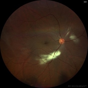
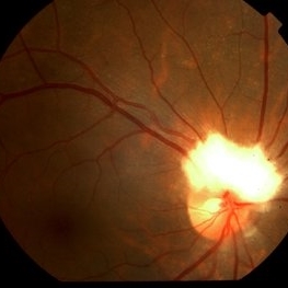
Fundus photo of an 80-year-old woman with myelinated RNFL, 20/20 vision, and mild hyperopia
Photographer: Pueyo-Bestue, I.L., MD, Universite Libre de Bruxelles, Ophthalmology Department
Condition/keywords: myelinated nerve fiber layer
-
 Myelinated Nerve Fibre (MNF)
Jun 17 2023 by Harsh Vardhan Singh, MS
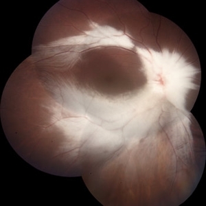
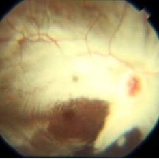
Fundus photograph of 32-year-old male having good best corrected visual acuity in both eyes with right eye having high myopia & MNF as incidental finding
Photographer: Dr Harsh Vardhan Singh, Assistant Professor, AIIMS, Guwahati
Condition/keywords: medullated nerve fibers, MNF, myelinated nerve fiber layer, myelinated nerve fibers, Nerve fiber layer arrangements, NFL
-
 Isolated myelinated nerve fiber layers
Mar 5 2023 by Niloofar Piri, MD
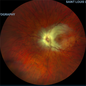
Fundus photograph of the right eye demonstrating patches of isolated myelinated nerve fiber layers along inferior arcade as well as nasal retina
Photographer: Sean Kelso, Saint Louis University
Condition/keywords: myelinated nerve fiber layer, myelinated nerve fibers
-
 Myelinated Nerve Fiber Layer
Nov 24 2022 by Eder Díaz Dorado
Fundus photograph of an 35-year-old woman with myelinated nerve fiber layer
Photographer: Eder Díaz Dorado, Hospital Central Militar CDMX
Imaging device: Smartphone
Condition/keywords: myelinated nerve fibers
-
 Normal Fundus Photo, OD in Pt. with Myelinated NFL, OS
Oct 27 2021 by Charles Hurth
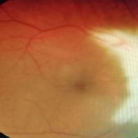
Fundus photograph of a 35-year-old woman with myelinated nerve fiber layer in her left eye who presented to the adult strabismus clinic with exotropia of her left eye.
Photographer: Charles Hurth, IV, DO, Saint Louis University
Condition/keywords: normal eye
-
 Myelinated Nerve Fiber Layer, OS
Oct 27 2021 by Charles Hurth
Fundus photograph of a 35-year-old woman with myelinated nerve fiber layer in her left eye who presented to the adult strabismus clinic with exotropia of her left eye.
Photographer: Charles Hurth, IV, DO, Saint Louis University
Condition/keywords: myelinated nerve fiber layer
-
 Myelinated Nerve Fiber Layer
Aug 2 2020 by Kelly Hannan
Myelinated nerve fiber layer.
Imaging device: heidelberg spectralis
Condition/keywords: myelinated nerve fiber layer
-
 Myelinated Nerve Fiber (mNFL)
Jun 21 2020 by Dhaivat Shah
Myelinated nerve fiber layer (mNFL) is a benign clinical entity that results from an embryologic developmental anomaly. Myelination along the visual pathway is noted around the eighth month of gestation, and typically reaches the posterior globe around the time of birth with virtually all fibers reaching complete myelination by age 7 months till the lamina cribrosa. Sometimes, due to altered neuro hormonal signals, this process of myelination extends past the lamina cribrosa and is visible on fundus examination as distinct white patches on the inner retinal surface. On infrared and red-free imaging, mNFL appears white, which is likely due to the high lipid content of myelin. Myelin blocks detection of underlying fluorescent material, thus appearing dark on fundus autofluorescence. On optical coherence tomography , it appears as a thickened and hyperreflective retinal nerve fiber layer. mNFL is typically benign but can be mistaken for other potentially serious conditions like neoplastic infiltration or infection. Hence, it is crucial to recognize the benign nature of mNFL to avoid superfluous medical testing.
Photographer: Ms Srishti Sharma
Imaging device: Choithram Netralaya
Condition/keywords: myelinated nerve fibers
-
 Plateau Fovea with Inner Retinal Thinning
May 27 2020 by Olivia Rainey
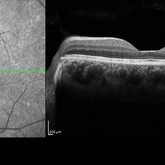
Optical coherence tomography of the left eye of a 20-year-old male with Alport Syndrome. The patient did not present with any ocular or visual symptoms, yet the distinct "plateau contour" of his fovea was noted on OCT during his visit. The patient presented with 20/25 vision at the time of his visit. There was myelinated nerve fiber layer noted in both eyes, but these features had remained stable from his appointment three years prior. The physician noted that myelinated nerve fiber was a congenital change, and had not affected his vision or health of the eye, nor is a feature of Alport Syndrome.
Photographer: Olivia Rainey, OCT-C, COA
Imaging device: Heidelberg Spectralis
Condition/keywords: Alports disease, Heidelburg Spectralis, inner retinal thinning, left eye, optical coherence tomography (OCT), plateau fovea
-
 CHRPE & Myelinated RNFL
May 21 2020 by John S. King, MD
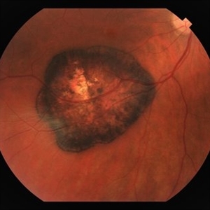
47-year-old white female, asymptomatic, sent to evaluate a scar OD. 20/40 cc, normotensive, examination significant for a flat, solitary lesion with pigmented borders and depigmented center with early lacunae forming, along with myeliated RNFL at the temporal edge of the lesion.
Photographer: Kay Dalby
Imaging device: Topcon
Condition/keywords: congenital hypertrophy of the retinal pigment epithelium (CHRPE), myelinated nerve fiber layer
-
 Multiple Areas of Myelinated RNFL OD
Sep 18 2019 by John S. King, MD
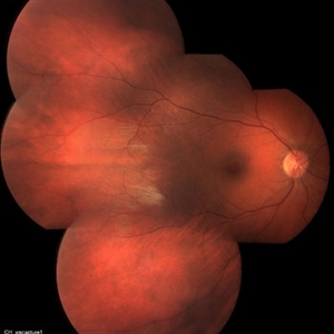
68-year-old African American male presented with an acute PVD in the fellow eye. Fellow eye had similar findings, but the pics were not as good as OD.
Photographer: Brittany Dewberry
Imaging device: Optos CA
Condition/keywords: myelinated nerve fiber layer, myelinated nerve fibers
-
 Myelinated Nerve Fiber Layer
Mar 26 2019 by Gary R. Cook, MD, FACS
39-year-old white male with persistent myelination of the nerve fiber layer superiorly in his left eye; VA=20/20.
Imaging device: Topcon VT-50
Condition/keywords: myelinated nerve fiber layer
-
 Macular Pucker With Myelinated Nerve Fiber Layer
Nov 1 2018 by Kevin J. Blinder, MD, FASRS
Multi-color photo of macular pucker with myelinated nerve fiber layer.
Photographer: Jarrod Wehmeier
Imaging device: Heidelberg Spectralis
Condition/keywords: macular pucker
-
 Myelinated Nerve Fiber Layer
May 3 2018 by Nichole Lewis
Myelinated nerve fiber layer.
Photographer: Nichole Lewis
Condition/keywords: myelinated nerve fiber layer
-
 Myelinated Nerve Fiber Layer
Apr 3 2018 by Mitzy E Torres Soriano, MD
Myelinated nerve fiber layer, left eye.
Photographer: Mitzy Torres Soriano
Condition/keywords: myelinated nerve fiber layer, myelinated nerve fibers
-
 Myelinated NFL
Dec 6 2017 by John S. King, MD
Myelinated NFL.
Imaging device: Topcon
Condition/keywords: background diabetic retinopathy (BDR), myelinated nerve fiber layer, myelinated nerve fibers
-
 Myelinated NFL
Dec 2 2017 by John S. King, MD
Myelinated NFL.
Imaging device: Topcon
Condition/keywords: myelinated nerve fiber layer, myelinated nerve fibers
-
 Myelinated Nerve Fiber Layer
Aug 19 2017 by Mitzy E Torres Soriano, MD
Myelinated Nerve Fiber Layer (Right eye)
Photographer: Mitzy E. Torres Soriano
Condition/keywords: myelinated nerve fiber layer, myelinated nerve fibers
-
 MNFL
MNFL
Sep 5 2015 by Ali Tavallali, MD, FASRS
A 16-year-old malewith diffuse MNFL of OD.
Photographer: Neda Shaibani
Condition/keywords: myelinated nerve fiber layer
-
 MNFL
MNFL
Sep 5 2015 by Ali Tavallali, MD, FASRS
A 16-year-old male with diffuse MNFL of OD.
Photographer: Neda Shaibani
Condition/keywords: myelinated nerve fiber layer
-
 Myelinated nerve fibres
Apr 29 2015 by Neha Goel, MS DNB FRCS (Glasg)
Montage fundus photograph of the left eye of a 16-year-old boy.
Photographer: Neha Goel
Imaging device: Zeiss visucam
Condition/keywords: myelinated nerve fiber layer, myelinated nerve fibers
-
 Myelinated Nerve Fibers NFL / Pseudotumor Cerebri
Feb 17 2015 by David Callanan, MD
Male patient, myelinated nerve fibers NFL / pseudo tumor cerebri.
Condition/keywords: myelinated nerve fiber layer, pseudotumor cerebri
-
 Myelinated Nerve Fiber Layer Causing Leukocoria
Jan 30 2015 by H. Michael Lambert, MD
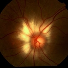
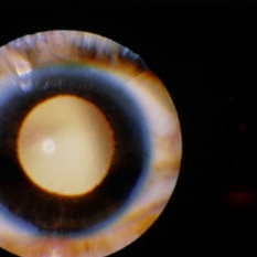
Leukocoria due to extensive myelination of optic nerve head.
Condition/keywords: leukocoria, myelinated nerve fiber layer
 Loading…