Search results (7 results)
-
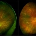
 T-Cell Lymphoma
Jul 3 2025 by Virginia Gebhart
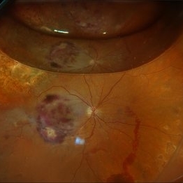
78 year old male s/p vitreous biopsy for T-Cell lymphoma. Pt presented with peripheral blot hemorrhages and numerous white subretinal infiltrates. Retinal pallor and thickening temporally. History of cutaneous T-cell lymphoma. PPV/vitreous biopsy performed to find differential diagnosis. Silicone oil was placed for 6 weeks, then removed and exchanged with a gas bubble. Hematology pathologist and Emory reviewed path report and agrees it is consistent with T-cell lymphoma. Pt received intravitreal Methotrexate and will be scheduled for weekly treatments. BCVA CF
Photographer: Virginia Gebhart, Retina Consultants of Carolina
Imaging device: Optos California
Condition/keywords: biopsy, gas bubble, lymphoma
-
 Birdshot Retinochoroiditis
Aug 8 2024 by Virginia Gebhart
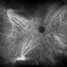
ICG angiogram of 45 year old male with Birdshot Retinochoroiditis. Has been improving on Humira and Methotrexate.
Photographer: Virginia Gebhart
Imaging device: Optos California
Condition/keywords: birdshot, indocyanine green (ICG) angiography
-
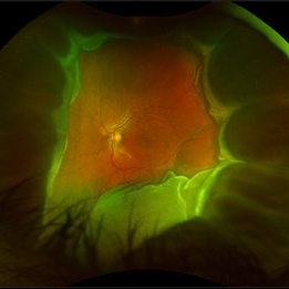
 Methotrexate Bubble following Intravitreal Injection for PVR
Sep 21 2022 by Zach Seim
Ultra-widefield fundus photograph of an 81 year old female with a Methotrexate bubble following an Intravitreal Injection for Proliferative Vitreoretinopathy. Patient has been presenting to the office for two week interval Methotrexate injections in her left eye. The image was taken prior to her eighth injection which revealed a residual Methotrexate bubble in her inferior retinal image. This patient was seeing "lots" of floaters, as well as having visual acuity of cc20/400 cc20/200 PH.
Photographer: Zach Seim
Imaging device: OPTOS California
Condition/keywords: bubble, fundus photograph, fundus photography, intravitreal injection, left eye, methotrexate, nasal retina, Optos, proliferative vitreoretinopathy (PVR), pseudocolor, ultra-wide field imaging
-
 Methotrexate Bubble in Silicone Oil Filled Eye: Proliferative Vitreoretinopathy Prevention
Jan 22 2022 by Yoshihiro Yonekawa, MD, FASRS
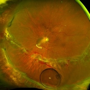
A middle-aged man underwent complex retinal detachment repair with vitrectomy, membrane peeling, relaxing retinectomy, and silicone oil tamponade. This is a wide-field image immediately after methotrexate injection during a postoperative clinic visit, for the prevention of proliferative vitreoretinopathy. The methotrexate bubble is seen inferiorly.
Photographer: Christina Rowland
Imaging device: Optos California
Condition/keywords: methotrexate, proliferative vitreoretinopathy (PVR), retinectomy, vitrectomy
-
 Bilateral Lymphoma Metastasis after Resolution with IVM
Sep 19 2018 by Olivia Rainey
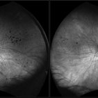
Ultra-wide field, autofluorescence images of an 86-year-old female treated with intravitreal methothrexate as a management of subretinal infiltrate in the macula of the right eye, as a manifestation of leukemia. Her last intravitreal methotrexate injection was 5/1/18.
Photographer: Olivia Rainey
Imaging device: Optos
Condition/keywords: bilateral, fundus autofluorescence (FAF), lymphoma, Optos, ultra-wide field imaging, uveitis
-
 Bilateral Lymphoma Metastasis after Resolution with IVM
Sep 19 2018 by Olivia Rainey
Ultra-wide field, pseudocolor fundus images of an 86-year-old female treated with intravitreal methothrexate as a management of subretinal infiltrate in the macula of the right eye, as a manifestation of leukemia. Her last intravitreal methotrexate injection was 5/1/18.
Photographer: Olivia Rainey
Imaging device: Optos
Condition/keywords: bilateral, leukemia, methotrexate, Optos, pseudocolor, ultra-wide field imaging, uveitis
-
 Disseminated Retinitis and Retinochoroiditis, Metastatic
May 16 2017 by Karen Panzegrau
Fundus photograph of 44-year-old male with plasmacytoma infiltation of the choroid confirmed by biopsy, associated with disseminated retinitis, and retinochoroiditis. Vision is LP. Patient treated with intravitreal methotrexate
Photographer: Karen Panzegrau
Imaging device: Optos
Condition/keywords: metastatic lesion, methotrexate, Optos, plasmacytoma, retinitis, retinochoroiditis, unilateral exudative retinal detachment
 Loading…