Search results (165 results)
-
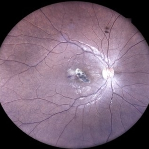
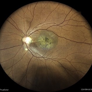
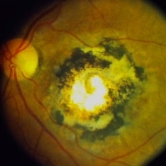
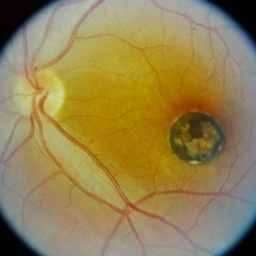
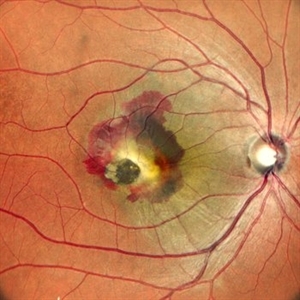 When the Macula Decides to Bleed... Artistically (Case of Macular Scar with Subretinal Bleed)
Jun 2 2025 by rohan jain
A case of 42 years old male. Color photograph showing macular scar with subretinal bleed.
Photographer: Dr. ROHAN JAIN
Imaging device: mirante
Condition/keywords: CNVM, macular scar, scar, subretinal hemorrhage, subretinal blood
-
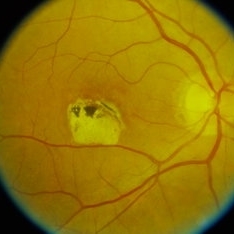
 Chorioretinal Macula Scar (Macula View)
May 12 2025 by Briana Hernandez
Zoomed in Macular View of Chorioretinal Macular Scar in 9-year-old female patient.
Photographer: Briana Hernandez, Hilton Head Retina Insitute
Imaging device: Optos
Condition/keywords: chorioretinal scar
-
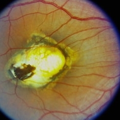
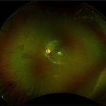 Chorioretinal Macula Scar (Ultrawide View)
May 12 2025 by Briana Hernandez
Ultra wide Optos image of Chorioretinal Macular Scar in 9-year-old female patient.
Photographer: Briana Hernandez, Hilton Head Retina Institute
Imaging device: Optos
Condition/keywords: chorioretinal scar, macular scar, ultra-wide field imaging
-
 Comets in the Eye (Retinopathy of Prematurity)
Apr 8 2025 by KANWALJEET HARJOT MADAN, M.S. (Ophthalmology); FAICO (Vitreous - Retina)
This is the fundus picture of right eye (RE) of a 4 years female child presented with outward deviation of right eye. Her parents also complained of diminution of vision in both eyes. On examination, her best corrected vision in RE was hand movements close to face and was 20/80 in LE. Posterior segment exam revealed presence of macular scar in RE and presence of dry retinal fold with dragging of retinal vessels. LE fundus revealed presence of nasal drag of optic disc. Parents gave history of untreated ROP as an infant. Retinopathy of Prematurity (ROP) is a Vaso proliferative disorder of Retina occurring in premature infants. Advances in neonatal care and ROP treatment has led these babies to live longer with this disease.
Photographer: Dr. Kanwaljeet Harjot Madan, Thind Eye Hospital, Jalandhar City (Punjab) INDIA.
Imaging device: Zeiss Fundus Camera
Condition/keywords: Retinopathy of Prematurity, Vaso proliferative disorder
-
 Comets in the Eye (Retinopathy of Prematurity)
Apr 8 2025 by KANWALJEET HARJOT MADAN, M.S. (Ophthalmology); FAICO (Vitreous - Retina)
This is the fundus picture of right eye (RE) of a 4 years female child presented with outward deviation of right eye. Her parents also complained of diminution of vision in both eyes. On examination, her best corrected vision in RE was hand movements close to face and was 20/80 in LE. Posterior segment exam revealed presence of macular scar in RE and presence of dry retinal fold with dragging of retinal vessels. LE fundus revealed presence of nasal drag of optic disc. Parents gave history of untreated ROP as an infant. Retinopathy of Prematurity (ROP) is a Vaso proliferative disorder of Retina occurring in premature infants. Advances in neonatal care and ROP treatment has led these babies to live longer with this disease.
Photographer: Dr. Kanwaljeet Harjot Madan, Thind Eye Hospital, Jalandhar City (Punjab) INDIA.
Imaging device: Zeiss Fundus Camera
Condition/keywords: Retinopathy of Prematurity
-
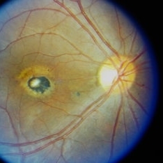
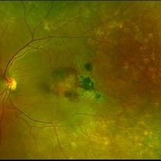
 Toxoplasma Macular Scar with CNVM
Mar 7 2025 by T. P . VIGNESH, MBBS,MS
Fundus photograph of the right eye of a 28-year-old woman with a macular toxoplasma scar and CNVM.
Photographer: Sivanath
Imaging device: EIDON
Condition/keywords: CNVM, ocular toxoplasmosis
-
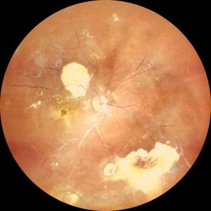
 Coat's Disease with Exudative RD
Feb 12 2025 by Tejaswita Verma
Fundus photo of a 7 year old boy with vision Counting fingers close to face in the right eye and intermittent outward deviation of the right eye observed by parents. Fundus examination shows subretinal exudates, telengiectatic vessels in superotemporal quadrant, intraretinal hemorrhages, macular scar, NVD.
Photographer: DR. TEJASWITA VERMA
Imaging device: MIRANTE
Condition/keywords: Coats' disease, exudative retinal detachment
-
 Gunshot Injury
Dec 19 2024 by Angela Rico
53 y/o M who suffered gunshot wound to OD. Picture shows macular scar and sub retinal hemorrhage
Photographer: Angela Rico M.D.
Condition/keywords: macular scar, penetrating trauma
-
 Coats Disease
Sep 29 2024 by Tejaswita Verma
Fundus photo of the RE of a 14 y/o female ,nil premorbid presented with reduced vision in the RE ,diagnosed incidentally on ophthalmological examination elsewhere .Vision was finger counting 3 meters in the RE . Fundus picture reveals macular scar , subretinal and intraretinal exudation ,with scattered hemorrhages esp. in STQ, sclerosed vessels in superior, superonasal quadrant ,nasal, inferonasal quadrant, CR scars inferiorly, Telengiectatic vessels S/O Coat's disease. She was advised RE anti VEGF x1 + laser PRP + PST kenacort under GA with guarded prognosis.
Photographer: DR. TEJASWITA VERMA
Imaging device: MIRANTE
Condition/keywords: Coats' disease
-
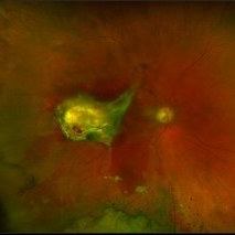
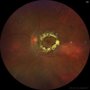
 Eyes Too Celebrate Valentine’s Day
Jul 28 2024 by KANWALJEET HARJOT MADAN, M.S. (Ophthalmology); FAICO (Vitreous - Retina)
A 53 years male patient presented with decrease in vision in left eye for 6 months. His vison in left eye was counting fingers 1 meter. His vison in right eye was 20/20. Fundus examination in left eye depicted presence of large orange shaped elevated subretinal mass superior to optic disc with scar in macula. We made clinical diagnosis of Choroidal Hemangioma with macular scar. Fundus Fluorescein Angiography (FFA) in left eye revealed early fluorescence in area corresponding to Choroidal Hemangioma which persisted in late phases. Macular scar was “HEART” shaped on FFA which was very unique incident finding.
Photographer: Dr. Kanwaljeet Harjot Madan
Imaging device: Ziess Clarus
Condition/keywords: Choroidal Hemangioma, Fundus examination, Fundus Fluorescein Angiography
-
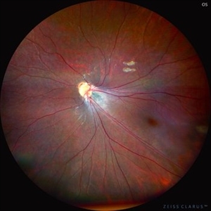
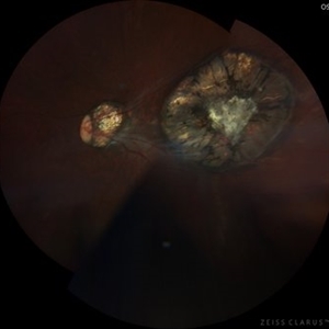
 Optic Disc Pit With Macular Scar
Jun 24 2024 by Akansha Sharma
Color fundus photograph of a 42 year old male with optic disc pit with macular scar.
Photographer: Dr. Akansha Sharma, Bharati Eye Hospital
Condition/keywords: macular scar, Optic disc pit
-
 Laser Photocoagulation Macular Fibrous Scar
May 25 2024 by Gustavo Del Castillo-Marquez, MD
Fundus photograph of an 55 year old man who received macular laser photocoagulation 12 years earlier.
Photographer: Gustavo Del Castillo-Márquez, Asociación Para Evitar la Ceguera en México, CDMX
Imaging device: Zeiss Clarus
Condition/keywords: fibrous macular scar, laser photocoagulation, macular laser
-
 Laser Photocoagulation Macular Scar 12 Years Later
May 25 2024 by Gustavo Del Castillo-Marquez, MD
Actual Fundus photograph of a 55 year old man who received macular laser photocoagulation 12 years earlier.
Photographer: Gustavo Del Castillo-Márquez, Asociación Para Evitar la Ceguera en México, CDMX
Imaging device: Zeiss Clarus
Condition/keywords: fibrous macular scar, laser macula, laser photocoagulation
-
 Toxoplasmosis Disease
Mar 30 2024 by Karen Flores Guevara
Fundus photograph of a 7-year-old-child with a macular scar observed over time for growth.
Photographer: Diana Elizabeth Arellano Acosta MD Pediatric Retina,Asociación para Evitar la Ceguera en México IAP. México
Condition/keywords: toxoplasmosis chorioretinitis
-
 Macular Telangiectasia Type 2
Mar 29 2024 by Lucy V Cobbs, M.D.
Color fundus photograph of the left eye of a 70-year-old male with a disciform scar resulting from a neovascular membrane. A minority of MacTel type 2 patients develop neovascular disease, and the gold standard treatment is anti-VEGF intravitreal therapy. Without treatment, membranes may progress to severe central macular scarring. Late stages of proliferative MacTel type 2 may be confused with AMD, and a differentiating aspect is that MacTel type 2 typically lacks pigment epithelial detachments and drusen.
Condition/keywords: Mac Tel type 2
-
 Toxoplasmosis Macular Scar
Mar 8 2024 by Andre Beckenkamp
Optos image of a patient with extensive choriorretinal scar due to toxoplasmosis infection.
Photographer: Andre Beckenkamp,MD , Prevent Senior
Condition/keywords: toxoplasmosis
-
 Ocular toxoplasmosis
Sep 21 2023 by Ben Serar
Fundus photograph of LE showing a scarred lesion at the macula in a case of Ocular Toxoplasmosis.
Condition/keywords: macular scarring, ocular toxoplasmosis
-
 Macular scar
Sep 21 2023 by Ben Serar
Fundus photograph of the RE showing scarring at the macula with hyperpigmention.
Condition/keywords: macular scar
-
 Subretinal fibrosis
Sep 14 2023 by Ben Serar
Fundus photograph of LE showing a scarred lesion at the macula, with sub retinal fibrosis.
Condition/keywords: macular scar, Subretinal fibrosis
-
 Macular scar
Sep 14 2023 by Ben Serar
Fundus photograph of RE showing hyper pigmented lesion at the posterior pole indicative of scarring at the macula.
Condition/keywords: macular scar
-
 Ocular toxoplasmosis
Sep 12 2023 by Ben Serar
Fundus photograph of RE showing Scarred Toxoplasma lesion at the macula.
Condition/keywords: macular scarring, ocular toxoplasmosis
-
 Ocular Toxoplasmosis
Sep 12 2023 by Ben Serar
Fundus photograph of LE showing scarring at macula in a case of Ocular Toxoplasmosis.
Condition/keywords: macular scarring, ocular toxoplasmosis
-
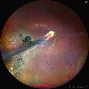
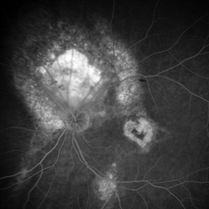
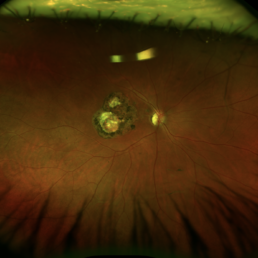
 Subretinal Hemorrhage with Chorioretinal Macular Scars
Sep 28 2022 by Denica Rodriguez
Ultra-widefield pseudocolor fundus photograph of a 59 year old female with Subretinal Hemorrhage with Chorioretinal Macular Scars affecting her left eye. The physician presumes the etiology is CNV from adjacent scarring/choroidal rupture. Patient has history of ocular trauma with cricket ball at age 10-12 years old. She suspects that she might have suffered choroidal rupture, which has resulted in secondary CNV and hemorrhage that we are seeing today. She recommends treatment with Eylea sample injection in a series of 4 at a 4-5 week interval. The patient's vision at the time of her appointment was Dcc20/40-2 PHNI.
Photographer: Denica Rodriguez, COA
Imaging device: Optos California
Condition/keywords: antiVEGF therapy, chorioretinal scar, choroidal neovascular membrane (CNVM), fundus photography, left eye, macular scar, Optos, peripheral drusen, pseudocolor, secondary CNV, subretinal hemorrhage, ULTRA WIDE FIELD, ultra-wide field imaging
-
 Ocular Toxoplasmosis
May 6 2022 by Eder Díaz Dorado
Fundus photograph of an 31-year-old man with a macular scar of toxoplasmosis and a review with OCT
Photographer: Eder Díaz Dorado, Hospital Central Militar, Ciudad de México
Imaging device: Heidelberg Spectralis /Smartphone Photography
Condition/keywords: inactive toxoplasmosis, ocular toxoplasmosis, toxoplasmosis chorioretinitis
-
 Congenital Toxoplasmosis Macular Scarring
Nov 6 2021 by Emmanouil Gavalas, MD
Fundus photographs of an 26-year-old female showing right eye macular scarring Incidental diagnosis Visual Acuity OD 6/12 OS 6/6
Photographer: Emmanouil Gavalas MD, Ophthalmos Reseach and Educational Institute,Nicosia,Cyprus
Imaging device: Zeiss Clarus 500
Condition/keywords: congenital toxoplasmosis, macular scar, ocular toxoplasmosis
 Loading…