Search results (608 results)
-
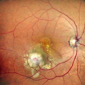
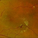
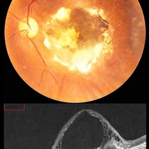
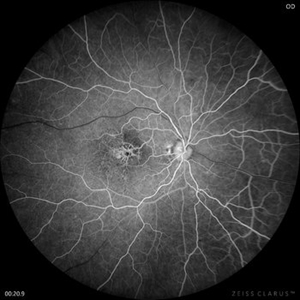
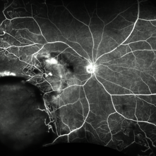
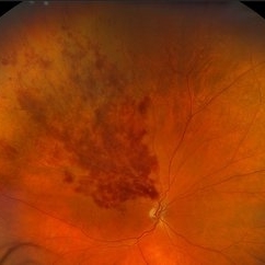
 Leber’s Miliary Aneurysm
Dec 12 2025 by KANWALJEET HARJOT MADAN, M.S. (Ophthalmology); FAICO (Vitreous - Retina)
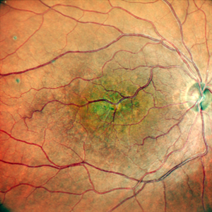
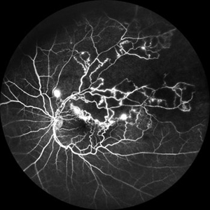
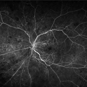
A 34 year-old male presented with decrease vision in right eye for 3 months. Anterior segment exam was normal. Fundus exam in RE revealed presence of macular edema which was evident on OCT. Multiple retinal vascular aneurysmal dilatations with telangiectasia of the retina blood vessels noted superiorly which was evident on FFA. These aneurysms were multiple, tiny and leaky on FFA. He was diagnosed to have Leber’s miliary aneurysms. It is a rare, typically unilateral eye condition, often seen in young males, characterized by multiple tiny, leaky aneurysms in the retinal blood vessels, leading to deposits of hard exudates and potential vision loss, especially if it affects the macula. It is considered a milder form of Coats' disease.
Photographer: Dr. Kanwaljeet Harjot Madan, Thind Eye Hospital, Jalandhar City (Punjab) INDIA.
Imaging device: Zeiss Fundus Camera
Condition/keywords: FFA, Leber's miliary aneurysm
-
 Ozurdex
Nov 22 2025 by Gabriel Costa Andrade, PhD
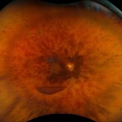
Anterior segment photograph of a Ozurdex implant in a 53-year-old man with macular edema due to intermediate uveitis.
Photographer: Gabriel Andrade
Condition/keywords: Ozurdex implant
-
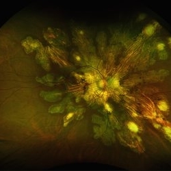
 Gyrate Atrophy
Nov 22 2025 by Gaurav Kamble
A 12-year-old female presented with progressive blurring of vision for distance and had a known history of convulsions. Ocular examination revealed bilateral proptosis and megalocornea. Fundus evaluation showed well-defined scalloped areas of peripheral chorioretinal degeneration characteristic of gyrate atrophy, along with cystoid macular edema involving the macular region. The overall clinical picture was consistent with gyrate atrophy.
Photographer: Ms. Vishaka Shah , Isha Eye Care Pvt Ltd ,Khadakpada, Kalyan
Imaging device: Optos Imaging Daytona
Condition/keywords: gyrate atrophy
-
 CRAO
CRAO
Oct 29 2025 by Jeffrey Barker
94 year old female with a CRAO with macular edema.
Photographer: Jeffrey P. Barker, B.S.
Condition/keywords: color fundus photograph, CRAO
-
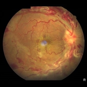
 The Great Vascular Traffic Jam: Combined Retinal Vein and Artery Occlusion
Oct 29 2025 by SHRADDHA RAJ SHRIVASTAVA
57 year old female, recently diagnosed with accelerated hypertension, developed Right eye Combined Retinal Vein and Artery Occlusion. Posterior pole image showed severe disc edema with peri-papillary haemorrhages. There is significant retinal whitening suggestive of edema leading to the classic cherry red spot at the macula. We can also see segmented flow of blood in retinal arterioles, which is the characteristic cattle-trucking seen in central retinal artery occlusion (CRAO). Widefield image revealed multiple intra-retinal blot hemorrhages in all quadrants with tortuous dilated vessels suggestive of central retinal vein occlusion (CRVO).
Photographer: Dr. Shraddha Raj Shrivastava
Imaging device: Nidek Mirante SLO/OCT (Confocal scanning/Spectral domain OCT)
Condition/keywords: central retinal vascular obstruction, central retinal vein occlusion (CRVO), CRVO with macular edema
-
 The Great Vascular Traffic Jam: Combined Retinal Vein and Artery Occlusion
Oct 29 2025 by SHRADDHA RAJ SHRIVASTAVA
57 year old female, recently diagnosed with accelerated hypertension, developed Right eye Combined Retinal Vein and Artery Occlusion. Posterior pole image showed severe disc edema with peri-papillary hemorrhages. There is significant retinal whitening suggestive of edema leading to the classic cherry red spot at the macula. We can also see segmented flow of blood in retinal arterioles, which is the characteristic cattle-trucking seen in central retinal artery occlusion (CRAO). Widefield image revealed multiple intra-retinal blot hemorrhages in all quadrants with tortuous dilated vessels suggestive of central retinal vein occlusion (CRVO).
Photographer: Dr. Shraddha Raj Shrivastava
Imaging device: Nidek Mirante SLO/OCT (Confocal scanning/Spectral domain OCT)
Condition/keywords: central retinal artery occlusion (CRAO), central retinal vascular obstruction, central retinal vein occlusion (CRVO), CRVO with macular edema
-
 Lasered Retinal Artery Macroaneurysm
Sep 22 2025 by Tejaswita Verma
Fundus image of a 73 year old hypertensive female status post focal laser for exudative RAM. There was associated macular edema on OCT. Vision was 6/18.Patient was also planned for intravitreal anti VEGF injection on the same day.
Photographer: DR. TEJASWITA VERMA
Imaging device: MIRANTE
Condition/keywords: focal laser, RAM, retinal artery macroaneurysm
-
 Retinal Artery Macroaneurysm With Macular Edema
Sep 12 2025 by Tejaswita Verma
Fundus photo of a 73 year old hypertensive female with 6/18 vision, presenting with RAM ,with surrounding hard exudates and macular edema. She was advised focal laser, anti VEGF injection.
Photographer: DR. TEJASWITA VERMA
Imaging device: MIRANTE
Condition/keywords: RAM, retinal arterial macroaneurysm
-
 Retinal Macroaneurysm OS (RAM)
Aug 20 2025 by Drew Mitchell
Optos Color photograph of a 79 year old woman with non central macular edema and exudates around RAM inferotemporally.
Photographer: Drew Mitchell OCT-C
Imaging device: Optos Silverstone
Condition/keywords: color photo, exudates, OPTOS, RAM, retinal macroaneurysm
-
 Macular Mount Everest
Aug 8 2025 by Anand Temkar
A 75 yrs old male came with the chief complains of DOV in LE since past 20 yrs. His BCVA in RE was 6/9 and in LE, it was CF 1 meter. His IOP was 13 mm of Hg in RE and 15 mm of Hg in LE. Patient is a k/c/o DM type 2 since past 20 yrs and is on regular medication. Patient is a k/c/o solitary kidney. Patient gives h/o ( LE ) Intravitreal injection Avastin 3 times 13 yrs ago i/c/o CNVM. In the LE color photo we can see the scarred CNVM along with altered foveal contour. LE OCT also shows cystic spaces with large elevation and scarring.
Photographer: Dr.Anand Temkar- Vasan Eye Hospital, Tiruchirapalli
Condition/keywords: CNVM, macular edema, scarred cnvm
-
 Congenital Retinal Macrovessel
Aug 7 2025 by Cesar Valdez, MD
Fundus photograph and fluorescein angiography of a 43-year-old man with typical findings of congenital retinal macrovessel and RPE atrophy due to chronic macular edema.
Photographer: César Valdez, Instituto Mexicano de Oftalmología, IAP. Querétaro, México.
Imaging device: Zeiss Clarus 700
Condition/keywords: congenital retinal macrovessel
-
 Congenital Retinal Macrovessel
Aug 7 2025 by Cesar Valdez, MD
Fundus photograph and fluorescein angiography of a 43-year-old man with typical findings of congenital retinal macrovessel and RPE atrophy due to chronic macular edema.
Photographer: César Valdez, Instituto Mexicano de Oftalmología, IAP. Querétaro, México.
Imaging device: Nidek Mirante
Condition/keywords: Retina
-
 Central Retinal Vein Occlusion with Macular Edema
Aug 4 2025 by Virginia Gebhart
58 year old female with Central Retinal Vein Occlusion with Macular Edema. Diffuse retinal hemorrhages, central SRF and sub-hyaloid/vitreous hemorrhage, no obvious active NV. Will start patient on series of 4 monthly intravitreal anti-VEGF injections, followed by T&E protocol once stable.
Photographer: Virginia Gebhart, Retina Consultants of Carolina
Imaging device: Optos California
Condition/keywords: central retinal vein occlusion, CRVO, CRVO with macular edema, retinal hemorrhage, vitreous hemorrhage
-
 Branch Retinal Vein Occlusion
Jul 23 2025 by Malvika Singh
Fluorescein angiogram of a 52 year old man showing capillary non perfusion areas and leakages along the superotemporal arcade and at the macula.
Photographer: Dr Malvika Singh, Retina Foundation, Ahmedabad, India
Imaging device: Mirante SLO/OCT
Condition/keywords: branch retinal vein occlusion (BRVO), CNP areas, FLUORESCEIN ANGIOGRAPHY, fluorescein leakage, macular edema
-
 Retinal Vein Occlusion
Jul 5 2025 by T. P . VIGNESH, MBBS,MS
Fundus photograph of a 52-year-old man with a macular branch retinal vein occlusion and macular edema.
Photographer: Sivanath
Imaging device: EIDON
Condition/keywords: branch retinal vein occlusion (BRVO)
-
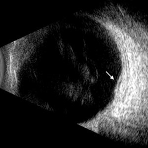 Diabetic Macular Edema
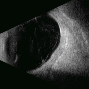
Jul 3 2025 by Gustavo Uriel Fonseca Aguirre
This B-mode longitudinal ultrasound scan demonstrates diabetic macular edema with mild subretinal fluid accumulation, appearing as a subtle hypoechoic space beneath the neurosensory retina. The macular region shows retinal thickening and heterogeneous medium reflectivity, consistent with active exudative changes (arrow). No vitreomacular traction is observed.
Photographer: Gustavo U. Fonseca Aguirre, Hospital Conde de Valenciana, Ciudad de México
Condition/keywords: diabetic macular edema
-
 Fluorescein Angiography (FA) of a Primary Retinal Vasoproliferative Tumor
Jun 29 2025 by Marcelo Zas, MD PhD
We present a case of a 33-year-old male patient, who presented with decreased visual acuity in his right eye with 20/80, presenting a primary retinal vasoproliferative tumor in the lower temporal quadrant. The fluorescein angiography findings are: 1. Early hyperfluorescence due to its rich intrinsic vascularity and often has dilated feeding arterioles and draining venules. 2. Marked progressive leakage from the tumor vessels. 3. The late leakage often obscures fine vascular details in the late phase and corresponds to exudation and macular edema seen clinically. 4. Staining of surrounding exudates, RPE disturbances and gliosis. 5. In our case also a marked peripheral capillary closure in the areas adjacent to the tumor and in other quadrants as well.
Photographer: Marcelo Zas MD PhD
Condition/keywords: RETINAL VASOPROLIFERATIVE TUMOR
-
 Serpiginous Choroidopathy
Jun 23 2025 by César Adrián Gómez Valdivia, MD
Fundus photograph of a 29 year-old female patient diagnosed with Serpiginous Choroidopathy. Finings were bilateral. The most common complication of SC is choroidal neovascularization affecting up to 35% of patients. Other reported complications are subretinal fibrosis, cystoid macular edema, branch vein occlusion, serous retinal detachment, optic disc neovascularization ,and anterior uveitis.
Photographer: @eyemissu2
Imaging device: TOPCON TRC-50DX
Condition/keywords: serpiginous choroiditis
-
 Serpiginous Choroidopathy
Jun 23 2025 by César Adrián Gómez Valdivia, MD
Fundus photograph of a 29 year-old female patient diagnosed with Serpiginous Choroidopathy. Finings were bilateral. The most common complication of SC is choroidal neovascularization affecting up to 35% of patients. Other reported complications are subretinal fibrosis, cystoid macular edema, branch vein occlusion, serous retinal detachment, optic disc neovascularization, and anterior uveitis.
Photographer: @eyemissu2
Imaging device: California ICG OPTOS
Condition/keywords: serpiginous choroiditis
-
 Central Retinal Vein Occlusion
Jun 21 2025 by Moazzam Parvez
Fundus photograph of a 56 year old male presenting with dilated tortuous vessels with adjoining Hard exudates and macular star.
Photographer: Moazzam Parvez , Netralayam , Kolkata
Imaging device: Topcon Maestro 2
Condition/keywords: CRVO with macular edema, hard exudates, macular star
-
 Central Retinal Vein Occlusion With Waldenstroms macroglobulinemia
Jun 18 2025 by Korey Starkey
64-year-old patient presents with CRVO with secondary macular edema in both eyes. Venous beading present in 2/4 quadrants OU. Patient diagnosed with Waldenstroms macroglobulinemia, found on SPEP and bone marrow biopsy. Treatment recommended of anti-vegF intravitreal injections OU.
Photographer: Korey Starkey
Imaging device: Optos
Condition/keywords: attenuated vessels, central retinal vein occlusion (CRVO), CRVO, FA early phase, FLUORESCEIN ANGIOGRAPHY, macular edema, Optomap, OPTOS CALIFORNIA, severe NPDR, venous beading, Waldenstroms macroglobulinemia
-
 Macular Edema
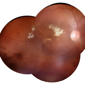
Jun 4 2025 by Paulina Araujo
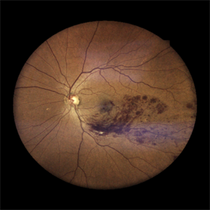
The composite fundus photograph of the right eye demonstrates circinate hard exudates in the thickened macular area, along with flame-shaped intraretinal hemorrhages along the inferior temporal arcade.
Photographer: Paulina D.Araujo Martínez, Asociación para Evitar la Ceguera en México I.A.P., Hospital Dr Luis Sánchez Bulnes.
Condition/keywords: macular edema
-
 Diabetic Macular Edema
Apr 28 2025 by Gustavo Uriel Fonseca Aguirre
This B-mode longitudinal ultrasound scan demonstrates irregular macular thickening with homogeneous medium-to-high internal reflectivity, consistent with diabetic macular edema. The lesion shows poorly defined borders and absence of cystic spaces or subretinal fluid on dynamic evaluation.
Photographer: Gustavo U. Fonseca Aguirre, Hospital Conde de Valenciana, Ciudad de México
Condition/keywords: diabetic macular edema
-
 Retinitis Pigmentosa
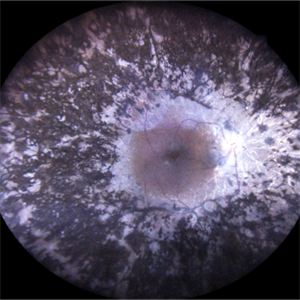
Mar 27 2025 by T. P . VIGNESH, MBBS,MS
Fundus photograph of a 52-year-old woman with retinitis pigmentosa with cystoid macular edema.
Photographer: Bharathi
Imaging device: EIDON
Condition/keywords: retinitis pigmentosa
-
 BRVO with Macular Edema
Mar 20 2025 by Virginia Gebhart
71 year old male with new branch retinal vein occlusion with macular edema. Mild central SRF, extensive superior CME and flame hemorrhages. Recommended series of anti-VEGF injections, completed first IVA
Photographer: Virginia Gebhart, Retina Consultants of Carolina
Imaging device: Optos California
Condition/keywords: branch retinal vein occlusion (BRVO), BRVO, macular edema
 Loading…