Search results (244 results)
-
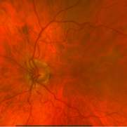
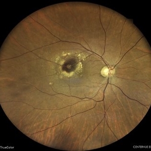
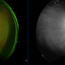 Retinal Macroaneurysm (Left Eye)
Apr 29 2025 by Daniela Bogenschutz
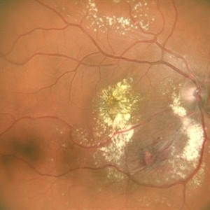
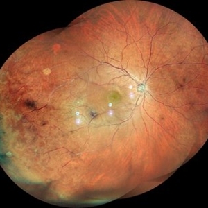
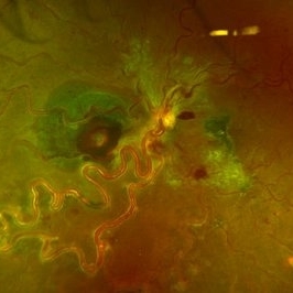
72 year-old female has visual complaints of central vision changes ongoing for 4 days. Patient was acutely symptomatic with an intraretinal hemorrhage due to the retinal macroaneurysm. We had a fun little laugh as this retinal macroaneurysm form a shape of a tick in her left eye. This photo is a side-by-side of the color photos and the autofluorescence done. She is being treated by her general doctor for elevated blood pressure.
Photographer: Daniela Bogenschutz, OSC; Retina Consultants of Carolina, P.A.
Imaging device: Optos
Condition/keywords: retinal macroaneurysm
-
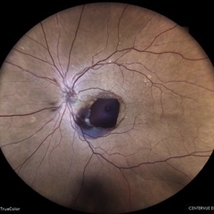
 MacroAneurysm - 1 Day After Rupture
Mar 31 2025 by Max Whitmeyer
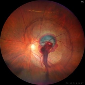
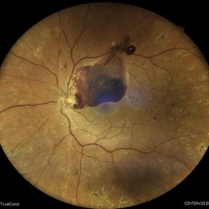
Fundus photograph of a macroaneurysm progression prior to and following rupture.
Photographer: Natasa Stankovich, Edward Hines Jr. VA Hospital
Imaging device: Zeiss Clarus 500
Condition/keywords: color fundus photograph, macroaneurysm
-
 MacroAneurysm - 1 Week Before Rupture
Mar 31 2025 by Max Whitmeyer
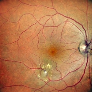
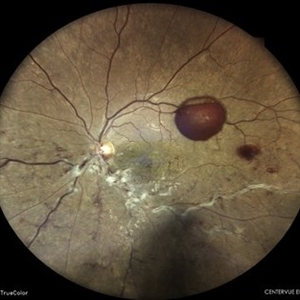
Fundus photograph of a macroaneurysm progression prior to and following rupture.
Photographer: Natasa Stankovich, Edward Hines Jr. VA Hospital
Imaging device: Zeiss Clarus 500
Condition/keywords: color fundus photograph, macroaneurysm
-
 MacroAneurysm - 2 Months Before Rupture
Mar 31 2025 by Max Whitmeyer
Fundus photograph of a macroaneurysm progression prior to and following rupture.
Photographer: Natasa Stankovich, Edward Hines Jr. VA Hospital
Imaging device: Zeiss Clarus 500
Condition/keywords: color fundus photograph, macroaneurysm
-
 MacroAneurysm - 3 Months Before Rupture
Mar 31 2025 by Max Whitmeyer
Fundus photograph of a macroaneurysm progression prior to and following rupture.
Photographer: Natasa Stankovich, Edward Hines Jr. VA Hospital
Imaging device: Zeiss Clarus 500
Condition/keywords: color fundus photograph, macroaneurysm
-
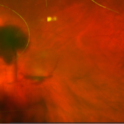
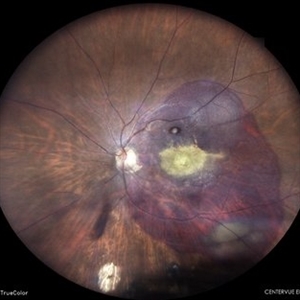
 The Pouring RAM
Mar 25 2025 by Shrishti mishra
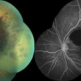
A 63 year old male with RAM lesion in the right eye associated with multilayered hemorrhage.
Imaging device: Optos nikon
Condition/keywords: FFA, retinal arterial macroaneurysm, subhyaloid hemorrhage
-
 Retinal Macroaneurysm (RAM)
Mar 19 2025 by Drew Mitchell
3x3 OCT-A of a Retinal Macroaneurysm in the left eye along the IT arcade that has surrounding edema and exudates
Photographer: Drew Mitchell, OCT-C
Imaging device: Zeiss Cirrus 5000
Condition/keywords: CIRRUS 5000 ANGIOPLEX, OCT Angiography, RAM, retinal macroaneurysm
-
 Retinal Macroaneurysm (RAM)
Mar 19 2025 by Drew Mitchell
3x3 OCT-A of a Retinal Macroaneurysm in the left eye along the IT arcade that has surrounding edema and exudates.
Photographer: Drew Mitchell OCT-C
Imaging device: Zeiss Cirrus 5000
Condition/keywords: OCT Angiography, RAM, retinal macroaneurysm
-
 Ruptured Retinal Arterial Macro-Aneurysm
Oct 27 2024 by César Adrián Gómez Valdivia, MD
Ruptured retinal arterial macro-aneurysm found in a 56 YO female patient with history of untreated hypertension. Round or fusiform dilation of a retinal arteriole is usually seen within a third degree branch of one of the four main arcade arteries. Most common location for a symptomatic macroaneurysm is from a branch of the superotemporal arcade.
Photographer: @eyemissu2
Imaging device: TOPCON TRC-50DX
Condition/keywords: ruptured macroaneurysm
-
 Retinal Artery Macro-aneurysms
Jul 19 2024 by Anjana Mirajkar, MS Ophthalmology
An intra operative still of LE showing a retinal artery macro aneurysm causing a sub hylaoid and sub ILM hemorrhage.
Photographer: Dr. Anjana Mirajkar -Retina Foundation, Ahmedabad
Imaging device: Mirante-Nidek
Condition/keywords: retinal arterial macroaneurysm, sub hyaloid hemorrhage, sub internal limiting membrane haemorrhage
-
 Retinal Artery Macroaneurysm
Jul 13 2024 by Tejaswita Verma
A 53 year old female presented with blurred vision in RE since a month ,with borderline DM and HTN not on medications .H/o highest BP recording was 160/90 mm Hg.Vision 6/60 .FFA revealed leakages. She was advised RE focal laser with intravitreal anti-VEGF injections
Photographer: DR. TEJASWITA VERMA
Imaging device: MIRANTE
Condition/keywords: RETINAL ARTERY MACROANEURYSM
-
 RAM
RAM
Jun 25 2024 by Tejaswita Verma
Right eye Fundus photo of a 73 year old female with 6/ 9 vision having retinal artery macroaneurysm.
Photographer: DR. TEJASWITA VERMA
Imaging device: MIRANTE
Condition/keywords: RETINAL ARTERY MACROANEURYSM
-
 Combined Retinal Artery Macro Aneurysm with Retinal Vein Occlusion
Jun 25 2024 by Aniruddh Soni, DO DNB FLVPEI
Color Fundus and Fluorescein Angiography Montage of a 55 year Lady with Combined Retinal Artery Macro Aneurysm with Retinal Vein Occlusion.
Photographer: Dr Aniruddh Soni, Anupam Eye Hospital, Jaipur, INDIA
Condition/keywords: macroaneurysm, Vein Occlusion
-
 Combined Retinal Artery Macro Aneurysm with Retinal Vein Occlusion
Jun 25 2024 by Aniruddh Soni, DO DNB FLVPEI
Left Eye Color Fundus and Fluorescein angiography Monatge of a 55 Year Female showing Retinal Artery Macro Aneurysm with Retinal Vein Occlusion.
Photographer: Dr Aniruddh Soni, Anupam Eye Hospital, Jaipur, INDIA
Condition/keywords: macroaneurysm, Vein Occlusion
-
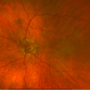
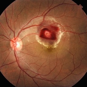
 Ruptured Retinal Artery Macroaneurysm
Jun 18 2024 by KANWALJEET HARJOT MADAN, M.S. (Ophthalmology); FAICO (Vitreous - Retina)
This is a fundus photo depicting ruptured Retinal Artery Macroaneurysm (RAM) in the left eye of a 63 years old female. RAM is an acquired saccular or fusiform dilatation of the retinal arterioles that usually occur within the first three orders of bifurcation. The Superotemporal artery is the most common location. RAM may be asymptomatic or cause a number of complications such as macular edema, serous macular detachment, and hemorrhages.
Photographer: Dr Kanwaljeet Harjot Madan
Condition/keywords: Haemorrhage, macroaneurysm, retinal arteriole
-
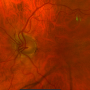
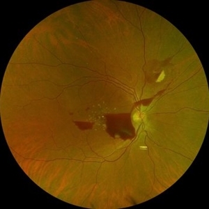
 Retinal Arterial Macroaneurysm
Jun 5 2024 by Akansha Sharma
Color fundus photograph of a 61 year old hypertensive male with retinal arterial macroaneurysm.
Photographer: Dr. Akansha Sharma, Bharati Eye Hospital
Condition/keywords: optic disc pallor, RAM
-
 Retinal Macroaneurysm
May 7 2024 by Akansha Sharma
Color fundus photograph of a 74 year old female with retinal artery macroaneurysm.
Photographer: Dr. Akansha Sharma, Bharati Eye Hospital
Condition/keywords: macroaneurysm, RAM
-
 Retinal Arterial Macroaneurysm
Apr 9 2024 by Akansha Sharma
Color fundus photograph of a 68 year old female patient with retinal arterial macroaneurysm with subretinal bleed.
Photographer: Dr. Akansha Sharma, Bharati Eye Hospital
Condition/keywords: macroaneurysm, subretinal hemorrhage
-
 Ruptured Macroaneurysm OCT
Mar 6 2024 by Mari Ann Z. Keithahn, MD, FASRS
OCT of 73 year-old female with ruptured macroaneurysm.
Photographer: JaTori Maxwell, Missouri Retina Consultants, PC
Imaging device: OPTOS Silverstone
Condition/keywords: Ruptured Macroaneurysm OCT
-
 Ruptured Macroaneurysm
Mar 6 2024 by Mari Ann Z. Keithahn, MD, FASRS
Fundus Photograph of 73 year-old female with hypertension.
Photographer: JaTori Maxwell, Missouri Retina Consultants, PC
Imaging device: OPTOS Silverstone
Condition/keywords: Rupture Macroaneurysm
-
 Choroidal Mass
Mar 4 2024 by ANKIT JAIN
RE color photo montage of right eye of 48 year old with sub retinal hemorrhage with sub retinal fluid at level of fovea.
Photographer: Dr Ankit Jain
Imaging device: MIRANTE
Condition/keywords: macroaneurysm, retinal arterial macroaneurysm
-
 Left Eye Arteriovenous Malformation, Vein Occlusion and Ruptured Macroaneurysm
Feb 9 2024 by Sandra R Montezuma, MD
47 year old female presented with acute changes in vision in the left eye, flashes of light and a new supero temporal scotoma. No history of trauma. She has history of retina bleeding in 1998 when she was pregnant and had pre-eclampsia. She was told had a retina scar. Her VA was 20/500. Fundus exam revealed an arteriovenous malformation along inferonasal vessels with prominent tortuous vessels. The optic nerve was hyperemic and there was peripapillary pre-retinal hemorrhage. There is a central macula scar and retina hemorrhage in the macula and mid periphery. In the nasal mid periphery, there is a ruptured macroaneurysm with hemorrhage in all layers of the retina. There are diffuse IRH. Her OCT revealed abnormal foveal contour with intraretinal fluid, Outer retinal atrophy and increased hyperreflectivity of the inner retina layers. The patient was treated with avastin injections with some improvement of the vision and resolution of the intraretinal fluid. Her MRI was normal.
Photographer: University of Minnnesota
Condition/keywords: arteriovenous malformation, macroaneurysm, vein occlusion
-
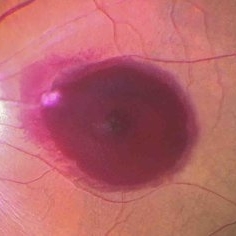
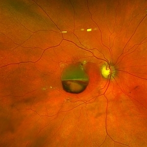
 Subhyaloid Hemorrhage From a Macroaneurysm
Jan 30 2024 by Akansha Sharma
Color fundus photograph of a 58 year old hypertensive and diabetic female patient with lasered proliferative diabetic retinopathy developing a subhyaloid hemorrhage from a macroaneurysm.
Photographer: Dr. Akansha Sharma, Bharati Eye Hospital
Condition/keywords: florid type PDR, macroaneurysm, proliferative diabetic retinopathy (PDR), SHH, Sub hyaloid haemorrhage
-
 Vasculitis
Jan 30 2024 by Akansha Sharma
Color fundus photograph of a 34 year old seronegative male patient with vasculitis presenting with macular edema associated with a macroaneurysm.
Photographer: Dr. Akansha Sharma, Bharati Eye Hospital
Condition/keywords: macroaneurysm, vasculitis
-
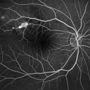 Macroaneurysms
Jan 28 2024 by Anjana Mirajkar, MS Ophthalmology
Fundus fluorescein angiography image (Late phase) in a 20 year old female showing leakage in a case of retinal artery macro-aneurysms.
Photographer: Dr. Anjana Mirajkar -Retina Foundation, Ahmedabad
Imaging device: Mirante-Nidek
Condition/keywords: retinal arterial macroaneurysm
 Loading…