Search results (26 results)
-
 Dislocation of the Crystalline Lens with a Retinal Detachment
Apr 21 2025 by Hrishikesh Naik, MS
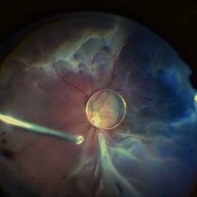
An intraoperative screen grab shows a dislocation of the crystalline lens along with an associated rhegmatogenous retinal detachment in a case of Marfan’s syndrome. The case was managed by a combined PPV-SB procedure. A vitrectomy cutter is seen at the left.
Photographer: Hrishikesh Naik
Condition/keywords: intraoperative, lens dislocation, Marfan's syndrome, Retinal Detachment, vitrectomy
-
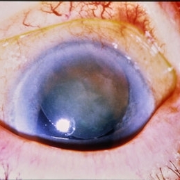
 Spontaneous Lens Subluxation
Sep 24 2024 by Christian A Leal, MD
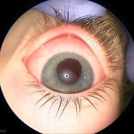
Anterior segment photo during exam under anesthesia in a child with spontaneous lens subluxation and history of ectopia lentis et pupillae. The pupil shown here has been pharmacologically dilated.
Photographer: Baker Hubbard, MD; Emory Eye Center
Condition/keywords: spontaneous lens dislocation
-
 Ectopia Lentis et Pupillae
Sep 24 2024 by Christian A Leal, MD
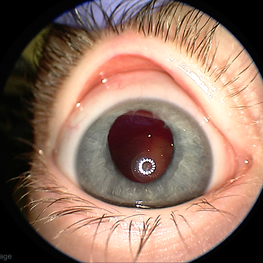
Anterior segment photo during exam under anesthesia in a child with spontaneous lens dislocation and history of ectopia lentis et pupillae. The lens is not visible. The pupil shown here has been pharmacologically dilated.
Photographer: Baker Hubbard, MD; Emory Eye Center
Condition/keywords: spontaneous lens dislocation
-
 Ectopia Lentis et Pupillae
Sep 24 2024 by Christian A Leal, MD
Fundus photograph of a 4 year old child with ectopia lentis et pupillae showing the crystalline lens dislocated into the vitreous cavity.
Photographer: Baker Hubbard, MD; Emory Eye Center
Condition/keywords: lens dislocation
-
 Traumatic Dislocation of the Crystalline Lens
Feb 20 2024 by Nikhil K Bommakanti, MD
Traumatic Dislocation of the Crystalline Lens
Condition/keywords: lens dislocation
-
 Tilted IOL
Dec 7 2023 by Virginia Gebhart
61 year old female with inferiorly tilted IOL. UBM shows haptic rubbing against the iris causing transillumination defect from 5 to 6 o'clock.
Photographer: Virginia Gebhart
Condition/keywords: lens dislocation, transillumination
-
 Lens Drop
Aug 21 2023 by Harsh Vardhan Singh, MS
Intra-operative image of Posterior lens dislocation as a complication of cataract surgery
Photographer: Harsh Vardhan Singh
Condition/keywords: lens dislocation, Lens Drop
-
Spontaneous lens dislocation (Weill Marchesani Syndrome)
Nov 9 2022 by Heitor Nogueira
A 9 year-old Male patient diagnosed with Weill Marchesani presented spontaneous bilateral lens dislocation. Weill-Marchesani syndrome, also known as spherophakia-brachymorphy syndrome and mesodermal dysmorphodystrophy, is an inherited connective tissue disorder characterized by eye lens abnormalities, secondary glaucoma, short stature, brachydactyly, joint stiffness, and cardiovascular defects.
Photographer: Heitor Nogueira, Insituto Penido Burnier, Campinas-SP, Brazil
Condition/keywords: mesodermal dysmorphodystrophy, spherophakia-brachymorphy syndrome, spontaneous lens dislocation, video, Weill Marchesani Syndrome
-
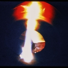
 Superiorly subluxed lens
Jun 16 2022 by Filipe Sampaio Carvalho
Seven-year-old boy presenting with superior nasal lens dislocation bilaterally.
Photographer: Filipe Sampaio Carvalho
Imaging device: iPhone 12
Condition/keywords: subluxation of lens
-
 Iris hooking the Intraocular Lens
Jun 16 2022 by Filipe Sampaio Carvalho
A fifty-year-old man with an intraocular lens immersed in the vitreous reports that for three days he no longer sees the shadow of the lens.
Photographer: Filipe Sampaio Carvalho
Condition/keywords: anterior dislocation of lens, dislocated intraocular lens (IOL), lens dislocation
-
 The Effects of Blunt Trauma
Feb 27 2022 by Jesus Lozano, MD
Axial Head Ct. 60 year old man with a history of blunt trauma and lost of vision after the event. VA HM. Iop 25mmhg. Cornea clear. Complete hyphema. BMode US: diffuse Vitreous Hemorrhage with a Dislocated Lens. Retina attached.
Photographer: Dr. Jesus Lozano. Retina Specialist. Hillel Yaffe Medical Center,Israel.
Imaging device: Axial Head CT
Condition/keywords: blunt trauma, hyphema, lens dislocation
-
 Plate-lens Dislocation
Feb 4 2022 by Tahsin Khundkar, MD
Fundus autofluorescence of the right eye of a 79-year-old male with loss of vision showing a dislocated plate lens in the vitreous cavity.
Condition/keywords: IOL dislocation, lens dislocation
-
 Dislocated Intraocular Lens
Aug 29 2021 by Jesus Lozano, MD
62 year-old man with a dislocated IOL.
Photographer: Dr. Jesús Lozano Gutiérrez. Hadassah Medical Center, Israel.
Imaging device: Slit lamp BI 900 camera
Condition/keywords: dislocated intraocular lens (IOL), lens dislocation, subluxation of lens
-
 Ectopia Lentis
Jan 21 2021 by Jamin S. Brown, MD
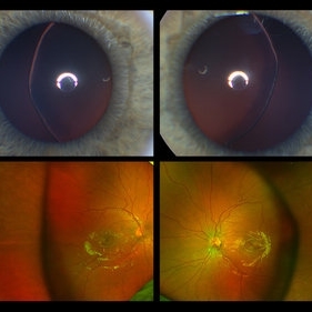
This image serial demonstrates a patient with simple ectopia lentis. Anterior segment photographs in the upper panel demonstrate nasally subluxated crystalline lenses. Widefield fundus photography shows a "pseudo-buckle" which is the result of an optical effect due to the lens subluxation (artifactual image enlargement). Also note the juvenile macular reflex in this young patient. Ectopia lentis can present isolated ("simple") or in combination with various systemic defects (Marfan's syndrome, Weil-Marchesani syndrome or Ehlers-Danlos syndrome to name a few). Isolated ectopia lentis can be hereditary and causative genes have been identified as ADAMTSL4 located on chromosome 4 and FBN1 gene located on chromosome 15. Defects in the genes cause weakness in the zonular fibers which can lead to lens dislocation. Lastly, various ocular disorders such as Aniridia, Axenfeld-Rieger, Pseudoexfoliation or Trauma may also result in lens dislocation or subluxation.
Photographer: Stefanie Palmer CRA, Retina Vitreous Surgeons of CNY
Condition/keywords: dislocated lens, ectopia lentis
-
 Traumatic Lens Drop in Vitreous
Dec 15 2020 by Manish Nagpal, MD, FRCS (UK), FASRS
Patient had come to us status post blunt trauma with the lens dislocated in inferior vitreous.
Photographer: Gayathri Mohan, Retina Fellow, Retina Foundation, Ahmedabad, India
Imaging device: Mirante CSLO
Condition/keywords: dropped nucleus, lens dislocation, traumatic cataract
-
 Dislocated Lens
Sep 7 2015 by Andrea Arriola-Lopez, MD MSc
Color fundus photography of right eye of a 54-year-old man, with history of blunt trauma seven month ago. VA HM. IOP 18mmHg. There is no peripherical lesions or traction.
Photographer: Andrea Elizabeth Arriola López, MD, MSc
Imaging device: OPTOS Dakota
Condition/keywords: blunt trauma, dislocated crystalline lens, lens dislocation
-
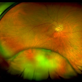
 Traumatic Lens Dislocation Over Retina
Aug 27 2015 by René Hernán Parada Vásquez
Fundus photograph of 82-year-old male with a traumatic lens dislocation over retina.
Photographer: Marroquín María José, ESO, Guatemala.
Imaging device: CANON CX-1
Condition/keywords: dislocated crystalline lens, posterior dislocation of lens
-
 Spontaneous Dislocation of Capsular Bag
Jul 26 2015 by Mehul A Shah
A 57-year-old male presented with complaint of loss of vision after 3 years of cataract surgery, on examination his lens bag found dislocated in anterior chamber.
Photographer: Mehul Shah, Drashti Netralaya
Condition/keywords: intraocular lens dislocation
-
 Trauma
-
 Trauma
-
 Traumatic Lens Dislocation, Extraocular
Oct 19 2012 by Larry Halperin, MD
Traumatic lens dislocation, extraocular
Condition/keywords: extraocular, lens dislocation
-
 Traumatic Lens Dislocation Over Disc
Oct 19 2012 by Larry Halperin, MD
Traumatic lens dislocation over disc
Condition/keywords: lens dislocation
-
 Traumatic Lens Dislocation
-
---thumb.jpg/image-square;max$300,300.ImageHandler) Traumatic Lens Dislocation
Traumatic Lens Dislocation
 Loading…