Search results (241 results)
-
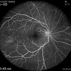
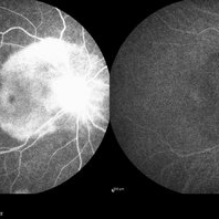
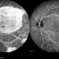
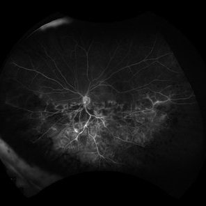
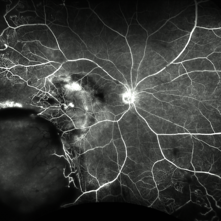 Fluorescein Angiography (FA) of a Primary Retinal Vasoproliferative Tumor
Jun 29 2025 by Marcelo Zas, MD PhD
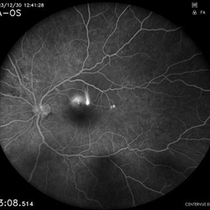
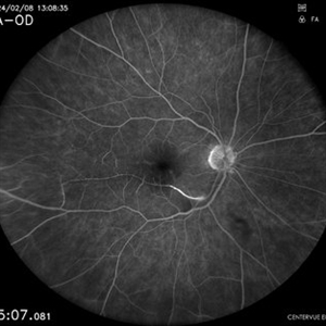
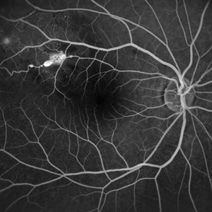
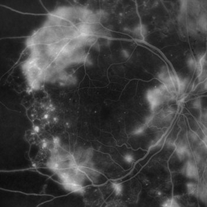
We present a case of a 33-year-old male patient, who presented with decreased visual acuity in his right eye with 20/80, presenting a primary retinal vasoproliferative tumor in the lower temporal quadrant. The fluorescein angiography findings are: 1. Early hyperfluorescence due to its rich intrinsic vascularity and often has dilated feeding arterioles and draining venules. 2. Marked progressive leakage from the tumor vessels. 3. The late leakage often obscures fine vascular details in the late phase and corresponds to exudation and macular edema seen clinically. 4. Staining of surrounding exudates, RPE disturbances and gliosis. 5. In our case also a marked peripheral capillary closure in the areas adjacent to the tumor and in other quadrants as well.
Photographer: Marcelo Zas MD PhD
Condition/keywords: RETINAL VASOPROLIFERATIVE TUMOR
-
Central Serous Chorioretinopathy
Apr 15 2025 by Filip Kecer
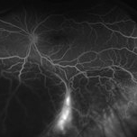
FA&ICG late phase of a young woman with CSCR
Photographer: Filip Kecer, Oftalmocentrum Betliarska, Bratislava, Slovakia
Imaging device: Spectralis, Heidelberg Engineering
Condition/keywords: central serous chorioretinopathy (CSCR), Central Serous Chorioretinopathy (CSR), FA late phase, indocyanine green (ICG) angiography
-
 Choroidal Hemangioma 4 Ways
Mar 13 2025 by Virginia Gebhart
Color fundus, FAF, late FA, late ICG of 64 year old male with choroidal hemangioma. Early hyperfluorescence with late leakage on FA, early hypercyanescence with late washout (25 min) on ICG.
Photographer: Virginia Gebhart, Retina Consultants of Carolina
Imaging device: Optos California
Condition/keywords: autofluorescence imaging, choroidal hemangioma, FA late phase, Fluorescein angiography, hemangioma, indocyanine green (ICG) angiography
-
 Uveal Effusion Syndrome
Jan 7 2025 by Drew Mitchell
Optos FA Late of Uveal Effusion Syndrome
Photographer: Drew Mitchell, OCT-C
Imaging device: Optos California
Condition/keywords: FA late phase, Optos, uveal effusion
-
 Branch Retinal Vein Occlusion
Aug 22 2024 by Virginia Gebhart
Fluorescein angiogram of branch retinal vein occlusion in 75 year old female. Scattered microaneurysms with late CME and persistent SRF. Pt will consider laser treatment but is hesitant for injections at this time due to possible side effects.
Photographer: Virginia Gebhart
Imaging device: Optos California
Condition/keywords: branch retinal vein occlusion (BRVO), BRVO, cystoid macular edema (CME), FA, FA late phase, fluorescein angiogram (FA), macular edema, microaneurysms, retinal microaneurysms
-
 Eyes Too Celebrate Valentine’s Day
Jul 28 2024 by KANWALJEET HARJOT MADAN, M.S. (Ophthalmology); FAICO (Vitreous - Retina)
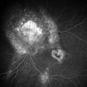
A 53 years male patient presented with decrease in vision in left eye for 6 months. His vison in left eye was counting fingers 1 meter. His vison in right eye was 20/20. Fundus examination in left eye depicted presence of large orange shaped elevated subretinal mass superior to optic disc with scar in macula. We made clinical diagnosis of Choroidal Hemangioma with macular scar. Fundus Fluorescein Angiography (FFA) in left eye revealed early fluorescence in area corresponding to Choroidal Hemangioma which persisted in late phases. Macular scar was “HEART” shaped on FFA which was very unique incident finding.
Photographer: Dr. Kanwaljeet Harjot Madan
Imaging device: Ziess Clarus
Condition/keywords: Choroidal Hemangioma, Fundus examination, Fundus Fluorescein Angiography
-
 Central Retinal Artery Occlusion
May 7 2024 by Akansha Sharma
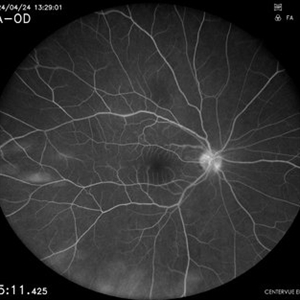
Late phase fluorescein angiography of a 40 year old female with central retinal artery occlusion.
Photographer: Dr. Akansha Sharma, Bharati Eye Hospital
Condition/keywords: central retinal artery occlusion (CRAO), CRAO
-
 Hemi Central Retinal Artery Occlusion
Apr 17 2024 by Akansha Sharma
Late phase fluorescein angiogram of a 30 year old female with complete perfusion.
Photographer: Dr. Akansha Sharma, Bharati Eye Hospital
Condition/keywords: central retinal artery occlusion (CRAO), CRAO, Hemi-Central Retinal Artery Occlusion (CRAO)
-
 Syphilitic Posterior Uveitis
Mar 22 2024 by Anjana Mirajkar, MS Ophthalmology
FA image of RE of a 36 year old female showing hyper-fluorescence (staining) from early to late phases of the angiogram in a case syphilitic posterior placoid chorioretinitis. ICG image depicts hypo-cyanence from early to late phases.
Photographer: Dr. Anjana Mirajkar -Retina Foundation, Ahmedabad
Imaging device: Heidelberg
Condition/keywords: acute syphilitic posterior placoid chorioretinitis
-
 Syphilitic Posterior Uveitis
Mar 22 2024 by Anjana Mirajkar, MS Ophthalmology
FA image of LE of a 36 year old female showing hyper-fluorescence (staining) from early to late phases of the angiogram in a case syphilitic posterior placoid chorioretinitis. ICG image depicts hypo-cyanence from early to late phases.
Photographer: Dr. Anjana Mirajkar -Retina Foundation, Ahmedabad
Condition/keywords: acute syphilitic posterior placoid chorioretinitis
-
 Ischemic HRVO with Macular Edema
Mar 7 2024 by Jenn Geelan
Optos FA of an 80 year old female.
Photographer: Jenn Geelan
Imaging device: Optos California
Condition/keywords: FA late phase, hemicentral retinal vein occlusion, ischemic CRVO, macular edema
-
 Central Serous Retinopathy
Mar 6 2024 by Akansha Sharma
Fluorescein angiography of a 34 year old male with smoke stack leak in the late phase in a case of central serous retinopathy.
Photographer: Dr. Akansha Sharma, Bharati Eye Hospital
Condition/keywords: Central Serous Chorioretinopathy (CSR), central serous retinopathy (CSR)
-
 Branch Retinal Artery Occlusion
Mar 6 2024 by Akansha Sharma
Fluorescein angiography of a 65 year old male showing delayed filling of the retinal artery in the late phase.
Photographer: Dr. Akansha Sharma, Bharati Eye Hospital
Condition/keywords: branch retinal artery occlusion
-
 Acute Posterior Multifocal Placoid Pigment Epitheliopathy
Feb 20 2024 by Soobien Lee
Fluorescein angiogram of a 20-year-old caucasian female with viral prodrome and vision loss OS>OD secondary to Acute Posterior Multifocal Placoid Pigment Epitheliopathy (APPME). Early blockage with late hyperfluorescent leakage can be seen on fluorescein angiography of the left eye.
Photographer: Ashley Metzger, Elman Retina Group
Imaging device: Optos Ultra-Widefield Fluorescein Angiography
Condition/keywords: acute posterior multifocal placoid pigment epitheliopathy (APMPPE), bacilliary layer detachment, FA, FA late phase, FA late phase leakage, fluorescein angiogram (FA), Optos, uveitis, white dot syndrome
-
 Flourescein Angiography of Cloroquine Toxicity
Feb 12 2024 by BENITO VERGARA, MD
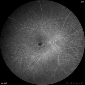
Image of a late phase fluorescein angiography at 4 minutes and 30 seconds of a 58-year-old woman treated with chloroquine at a daily dose of 3mg/kg, (recommended dose >2.3 mg/kg) that shows circular window defect suggestive of bullseye maculopathy.
Photographer: Benito Vergara Flores.
Imaging device: Clarus 700
Condition/keywords: chloroquine toxicity
-
 Macroaneurysms
Jan 28 2024 by Anjana Mirajkar, MS Ophthalmology
Fundus fluorescein angiography image (Late phase) in a 20 year old female showing leakage in a case of retinal artery macro-aneurysms.
Photographer: Dr. Anjana Mirajkar -Retina Foundation, Ahmedabad
Imaging device: Mirante-Nidek
Condition/keywords: retinal arterial macroaneurysm
-
 Impending STBRVO
Jan 7 2024 by MEENAL SONI
A middle aged female presented to the OPD with diminution of vision in right eye for past 7 days. Fundus examination findings depict supero-temporal AV crossing changes with macular hard exudates and oedema. On FFA we could clearly visualise the artery compressing the vein with leakage of dye in late phase extending into the macular region. On systemic evaluation the patient was found to be hypertensive with deranged lipid profile. She was advised injection anti VEGF for macular oedema and a physician consult for commencing the treatment for systemic condition. Despite a physician reference patient was not started on anti hypertensives and later presented with frank STBRVO with macular oedema after 3 months.
Photographer: Dr. Meenal Soni, VR fellow ASG eye hospital, Jodhpur (Raj)
Imaging device: Visucam
Condition/keywords: Impending BRVO with macular edema
-
 Choroidal Melanoma
Oct 27 2023 by Virginia Gebhart
76 year old male with suspicious pigmented choroidal lesion with new collar button growth. Blocking defect and vascularity noted on FA
Photographer: Virginia Gebhart
Condition/keywords: FA late phase, fluorescein angiogram (FA), Fluorescein angiography, melanoma
-
 Central Artery Occlusion with Cilio Retinal Artery Sparing
Aug 6 2023 by Anjana Mirajkar, MS Ophthalmology
Central FA frame (Late phase) of a 42 year old male in a case of central artery occlusion with cilio retinal artery sparing
Photographer: Dr. Anjana Mirajkar -Retina Foundation, Ahmedabad
Condition/keywords: central retinal artery occlusion (CRAO)
-
 Central Artery Occlusion with Cilio Retinal Artery Sparing
Aug 6 2023 by Anjana Mirajkar, MS Ophthalmology
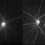
Wide field view of FA (Late phase) of a 42 year old male in a case of central artery occlusion with cilio retinal artery sparing showing delayed arterial filling with choroidal filling
Photographer: Dr. Anjana Mirajkar -Retina Foundation, Ahmedabad
Condition/keywords: central retinal artery occlusion (CRAO)
-
 Polypoidal Choroidal Vasculopathy
Jul 20 2023 by Gregg T. Kokame, MD, MMM, FASRS
64 Year Old Male, with Polypoidal Choroidal Vasculopathy. Pre-op and Post-op PDT/Vabysmo Injection
Photographer: Jaclyn Pisano
Imaging device: Heidelberg Spectralis
Condition/keywords: FA late phase, indocyanine green (ICG) angiography, OCT, PDT, polypoidal choroidal vasculopathy (PCV), subretinal, subretinal fluid
-
 Peripapillary Congenital Hypertrophy of the Retinal Pigment Epithelium
May 31 2023 by Luis Pimentel Silva, MD
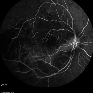
Late phase Fluorescein Angiography of an 51 years old man with Peripapillary Congenital Hypertrophy of the Retinal Pigment Epithelium
Photographer: Luis Pimentel Silva, University of São Paulo
Condition/keywords: Peripapillary Congenital Hypertrophy
-
 Peripapillary Congenital Hypertrophy of the Retinal Pigment Epithelium
May 31 2023 by Luis Pimentel Silva, MD
Late phase Fluorescein Angiography of an 51 years old man with Peripapillary Congenital Hypertrophy of the Retinal Pigment Epithelium
Photographer: Luis Pimentel Silva, University of São Paulo
Condition/keywords: Peripapillary Congenital Hypertrophy
-
 Cystoid Macular Degeneration
Feb 1 2023 by Kachelle Brown
Fluorescein Angiogram of a 56 year old woman with bilateral Cystoid Macular Degeneration. Patient vision was 20/60 OU.
Photographer: Kachelle Brown OMA, Retina Specialist of Michigan
Condition/keywords: cystoid macular degeneration, cystoid macular edema (CME), FA late phase, fluorescein angiogram (FA)
-
 PROLIFERATIVE DIABETIC RETINOPATHY
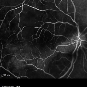
Oct 31 2022 by Akansha Sharma
LATE PHASE FLUORESCEIN ANGIOGRAPHY OF A 50 YEAR OLD MALE WITH PROLIFERATIVE DIABETIC RETINOPATHY
Photographer: Dr. Akansha Sharma-Retina Foundation, Ahmedabad
Condition/keywords: florid type PDR, proliferative diabetic retinopathy (PDR)
 Loading…