Search results (119 results)
-
 Subhyaloid Hemorrhage With Dispersed Vitreous Hemorrhage in a Case of Old Lasered Branch Retinal Vein Occlusion
Jul 12 2025 by Akansha Sharma
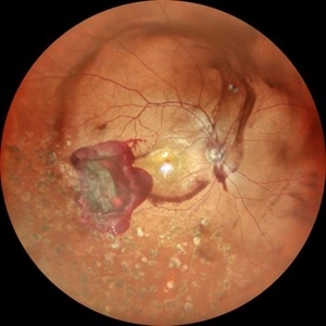
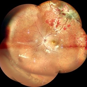
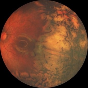
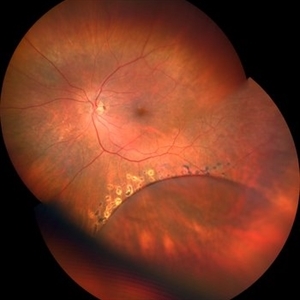
Color fundus photograph of a 32 year old hypertensive and diabetic male with subhyaloid hemorrhage with dispersed vitreous hemorrhage in a case of old lasered branch retinal vein occlusion.
Photographer: DR. AKANSHA SHARMA
Condition/keywords: branch retinal vein occlusion (BRVO), laser photocoagulation, SHH, subhyaloid hemorrhage, VH, vitreous hemorrhage
-
 Subhyaloid Hemorrhage With Dispersed Vitreous Hemorrhage in a Case of Old Lasered Branch Retinal Vein Occlusion
Jul 12 2025 by Akansha Sharma
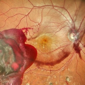
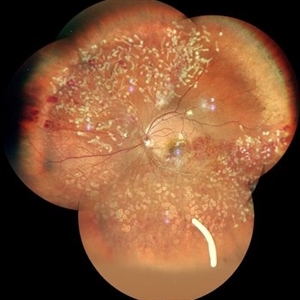
Color fundus photograph of a 32 year old hypertensive and diabetic male with subhyaloid hemorrhage with dispersed vitreous hemorrhage in a case of old lasered branch retinal vein occlusion.
Photographer: DR. AKANSHA SHARMA
Condition/keywords: branch retinal vein occlusion (BRVO), laser photocoagulation, SHH, subhyaloid hemorrhage, VH, vitreous hemorrhage
-
 Aggressive Posterior Retinopathy of Prematurity (APROP)
May 16 2025 by KANWALJEET HARJOT MADAN, M.S. (Ophthalmology); FAICO (Vitreous - Retina)
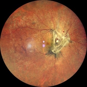
This is the fundus picture of right eye of a premature neonate depicting Aggressive Posterior Retinopathy of Prematurity (APROP). It is a severe rapidly progressing form of retinopathy that can lead to vision loss and blindness. It requires prompt diagnosis and treatment in the form of anti-VEGF agents and laser photocoagulation.
Photographer: Dr. Kanwaljeet Harjot Madan, Thind Eye Hospital, Jalandhar City (Punjab) INDIA.
Imaging device: Zeiss Clarus
Condition/keywords: Oxygen Exposure, retinopathy of prematurity (ROP)
-
 Eales Disease
Jan 31 2025 by Thirumalesh Mochi Basavaraj, MD
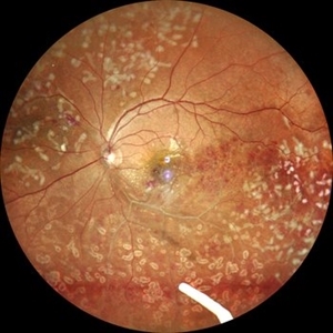
Ultra wide field image of a 24 year-old young healthy adult male with a visible sea fan neovascularization with partial PVD secondary to Scatter LASER photocoagulation with Vitreous and subhyaloid hemorrhage.
Photographer: Puttaswamy N K
Condition/keywords: Eales disease, Neovascularisation elsewhere (NVE), sea fan
-
 Pneumatic Retinopexy
Jan 6 2025 by Mateus Queiroz Corrêa, MD
Fundus photograph of a pneumatic retinopexy. The upper photo taken just 30 minutes after C3F8 gas injection shows rhegmatogenous retinal detachment with superior temporal horseshoe tear and gas bubbles resembling fish-eggs. After two days with appropriated head position (botton photo), the retina is attached and laser photocoagulation was performed on the border of the break. A Single great gas bubble was formed.
Photographer: Mateus Queiroz Corrêa, Sorocada Eye Bank Hospital
Imaging device: Optos California
Condition/keywords: pneumatic retinopexy
-
 TRD
TRD
Jun 25 2024 by Tejaswita Verma
Right eye fundus photo of an elderly male having diabetic tractional retinal detachment in a case of lasered Proliferative diabetic retinopathy.
Photographer: DR. TEJASWITA VERMA
Imaging device: MIRANTE
Condition/keywords: laser photocoagulation, proliferative diabetic retinopathy (PDR), tractional retinal detachment
-
 Von Hippel Lindau Syndrome
Jun 9 2024 by Anjana Mirajkar, MS Ophthalmology
A widefield montage of a 23 year old female of LE case of VHL syndrome showing some hemorrhages with traction superiorly in a silicon oil filled eye with central settled retina. Cryo and laser marks are noted in periphery.
Photographer: Dr. Anjana Mirajkar -Retina Foundation, Ahmedabad
Imaging device: Mirante-Nidek
Condition/keywords: cryotherapy, exudative detachment, laser photocoagulation, vitreous hemorrhage, Von Hippel-Lindau
-
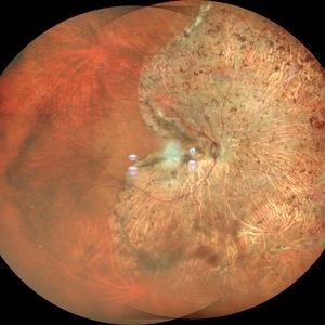
 Idiopathic Retinal Vasculitis
Jun 9 2024 by Anjana Mirajkar, MS Ophthalmology
A color photo montage of an 32 year old male of LE showing laser marks in inferior and superior half with an floating ozurdex implant (inferiorly) in a case of idiopathic retinal vasculitis.
Photographer: Dr. Anjana Mirajkar -Retina Foundation, Ahmedabad
Imaging device: Mirante-Nidek
Condition/keywords: idiopathic retinal vasculitis, laser photocoagulation, Ozurdex implant
-
 Idiopathic Retinal Vasculitis
Jun 9 2024 by Anjana Mirajkar, MS Ophthalmology
A widefield image of a 32 year old male of LE showing laser marks in inferior and superior half with an floating ozurdex implant (inferiorly) in a case of idiopathic retinal vasculitis.
Photographer: Dr. Anjana Mirajkar -Retina Foundation, Ahmedabad
Imaging device: Mirante-Nidek
Condition/keywords: idiopathic retinal vasculitis, laser photocoagulation, Ozurdex implant, pan-retinal photocoagulation (PRP)
-
 Laser Photocoagulation Macular Fibrous Scar
May 25 2024 by Gustavo Del Castillo-Marquez, MD
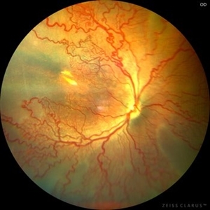
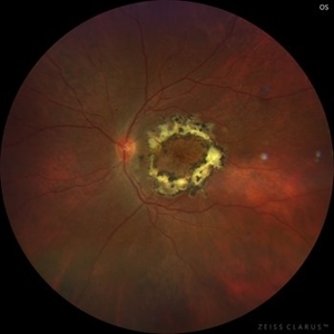
Fundus photograph of an 55 year old man who received macular laser photocoagulation 12 years earlier.
Photographer: Gustavo Del Castillo-Márquez, Asociación Para Evitar la Ceguera en México, CDMX
Imaging device: Zeiss Clarus
Condition/keywords: fibrous macular scar, laser photocoagulation, macular laser
-
 Laser Photocoagulation Macular Scar 12 Years Later
May 25 2024 by Gustavo Del Castillo-Marquez, MD
Actual Fundus photograph of a 55 year old man who received macular laser photocoagulation 12 years earlier.
Photographer: Gustavo Del Castillo-Márquez, Asociación Para Evitar la Ceguera en México, CDMX
Imaging device: Zeiss Clarus
Condition/keywords: fibrous macular scar, laser macula, laser photocoagulation
-
 Floating Ozurdex Implant
May 20 2024 by Tejaswita Verma
Fundus photograph of the left eye of a 73 year old female with ozurdex implant floating in the vitreous in a diabetic lasered patient.
Photographer: DR. TEJASWITA VERMA
Imaging device: MIRANTE
Condition/keywords: diabetic macular edema, laser photocoagulation, Ozurdex implant
-
 VH i/c/o Lasered PDR
May 8 2024 by Anand Temkar
Widefield fundus photograph of LE of a 58 years old male with Vitreous Hemorrhage inferiorly status post pan retinal photocogulation.
Photographer: Dr.Anand Temkar- Retina Foundation, Ahmedabad
Imaging device: Mirante
Condition/keywords: laser photocoagulation, proliferative diabetic retinopathy (PDR), vitreous hemorrhage
-
 Fight for Sight
Mar 26 2024 by Tushar Agrawal
Fundus photograph showing 28 weeker APROP; regressed well after ROP Laser photocoagulation as seen at age 3 months.
Imaging device: Retcam neo
Condition/keywords: aggressive posterior retinopathy of prematurity (APROP), pediatric retina, retinopathy of prematurity (ROP)
-
 Completed Pan-Retinal Fill-in Laser Photocoagulation in an Air filled eye at the end of Diabetic Vitrectomy Retina Surgery
Apr 28 2023 by Veer Singh, MS, FVRS, FMRF, FICO (Retina)
Completed Pan-Retinal Fill-in Laser Photocoagulation in an Air filled eye at the end of Diabetic Vitrectomy Retina Surgery
Photographer: Dr. Veer Singh
Condition/keywords: air-filled, pan-retinal photocoagulation (PRP), vitrectomy
-
 Intra-operative PRP in a diabetic vitrectomy case
Apr 28 2023 by Veer Singh, MS, FVRS, FMRF, FICO (Retina)
Pan-Retinal Fill-in Laser Photocoagulation in a Diabetic Vitrectomy Retina Surgery
Photographer: Dr. Veer Singh
Condition/keywords: diabetic, pan-retinal photocoagulation (PRP), vitrectomy
-
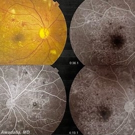 Grid macular photocoagulation for DME
Feb 17 2023 by Mohamed Awadalla
A case of aggressive grid macular photocoagulation treatment for DME. The treating ophthalmologist had done almost "pan-macular" photocoagulation
Condition/keywords: Grid macular photocoagulation, laser photocoagulation
-
 Type-1 ROP treated with laser
Nov 25 2022 by Alexandre Grandinetti, MD, PhD
This picture shows the case of a 8-year-old girl born with 26 weeks of gestation, who was treated with laser photocoagulation due to type-1 ROP
Photographer: Corina Szrek, Hospital de Olhos do Paraná
Imaging device: California
Condition/keywords: retinopathy of prematurity (ROP), rop laser
-
 Familial Exudative Vitreoretinopathy
Nov 25 2022 by Aditya S Kelkar, MS, FRCS, FASRS,FRCOphth
Colour fundus photograph of the right eye of a 56-year-old lady showing lasered FEVR with epiretinal membrane and vitreous band.
Photographer: Dr. Pranali Surawase. National Institute of Ophthalmology, Pune, Maharashtra, India
Imaging device: Zeiss Clarus 500
Condition/keywords: ERM, familial exudative vitreoretinopathy (FEVR), laser photocoagulation
-
 High risk Proliferative Diabetic Retinopathy treated with Pan Retinal Photocoagulation
Nov 5 2022 by Somnath Chakraborty, MD
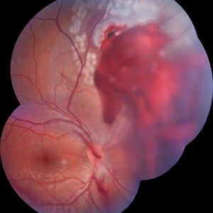
A Fundus Photo Montage of 43 year old Asian Male with Type 2 Diabetes Mellitus since 7 years who presented with sudden onset diminition of vision in his Left eye. BCVA OS was 20/200. He was diagnosed to have Pre retinal bleed due to Proliferative Diabetic Retinopathy and was treated with Pan Retinal Photocoagulation. This image shows a large neo-cascular frond at the disc and superior to it with Pre-retinal bleed and Fresh laser marks along
Photographer: Pulak Roy
Condition/keywords: diabetic blindness, diabetic retinopathy vitrectomy study (DRVS), fresh laser burns, laser photocoagulation, preretinal hemorrhage, proliferative diabetic retinopathy (PDR)
-
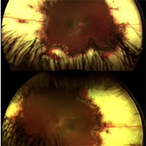
 STATUS POST PAN-RETINAL PHOTOCOAGULATION
Oct 11 2022 by Akansha Sharma
COLOUR FUNDUS PHOTO OF A 71 YEAR OLD MALE WITH SCARRING POST PAN-RETINAL PHOTOCOAGULATION
Photographer: Dr. Akansha Sharma-Retina Foundation, Ahmedabad
Condition/keywords: laser photocoagulation, pan-retinal photocoagulation (PRP)
-
 RETINOSCHISIS
Sep 7 2022 by JEFFERSON R SOUSA, Tecg.º (Biomedical Systems Technology)
Patient 55 years old, Female, progressive loss of vision. In funduscopic evaluation and photographic documentation, the presence of retinoschisis in the inferior temporal region surrounded with laser photocoagulation was observed.
Photographer: JEFFERSON ROCHA DE SOUSA - Departamento de Retina do Instituto Dr. Suel Abujamra São Paulo-Brasil.
Imaging device: Clarus 700 - Zeiss, 135-degree images.
Condition/keywords: fold in outer layer of retinoschisis, retinoschisis
-
 Intraocular Foreign Body
Jul 18 2022 by Nelson Chamma Capelanes, MD
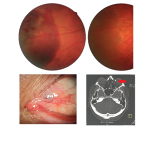
Intraocular foreign body after stone trauma. Foreign body is found in the choroid. - Fundus image on the upper left: one day after the trauma showing subretinal and intraretinal hemorrhage - Fundus image on the upper right: 40 days after laser photocoagulation. - Lower left image: 30 days after the trauma, showing part of the foreign body in the nasal region. - Lower right image showing CT scan and intraocular foreign body location.
Photographer: Nelson Chamma Capelanes, Promacula Indaiatuba, Brazil
Imaging device: Canon CX-2
Condition/keywords: intraocular foreign body
-
 Perforating Ocular Injury
Apr 18 2022 by Franco Benvenuto, MD
16 Y/O Male with history of perforating injury with a metal nail, days after nail extraction laser photocoagulation around the exit injury was performed.
Photographer: Franco Benvenuto, Universidad de Buenos Aires, Argentina
Condition/keywords: laser photocoagulation, retina surgery, Trauma
-
 Isolated Retinal Capillary Hemangioblastoma - Late phase IVFA
Mar 11 2022 by Bryon R McKay, MD, PhD, FRCSC, DRCPSC - Retina
Optos widefield fundus photograph and IVFA of a 23-year-old female with asymptomatic isolated retinal capillary hemangioblastoma without exudation. IVFA demonstrates some mild late leakage. The tumor measures 1.5mm and was effectively ablated with laser photocoagulation.
Imaging device: Optos
Condition/keywords: retinal capillary hemangioblastoma
 Loading…