Search results (57 results)
-
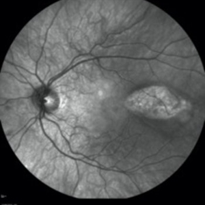
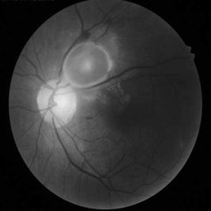
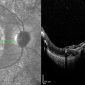
 PAMM-IR
Nov 29 2023 by Daniel Davis, OCT-C
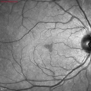
Infrared fundus of a 30 yo female with PAMM OD.
Photographer: Daniel Davis, OCT-C
Imaging device: Heidelberg Spectralis
Condition/keywords: infrared image
-
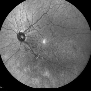
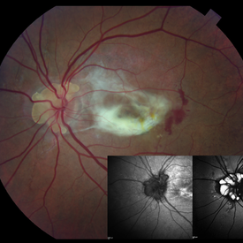
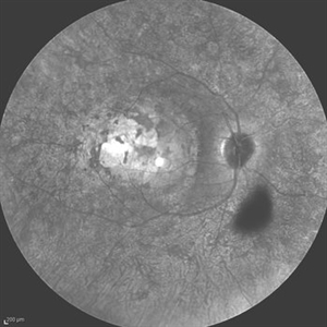
 "NVD Flower"
Oct 20 2023 by Daniel Davis, OCT-C
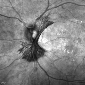
Infrared image of NVD (52F)
Imaging device: Heidelberg Spectralis
Condition/keywords: neovascularization of the disc (NVD)
-
 Iris Vascular Tuft
Jul 5 2022 by Olivia Rainey
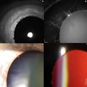
Anterior segment imaging of a 66-year-old male with Vascular Disorders of Iris and Ciliary Body affecting his right eye. The physician stated that the findings are most consistent with a benign vascular tuft at the pupillary margin. The patient presented at the office with 20/20 vision in both eyes and had no ocular complaints at the time of his appointment.
Photographer: Olivia Rainey, OCT-C, COA
Imaging device: Heidelberg Spectralis, Slit Lamp with Samsung Galaxy 7
Condition/keywords: anterior segment, fluorescein angiogram (FA), heidelberg spectralis, infrared image, near infrared autofluorescence (NIRAF), slit lamp photo, vascular anomaly, vascular disorders of iris and ciliary body, vascular tuft
-
 Mucopolysaccharidosis
Dec 9 2021 by Filip Kecer
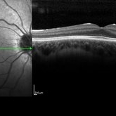
Infrared image of an 18-year-old boy with Mucopolysaccharidosis. His 24-year-old brother has exactly the same finding.
Photographer: Filip Kecer
Imaging device: Spectralis, Heidelberg Engineering
Condition/keywords: mucopolysaccharidoses
-
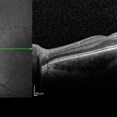
 CSNB-OCT-OD
Aug 23 2021 by Jennifer Carstens
OCT/infrared image showing myopic fundus with normal retinal structure in patient with CACNA1F associated X-linked CSNB (OD).
Photographer: Jing Zhang, Ophthalmic Photographer
Condition/keywords: congenital stationary night blindness (CSNB), infrared image, optical coherence tomography (OCT)
-
 CSNB-OCT-OS
Aug 23 2021 by Jennifer Carstens
OCT/infrared image showing myopic fundus with normal retinal structure in patient with CACNA1F associated X-linked CSNB (OS).
Photographer: Jing Zhang, Ophthalmic Photographer
Condition/keywords: congenital stationary night blindness (CSNB), infrared image, optical coherence tomography (OCT)
-
CSNB-OCT-OS
Aug 17 2021 by Christine Kay, MD
This is an OCT/infrared image of the left eye exhibiting normal fundus in a 16 year-old male with X-linked CSNB with proven mutation in CACNA1F.
Photographer: Christine Kay, MD
Condition/keywords: X-linked CSNB
-
CSNB-OCT-OD
Aug 17 2021 by Christine Kay, MD
This is an OCT/infrared image OD exhibiting normal fundus in a 16 year-old male with X-linked CSNB with proven mutation in CACNA1F.
Photographer: Christine Kay, MD
Condition/keywords: infrared image, X-linked CSNB
-
 Cuticular and soft drusen
Jun 14 2021 by Gerardo Garcia-Aguirre, MD
Fundus photograph (left) and Retro mode infrared image (right) of an eye with soft and cuticular drusen. Drusen are highlighted and better visualized with retro mode imaging.
Photographer: Gerardo Garcia-Aguirre
Imaging device: Nidek Mirante
Condition/keywords: drusen, dry age-related macular degeneration (dry AMD)
-
 Macular Edema
Jun 5 2021 by Aditya Verma, MBBS, MS, Post fellow
Multimodal imaging of a 36-year-old male with peripheral vasoproliferative tumor. Posterior pole showed exquisite pattern of hard exudate accumulation in petaloid pattern (A), fundus autofluorescence (B) and infrared image (C) portrayed the precise pattern of hard exudate distribution; Optical coherence tomography scan (D) showed a uniformly distributed parallel clumps of exudates in the outer plexiform layer.
Photographer: Aditya Verma
Imaging device: Heidelberg Spectralis
Condition/keywords: hard exudates, macular edema, macular exudates
-
 Combined Hamartoma of the Retina and Retinal Pigment Epithelium (CHRRPE)
Jan 21 2020 by Pierre-Henry Gabrielle, MD
IR imaging of a 17-year-old man with Combined hamartomas of the retina and retinal pigment epithelium (CHRRPE) at the posterior pole of the left eye.
Photographer: Pierre-Henry Gabrielle, Ophthalmology department, Dijon University Hospital, France
Imaging device: Heidelberg Spectralis
Condition/keywords: combined hamartoma, infrared image
-
 Torpedo Maculopathy
Jan 20 2020 by Pierre-Henry Gabrielle, MD
IR imaging photograph of an asymptomatic 12-year-old girl with torpedo maculopathy of the left eye.
Photographer: Pierre-Henry Gabrielle, Ophthalmology department, Dijon University Hospital, France
Imaging device: Heidelberg Spectralis
Condition/keywords: infrared image, torpedo maculopathy
-
 Acute Macular Neuroretinopathy
Dec 11 2019 by Lauren Whaley
34-year-old female patient presented with changes in vision after recent upper respiratory infection. Referring doctor originally thought it was a blood pressure issue. She noticed a "C" shape in her vision. Infrared image was captured showing exactly what patient was describing! Doctor confirmed with this image that it was AMN.
Photographer: Lauren R. Whaley, COA
Imaging device: Heidelberg Spectralis
Condition/keywords: 30 degrees, acute macular neuroretinopathy, Heidelburg Spectralis, left eye, macula, near infrared autofluorescence (NIRAF)
-
 Cysticercosis Cyst
Apr 29 2019 by Chintan Sarvaiya, MS
Infrared image of a 43-year-old male with Cysticercosis cyst
Photographer: Dr. Chintan Sarvaiya, Banker's Retina Clinic
Condition/keywords: cysticercosis
-
 Retinal Detachment Sparing Fovea By Microns
Sep 25 2018 by samarth mishra
A 29-year-old young female presented with complain of blurring of vision in the right eye since one year. Best corrected visual acuity was 20/40. On routine examination inferior retinal detachment was noted. Optical coherence tomography (OCT) showed the retinal detachment sparing the fovea by few microns.
Photographer: Aditya Birla Sankara Nethralaya, Kolkata, West Bengal , India
Condition/keywords: color fundus photograph, infrared image, multicolor, optical coherence tomography (OCT), red-free
-
 Angioid Streaks With Associated Disc Drusen and CNV
Sep 21 2018 by Sarah Oelrich
Angioid streaks with associated disc drusen and CNV.
Photographer: Sarah Oelrich CRA COT, Southeastern Retina Associates Knoxville Tn
Condition/keywords: angioid streaks, autofluorescence imaging, choroidal neovascularization (CNV), disc drusen, infrared image
-
 Multiple Astrocytic Hamartomas
Jul 26 2018 by Olivia Rainey
Optical coherence tomography of a 7-year-old female with multiple astrocytic harmartomas as a retinal manifestation of tuberous sclerosis. Patient came to our office to rule out possible drug toxicity from Sabril, a an anticonvulsant. There were no signs of retinal toxicity by extended ophthalmoscopy or imaging, yet she will be monitored every 6 months.
Photographer: Olivia Rainey
Imaging device: Heidelberg Spectralis
Condition/keywords: astrocytic hamartoma, Heidelburg Spectralis, infrared image, left eye, optical coherence tomography (OCT), tuberous sclerosis
-
 Silicon Oil
Apr 27 2018 by Giselle DeOliveira
Infrared photo of 75-year-old male with retinal detachment.
Photographer: Giselle DeOliveira, University of Miami, Bascom Palmer Eye Institute
Imaging device: Heidelberg Spectralis
Condition/keywords: infrared image, silicone oil
-
 Severe PDR
Apr 10 2018 by MOHAMED AHMED ALI, MD
Infrared image of case of severe PDR.
Photographer: Mohamed A.Tawfik MD,FRCSed
Condition/keywords: proliferative diabetic retinopathy (PDR)
-
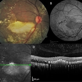
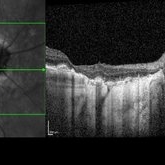 Optic Nerve Head Drusen with OCT
Feb 2 2018 by Olivia Rainey
Optical coherence tomography with enhanced depth imaging of a 86-year-old male with optic nerve head drusen affecting his right eye. This patient has also been diagnosed with pseudoxanthoma elasticum and macular degeneration.
Photographer: Olivia Rainey
Imaging device: Heidelberg Spectralis
Condition/keywords: enhanced depth imaging, infrared image, macular degeneration, optic disc drusen, optic nerve, optical coherence tomography (OCT), pseudoxanthoma elasticum (PXE)
-
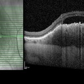 Choroidal Melanoma
Feb 2 2018 by Olivia Rainey
Optical coherence tomography with enhanced depth imaging of a 78-year-old female with choroidal melanoma with subretinal fluid affecting her right eye.
Photographer: Olivia Rainey
Imaging device: Heidelberg Spectralis
Condition/keywords: enhanced depth imaging, infrared image, optical coherence tomography (OCT), subretinal fluid, superior retina
-
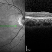 Cone-Rod Dystrophy
Mar 15 2017 by Hamid Ahmadieh, MD
Infrared and OCT images of the right eye of a 16-year-old boy with decreased visual acuity and color vision deficiency due to cone-rod dystrophy.
Photographer: Abazarnezhad , Negah Eye Center, Tehran, Iran
Imaging device: Spectralis OCT
Condition/keywords: cone dystrophy, infrared image, optical coherence tomography (OCT)
-
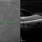 Cone-Rod Dystrophy
Mar 15 2017 by Hamid Ahmadieh, MD
Infrared and OCT images of the left eye of a 16-year-old boy with decreased visual acuity and color vision deficiency due to cone-rod dystrophy.
Photographer: Abazarnezhad , Negah Eye Center, Tehran, Iran
Imaging device: Spectralis OCT
Condition/keywords: cone dystrophy, infrared image, optical coherence tomography (OCT)
-
 Macular Coloboma and Pigmentary Retinopathy
Feb 25 2017 by Hamid Ahmadieh, MD
Infrared and OCT images of the right eye of a 25-year-old woman with bilateral macular colobomata and pigmentary retinopathy similar to Leber's congenital amaurosis.
Photographer: Shabnam Poureh, Negah Eye Center, Tehran, Iran
Condition/keywords: infrared image, macular coloboma, optical coherence tomography (OCT)
-
 Leber's Congenital Amaurosis
Feb 25 2017 by Hamid Ahmadieh, MD
Infrared image of the right eye of a 25-year-old woman with bilateral macular colobomata and pigmentary retinopathy similar to Leber's congenital amaurosis.
Photographer: Shabnam Poureh, Negah Eye Center, Tehran, Iran
Condition/keywords: bilateral pigmentary retinopathy, infrared image, macular coloboma
 Loading…