Search results (18 results)
-
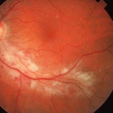
 Cilioretinal Artery Occlusion
May 14 2024 by Eloy Mata-Cortes, MD
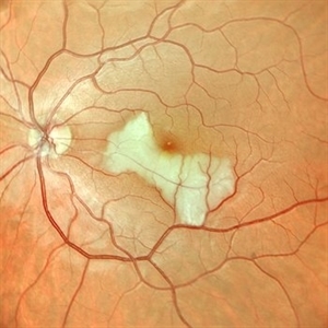
Color image capturing the left eye of a 32-year-old female. Despite a negative ophthalmological and medical history, she reported three days of blurred vision and a paracentral scotoma in her left eye, while maintaining central vision. The image reveals retinal whitening, extends from the parafoveal region to the inferotemporal arcade indicative of cilioretinal artery occlusion. Following this observation, the patient was referred for systemic assessment to explore the underlying etiology of the occlusion.
Photographer: Eloy Mata-Cortes, MD, Instituto Mexicano de Oftalmología, Querétaro, México
Imaging device: Nidek Mirante
Condition/keywords: cilioretinal artery occlusion, oclussion, retinal whitening
-
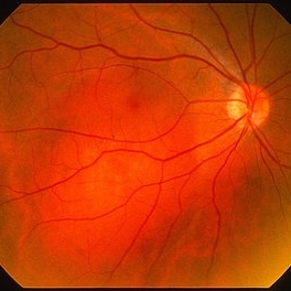
 Toxoplasmosis Chorioretinitis
Mar 2 2024 by James P Dossett, MD
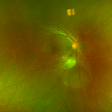
Pseudocolor fundus photograph of the right eye of a 34-year-old man with retinitis along the inferotemporal arcade with associated subretinal fluid and overlying vitritis. Aqueous paracentesis was performed and PCR was positive for Toxoplasma gondii. He was administered intravitreal clindamycin.
Imaging device: Optos
Condition/keywords: posterior uveitis, toxoplasmosis chorioretinitis
-
 Branch Retinal Vein Occlusion (BRVO)
Sep 14 2023 by Ben Serar
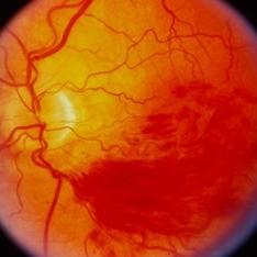
Fundus photograph of LE showing superficial retinal haemorrhages along the inferotemporal arcade in a case of inferotemporal Branch Retinal Vein Occlusion (BRVO).
Condition/keywords: branch retinal vein occlusion (BRVO)
-
 CSR FA
May 15 2021 by Deepak Bhojwani, MS
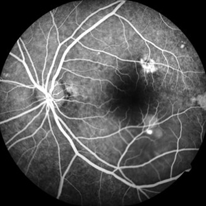
Fundus fluoroscein angiography image of a 38-year-old gentlemen with features of chronic CSR with multiple old areas of RPE mottling and staining. Also note there is a single active leakage site along inferotemporal arcade.
Photographer: Deepak Bhojwani
Condition/keywords: central serous retinopathy (CSR)
-
 Proliferative Diabetic Retinopathy with Macular Hole IVFA
Jul 26 2020 by Gareth Lema, MD, PhD
IVFA shows NVE along the inferotemporal arcade with ischemia immediately inferior to the NV.
Condition/keywords: macular hole, proliferative diabetic retinopathy (PDR)
-
 Early Venous Phase
Aug 26 2019 by Narciso F. Atienza, MD, MBA, FASRS, FPCS, FPAO.
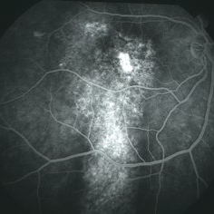
Early venous phase shows asymmetrical transit of dye perfusion of the infero-temporal arcade. Infero-temporal arcade shows beginning perfusion. Areas of non perfusion are also more prominent.
Photographer: Narciso F Atienza, Jr. MD, MBA
Imaging device: Topcon TRC
Condition/keywords: capillary nonperfusion, inferotemporal arcade
-
 02123-20190508-171643-Fluorescein-R-001
Aug 26 2019 by Narciso F. Atienza, MD, MBA, FASRS, FPCS, FPAO.
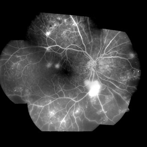
47-year-old female who came in with blurring of vision of the right eye of 2 weeks duration. She is hypertensive with poor control, taking Amlodipine irregularly. Denies any cardiac problem non-diabetic. Vision upon presentation was 20/400 (OD), 20/20 (OS) . Early arterial phase shows beginning asymmetrical perfusion of the supero-temporal arcade supplying the macula. Infero-temporal arcade shows no perfusion.
Photographer: Narciso F Atienza, Jr. MD, MBA
Imaging device: Topcon TRC
Condition/keywords: asymmetrical perfusion, inferotemporal arcade, superotemporal arcade
-
 Retinal Ischemia, Edema, and Hemorrhages on the Infero-Temporal Macula
Aug 26 2019 by Narciso F. Atienza, MD, MBA, FASRS, FPCS, FPAO.
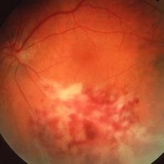
47-year-old female who came in with blurring of vision of the right eye of 2 weeks duration. She is hypertensive with poor control, taking Amlodipine irregularly. Denies any cardiac problem non-diabetic. Vision upon presentation was 20/400 (OD), 20/20 (OS) colored fundus photo of the right eye showing areas of retinal ischemia, edema and hemorrhages on the infero-temporal macula extending to the arcade.
Photographer: Narciso F Atienza, Jr. MD, MBA
Imaging device: Topcon TRC
Condition/keywords: edema, hemorrhage, inferotemporal arcade, retinal ischemia
-
 Acute Toxoplasmosis
Apr 8 2019 by Gary R. Cook, MD, FACS
14-year-old white male teenager with acute toxoplasmosis lesion along the inferotemporal arcade OD; V.A. = 20/20-2; toxo titer = 1:128
Imaging device: Topcon VT-50
Condition/keywords: toxo chorioretinitis, toxoplasmosis
-
 CMV Retinitis
Mar 26 2019 by Gary R. Cook, MD, FACS
39-year-old white male with HIV/AIDS and active CMV retinitis along inferotemporal arcade.
Imaging device: Topcon VT-50
Condition/keywords: CMV retinitis
-
 Central Serous Retinopathy
Mar 26 2019 by Gary R. Cook, MD, FACS
Late-phase FA frame of the right eye of a 45-year-old white male with a history of CSR showing pooling of dye beneath a small chronic RPED near fovea and stippled late staining of the RPE track in macula and beneath the inferotemporal arcade OD; VA = 20/200.
Imaging device: Topcon VT-50
Condition/keywords: central serous retinopathy (CSR), FA late phase
-
 Central Serous Retinopathy
Mar 26 2019 by Gary R. Cook, MD, FACS
Mid-phase FA frame of right eye of a 45-year-old white male with a history of bilateral CSR showing stippled hyperfluorescence in an area of an RPE track beneath the inferotemporal arcade OD; no active dye leakage is present; VA = 20/200.
Imaging device: Topcon VT-50
Condition/keywords: central serous retinopathy (CSR), FA mid phase
-
 Cytomegalovirus (CMV) Retinitis
Mar 26 2019 by Gary R. Cook, MD, FACS
31-year-old white male with active CMV retinitis along inferotemporal arcade OS; VA = 20/20.
Imaging device: Topcon VT-50
Condition/keywords: CMV retinitis
-
 CMV Retinitis/ Before Treatment
Mar 13 2015 by Niloofar Piri, MD
Fundus photograph of the left eye of a 40-year-old Caucasian female with history of positive HIV test for 23 years. She has been off HAART therapy for the past 2 years and presented with decreased vision OS and upper visual field defect. On examination, she had trace cells in anterior vitreous , hemorrhagic retinitis which starts around the optic nerve and extending to inferotemporal arcade with secondary inferotemporal BRVO; in temporal periphery , she had granular pattern of CMV retinitis which is a manifestation of outer retina involvement.
Photographer: Angela Anderson
Condition/keywords: CMV retinitis, HIV
-
---thumb.jpg/image-square;max$300,300.ImageHandler) Proliferative Diabetic Retinopathy - Neovascularization Elsewhere
Proliferative Diabetic Retinopathy - Neovascularization Elsewhere
Nov 11 2013 by Gerardo Garcia-Aguirre, MD
Fundus photograph showing neovascularization in the inferotemporal arcade.
Condition/keywords: fundus photograph, neovascularization (NV)
-
 Choroidal Hemangioma
Mar 29 2013 by Henry J. Kaplan, MD
Orange-red choroidal hemangioma extending from the optic nerve toward inferotemporal arcade involving the macula.#1
-
 Cytomegalovirus Retinitis, Active, with Papillary Involvement
Sep 27 2012 by Jeffrey G. Gross, MD, FASRS
CMV retinitis active with papilary involvement, inferotemporal arcade.
Condition/keywords: inferotemporal arcade
-
 HIV Retinopathy with Very Early Cytomegalovirus Retinitis
Sep 27 2012 by Jeffrey G. Gross, MD, FASRS
HIV retinopathy with very early CMV retinitis, inferotemporal arcade.
Condition/keywords: HIV, inferotemporal arcade, retinopathy
 Loading…