Search results (603 results)
-
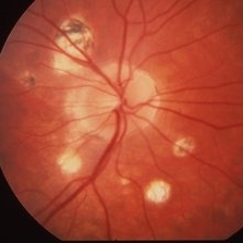
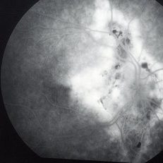
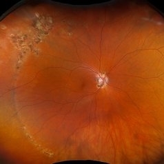 POHS/Schlaegel Lines
Sep 19 2024 by Virginia Gebhart
46 year old female with h/o Histoplasmosis. Multiple punched out chorioretinal scars with Schlaegel lines. No evidence of CNV or active inflammation. VA 20/20
Photographer: Virginia Gebhart, Retina Consultants of Carolina
Imaging device: Optos California
Condition/keywords: chorioretinal scar, histoplasmosis, presumed ocular histoplasmosis syndrome (POHS), Schlaegel
-
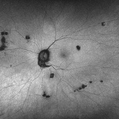 Histoplasmosis Capsulatum Retinitis OS
Dec 20 2021 by Brad Lovelace
Autofluorescence image of a 63-year-old woman with presumed ocular histoplasmosis syndrome OS observed for change.
Photographer: Cathy Harsma, COA
Imaging device: Optos Ultra-Widefield
Condition/keywords: histoplasmosis, presumed ocular histoplasmosis syndrome (POHS)
-
 Histoplasmosis Capsulatum Retinitis OD
Dec 20 2021 by Brad Lovelace
Autofluorescence image of a 63-year-old woman with presumed ocular histoplasmosis syndrome OD observed for change.
Photographer: Cathy Harsma, COA
Condition/keywords: histoplasmosis, presumed ocular histoplasmosis syndrome (POHS)
-
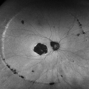
 Histoplasmosis Capsulatum Retinitis OS
Dec 20 2021 by Brad Lovelace
Color SLO image OD of a 63-year-old woman with presumed ocular histoplasmosis syndrome OS observed for change.
Photographer: Cathy Harsma, COA
Imaging device: Optos Ultra-Widefield
Condition/keywords: histoplasmosis, presumed ocular histoplasmosis syndrome (POHS)
-
 Histoplasmosis Capsulatum Retinitis OD
Dec 20 2021 by Brad Lovelace
Color SLO image of a 63-year-old woman with presumed ocular histoplasmosis syndrome OD observed for change.
Photographer: Cathy Harsma, COA
Imaging device: Optos Ultra-Widefield
Condition/keywords: histoplasmosis, presumed ocular histoplasmosis syndrome (POHS)
-
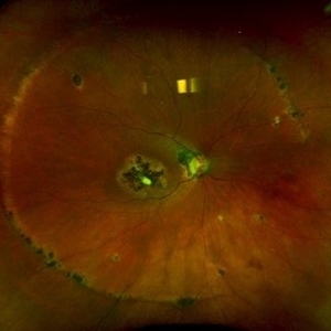
 Subretinal Fibrosis (PPCNVM and POHS) OS
Sep 18 2019 by John S. King, MD
57-year-old white male with history of PPCNVM OS and POHS OU here for a routine visit. History of avastin in 2014, and stable since then. Va OS 20/20. PP scar with macular subretinal fibrosis. No heme or exudates. CR spot supero-nasally.
Photographer: Shelly Blair
Imaging device: Topcon 50
Condition/keywords: choroidal neovascular membrane (CNVM), ocular histoplasmosis syndrome (OHS), peripapillary choroidal neovascularization (PPCNVM), presumed ocular histoplasmosis syndrome (POHS)
-
 Histoplasmosis and Idiopathic Macular Pucker
Mar 27 2019 by Gary R. Cook, MD, FACS
62-year-old female with presumed ocular histoplasmosis and an idiopathic macular pucker in her right eye; no Histo spots or CNV present in the macula; V.A.= 20/60.
Imaging device: Topcon VT-50
Condition/keywords: epiretinal membrane (ERM), macular pucker, presumed ocular histoplasmosis syndrome (POHS)
-
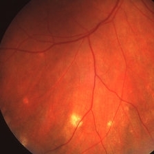
 Linear Histo Streak
Mar 27 2019 by Gary R. Cook, MD, FACS
53-year-old female with presumed ocular histoplasmosis demonstrating a depigmented linear histoplasmosis streak in the temporal periphery; V.A. = 20/20.
Imaging device: Topcon VT-50
Condition/keywords: presumed ocular histoplasmosis syndrome (POHS)
-
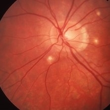 Histoplasmosis
Mar 27 2019 by Gary R. Cook, MD, FACS
24-year-old white female with presumed ocular histoplasmosis (POHS) demonstrating minimal peripapillary atrophy but 3 atrophic histo spots around the optic nerve of her left eye; patient was asymptomatic; V.A.= 20/20.
Imaging device: Topcon VT-50
Condition/keywords: atrophic spot, ocular histoplasmosis syndrome (OHS), presumed ocular histoplasmosis syndrome (POHS)
-
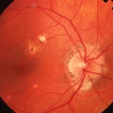
 Histoplasmosis
Mar 27 2019 by Gary R. Cook, MD, FACS
24-year-old white female with presumed ocular histoplasmosis (POHS) demonstrating some peripapillary atrophy and multiple atrophic histo spots around the optic nerve of her right eye; the patient was asymptomatic; V.A.= 20/20.
Imaging device: Topcon VT-50
Condition/keywords: atrophic spot, ocular histoplasmosis syndrome (OHS), peripapillary atrophy, presumed ocular histoplasmosis syndrome (POHS)
-
 Linear Histoplasmosis
Mar 27 2019 by Gary R. Cook, MD, FACS
Middle-aged white male with presumed ocular histoplasmosis (POHS) demonstration single astrophic histo spots and a linear histo streak in the superior periphery.
Imaging device: Topcon VT-50
Condition/keywords: atrophic spot, ocular histoplasmosis syndrome (OHS), presumed ocular histoplasmosis syndrome (POHS)
-
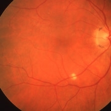
 Atrophic Histoplasmosis Spots
Mar 27 2019 by Gary R. Cook, MD, FACS
Atrophic histo spots in the mid-periphery OD of an adult white male.
Imaging device: Topcon VT-50
Condition/keywords: atrophic spot, ocular histoplasmosis syndrome (OHS), presumed ocular histoplasmosis syndrome (POHS)
-
 Linear Histoplasmosis Streaks
Mar 27 2019 by Gary R. Cook, MD, FACS
26-year-old white female with POHS showing multiple linear histo streaks in the periphery; V.A.= 20/200.
Imaging device: Topcon VT-50
Condition/keywords: atrophic, atrophic spot, ocular histoplasmosis syndrome (OHS), presumed ocular histoplasmosis syndrome (POHS)
-
 Histoplasmosis and Subfoveal Neovascular Membrane
Mar 27 2019 by Gary R. Cook, MD, FACS
26-year-old white female with new-onset subfoveal CNVM secondary to presumed ocular histoplasmosis (POHS) in her right eye; V.A.= 20/200.
Imaging device: Topcon VT-50
Condition/keywords: presumed ocular histoplasmosis syndrome (POHS), subfoveal choroidal neovascularization, subfoveal neovascular membrane
-
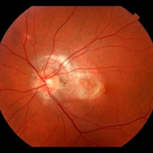
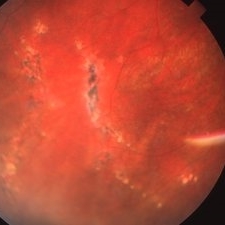
 Histoplasmosis and Old Disciform Macular Scar
Mar 27 2019 by Gary R. Cook, MD, FACS
Left eye of a 59-year-old white male with an old, inactive, disciform macular scar secondary to presumed ocular histoplasmosis (POHS); V.A.= counting fingers at 3 feet.
Imaging device: Topcon VT-50
Condition/keywords: central disciform scar, disciform scar, peripapillary atrophy, presumed ocular histoplasmosis syndrome (POHS)
-
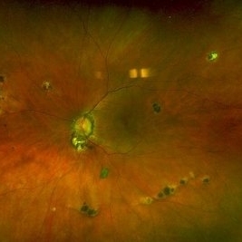
 Histoplasmosis and Subfoveal Neovascular Membrane
Mar 27 2019 by Gary R. Cook, MD, FACS
Late-phase fluorescein angiogram image of the right eye of a 59-year-old white male with ocular histoplasmosis and a subfoveal neovascular membrane showing late leakage and diffusion of dye from the membrane; V.A.= 20/80+2.
Imaging device: Topcon VT-50
Condition/keywords: FA late phase, fluorescein angiogram (FA), ocular histoplasmosis syndrome (OHS), peripapillary atrophy, presumed ocular histoplasmosis syndrome (POHS), subfoveal neovascular membrane
-
 Histoplasmosis and Subfoveal Neovascular Membrane
Mar 27 2019 by Gary R. Cook, MD, FACS
Mid-phase (20.4 seconds) fluorescein angiogram image of the right eye of 59-year-old white male with ocular histoplasmosis and a well-defined subfoveal CNVM OD; V.A.= 20/80+2
Imaging device: Topcon VT-50
Condition/keywords: FA mid phase, fluorescein angiogram (FA), ocular histoplasmosis syndrome (OHS), peripapillary atrophy, presumed ocular histoplasmosis syndrome (POHS), subfoveal choroidal neovascularization, subfoveal neovascular membrane
-
 Histoplasmosis with Choroidal Neovascularization
Mar 27 2019 by Gary R. Cook, MD, FACS
59-year-old white male with presumed ocular histoplasmosis (POHS) and a choroidal neovascular membrane (CNVM) along the temporal margins of the peripapillary atrophy; V.A.= 20/80+2.
Imaging device: Topcon VT-50
Condition/keywords: choroidal neovascular membrane (CNVM), peripapillary atrophy, presumed ocular histoplasmosis syndrome (POHS)
-
 Histo and Subfoveal Neovascular Membrane
Mar 27 2019 by Gary R. Cook, MD, FACS
41-year-old white female with a large subfoveal CNVM, subretinal fluid, and hemorrhage secondary to presumed ocular histoplasmosis (POHS) OS; V.A.= 20/400.
Imaging device: Topcon VT-50
Condition/keywords: hemorrhage, peripapillary atrophy, presumed ocular histoplasmosis syndrome (POHS), subfoveal choroidal neovascularization, subfoveal neovascular membrane
-
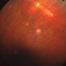
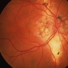
 Ocular Histoplasmosis
Mar 27 2019 by Gary R. Cook, MD, FACS
Fellow eye (OD) of a 41-year-old white female with ocular histoplasmosis showing peripapillary atrophy and several atrophic histo spots OD; no CNVM present; V.A.= 20/20.
Imaging device: Topcon VT-50
Condition/keywords: atrophic spot, histoplasmosis, peripapillary atrophy, presumed ocular histoplasmosis syndrome (POHS)
-
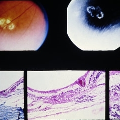
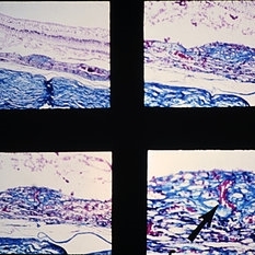 Slide 9-94
Feb 26 2019 by Lancaster Course in Ophthalmology
Macular disciform lesion in the ocular histoplasmosis syndrome. Note choroidal scar with vessels (arrow) extending through a break in Bruch's membrane.
Condition/keywords: Bruch's membrane, disciform macular lesion, ocular histoplasmosis syndrome (OHS)
-
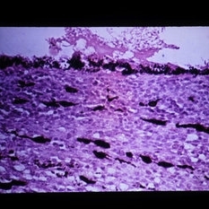
 Slide 9-66
Feb 26 2019 by Lancaster Course in Ophthalmology
Midperipheral punched-out lesions in the presumed ocular histoplasmosis syndrome. There is scarring in the choroid and retina, discontinuity in Bruch's membrane, and loss of the RPE. An infiltrate of lymphocytes is present in the subjacent choroid (lower middle and right).
Condition/keywords: Bruch's membrane, ocular histoplasmosis syndrome (OHS), retinal pigment epithelium, scar
-
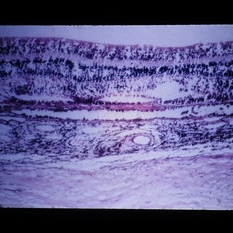
 Slide 3-15
Feb 20 2019 by Lancaster Course in Ophthalmology
Higher-power view of choroid in presumed histoplasmosis retinochoroiditis, showing marked epithelioid cell infiltrate ( x65).
Condition/keywords: choroid
-
 Slide 3-14
Feb 20 2019 by Lancaster Course in Ophthalmology
Adjacent area to that in Slide 3-13, showing degenerative change and inflammatory infiltrate in presumed histoplasmosis retinochoroiditis ( x25).
Condition/keywords: histoplasmosis, inflammatory infiltrate
-
 Slide 3-13
Feb 20 2019 by Lancaster Course in Ophthalmology
Cystoid macular change with granulomatous inflammation of choroid and retina in eye of patient with presumed histoplasmosis retinochoroiditis ( x25).
Condition/keywords: choroid, cystoid macular degeneration, granulomatous inflammation, histoplasmosis
 Loading…