Search results (50 results)
-
 Progressive Chorioretinal Degeneration
Jun 27 2024 by Natalia Moraes
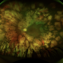
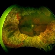
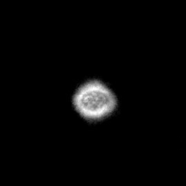
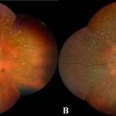
Funds photograph of a 63-year-old male with a progressive chorioretinal degeneration.
Photographer: Natália V. Moraes, Instituto Penido Burnier, Brazil
Imaging device: Daytona
Condition/keywords: gyrate atrophy
-
 Peripheral Retinal Degeneration (L-ORD)
Apr 17 2024 by Virginia Gebhart
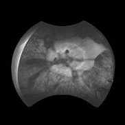
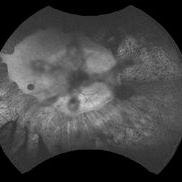
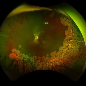
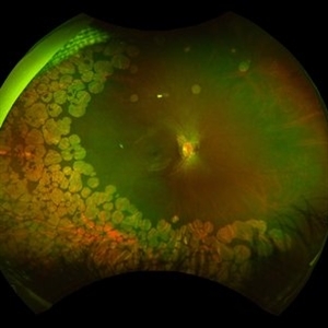
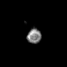
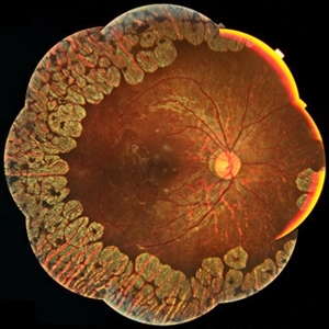
92 year old female with bilateral patchy, sharply demarcated circular areas of chorioretinal atrophy with hyperpigmented margins in the mid to far periphery. Labs showed normal plasma ornithine levels ruling out generalized gyrate atrophy. Also intermediate uveitis and CMD/CME. FTA-ABS, Quant gold, and HLA-A29 labs all negative.
Photographer: Virginia Gebhart
Imaging device: Optos California
Condition/keywords: cystoid macular degeneration, cystoid macular edema (CME), FA, Fluorescein angiography, peripheral retinal degeneration
-
 Gyrate Atrophy
Feb 5 2024 by Ali Al-Ani, M.B.Ch.B, FRCS, FRCOphth, FAAO
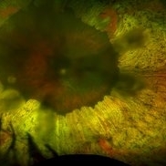
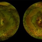
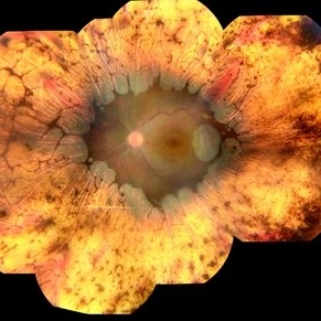
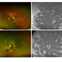
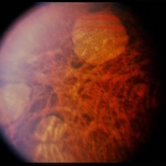
A 22-year old woman presenting with a 3 year history of nyctalopia. Investigations showed elevated ornithine levels in plasma, urine and CSF.
Imaging device: OPTOS
Condition/keywords: gyrate atrophy
-
 Gyrate Atrophy
Feb 5 2024 by Ali Al-Ani, M.B.Ch.B, FRCS, FRCOphth, FAAO
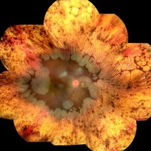
A 22-year old woman presenting with a 3 year history of nyctalopia. Investigations showed elevated ornithine levels in plasma, urine and CSF.
Imaging device: OPTOS
Condition/keywords: gyrate atrophy
-
 Gyrate Atrophy
Feb 5 2024 by Ali Al-Ani, M.B.Ch.B, FRCS, FRCOphth, FAAO
A 22-year old woman presenting with a 3 year history of nyctalopia. Investigations showed elevated ornithine levels in plasma, urine and CSF.
Imaging device: OPTOS
Condition/keywords: gyrate atrophy
-
 Gyrate Atrophy
Feb 5 2024 by Ali Al-Ani, M.B.Ch.B, FRCS, FRCOphth, FAAO
A 22-year old woman presenting with a 3 year history of nyctalopia. Investigations showed elevated ornithine levels in plasma, urine and CSF.
Imaging device: OPTOS
Condition/keywords: gyrate atrophy
-
 Gyrate Atrophy
Jan 25 2024 by Ricardo Leal Rodríguez, MD
Fundus photograph of an 11-year-old female with hyperornithinemia (932 µmol/L ) in plasma and mutation in the OAT gene (p.Asp242Argfs*6) confirming Gyrate Atrophy
Photographer: Dr Ricardo Leal Rodríguez, Instituto de Enfermedades y Cirugía Ocular, IECO , Mérida Yucatán, México
Imaging device: Triton Plus (Ver 10.16) TOPCON
Condition/keywords: gyrate atrophy, inherited retinal disease
-
 Gyrate Atrophy
Apr 12 2023 by Ahmed Abbas Hashmi, OD
Left eye fundus of a 53-year-old male patient with advanced gyrate atrophy of the choroid and retina with macular sparing. Optic nerve head is healthy.
Photographer: Ahmed Abbas Hashmi
Imaging device: Topcon TRC-NW8F
Condition/keywords: chorioretinal atrophy
-
 Gyrate Atrophy
Oct 15 2022 by Maxwell J Wingelaar, MD
Fundus photograph of a 15-year-old male with peripheral retinal changes consistent with gyrate atrophy
Photographer: Jarrod Wehmeier
Condition/keywords: gyrate atrophy
-
 Gyrate Atrophy
Oct 15 2022 by Maxwell J Wingelaar, MD
Fundus photograph of a 15-year-old male with peripheral retinal changes consistent with gyrate atrophy
Photographer: Jarrod Wehmeier
Condition/keywords: gyrate atrophy
-
 Gyrate Atrophy
Aug 20 2022 by Krushna Gopal Panda
Fundus photograph of 13-year-old child with gyrate atrophy
Photographer: Krushna Gopal Panda
Imaging device: Optos- California
Condition/keywords: gyrate atrophy
-
 PRP Marks
Apr 26 2021 by Priya Rasipuram Chandrasekaran, MBBS, DO, DNB, FRCS
This is the fundus photo montage of both eyes of a patient showing pan retinal photocoagulation marks. Theses marks can be confused with gyrate atrophy, cobble stone degeneration and myopic degeneration.
Condition/keywords: pan-retinal photocoagulation (PRP)
-
 Gyrate Atrophy
Oct 30 2020 by JEFFERSON R SOUSA, Tecg.º (Biomedical Systems Technology)
Female patient, 28-year-old, with low vision in both eyes since childhood. In routine examination, important changes were observed with atrophic, symmetrical and bilateral aspects with apparently preservation of the central retina.
Photographer: JEFFERSON R SOUSA - Study Center and Ophthalmological Research Dr. Andre M V Gomes, Institute Dr. Suel Abujamra São Paulo-Brazil
Imaging device: Topcon TRC-50 DX, Imaginet 5.0, angle de 50 graus. Flash 36 w-s
Condition/keywords: gyrate atrophy
-
 Gyrate Atrophy
Oct 30 2020 by JEFFERSON R SOUSA, Tecg.º (Biomedical Systems Technology)
Female patient, 28-year-old, with low vision in both eyes since childhood. In routine examination, important changes were observed with atrophic, symmetrical and bilateral aspects with apparently preservation of the central retina.
Condition/keywords: gyrate atrophy
-
 Gyrate Atrophy
Sep 23 2020 by Hashim Ali Khan, OD, FAAO
Widefield color fundus image of a young male with gyrate atrophy.
Imaging device: Optomap
Condition/keywords: gyrate atrophy
-
 Gyrate Atrophy
Sep 23 2020 by Hashim Ali Khan, OD, FAAO
Widefield choroidal image from a young male with gyrate atrophy.
Imaging device: Optomap
Condition/keywords: gyrate atrophy
-
 Gyrate Atrophy
Sep 23 2020 by Hashim Ali Khan, OD, FAAO
Widefield red-free image from a young male with gyrate atrophy.
Imaging device: Optomap
Condition/keywords: gyrate atrophy
-
 Gyrate Atrophy of the Choroid and Retina
May 1 2019 by Anmol Naik
A 34-year-old Indian male presented with gradual progressive bilateral diminution of peripheral vision since 6 years. His best corrected visual acuity was 6/60, N36 in right eye and 6/9, N6 in left. Wide-field fundus imaging demonstrated scalloped areas of chorioretinal atrophy with well-defined margins. His plasma ornithine levels were elevated.at 203.9 nmol/ml. Based on the typical features, a diagnosis of gyrate atrophy was made.
Photographer: Anmol Naik, Sankara Nethralaya, Chennai, India
Imaging device: Zeiss CLARUS 500
Condition/keywords: chorioretinal atrophy, gyrate atrophy
-
 Gyrate Atrophy
Jan 6 2019 by Hashim Ali Khan, OD, FAAO
Montage of Multiple Fundus Photographs from the right eye of a 25-year-old woman with gyrate atrophy.
Photographer: Ahmed Abbass
Imaging device: Topcon TRC-NW8F
Condition/keywords: gyrate atrophy, hereditary retinal dystrophy, retinal dystrophy
-
 Gyrate Atrophy
Oct 31 2018 by Dhaivat Shah
50-year-old male came in with complaint of daytime vision loss for a year and nighttime vision loss for more than 20 years, gradually increasing day by day. Fundus showed paving-stone like areas of atrophy of the RPE involving the macula which coalesces to form a characteristic scalloped border at the junction of normal and abnormal RPE. Gyrate atrophy is an autosomal recessive dystrophy caused by tenfold elevations of plasma ornithine, which is toxic to the RPE and choroid. Patients with gyrate atrophy have hyperpigmented fundi, with lobular loss of the RPE and choroid, normally sparing the fovea. The finding of generalized hyperpigmentation of the remaining RPE helps to clinically distinguish gyrate atrophy from choroideremia. Affected patients usually develop night blindness during the first decade of life and experience progressive loss of visual field and visual acuity later in the disease course. Early diagnosis is crucial because treatment in form of Arginine free diet and oral pyridoxine helps in slowing the progression of disease.
Imaging device: Optos
Condition/keywords: fundus autofluorescence (FAF), gyrate atrophy
-
 Gyrate Atrophy
Dec 22 2014 by H. Michael Lambert, MD
Depigmented areas and hyperpigmentation.
Condition/keywords: gyrate atrophy
-
 Gyrate Atrophy
-
 Gyrate Atrophy
Dec 22 2014 by H. Michael Lambert, MD
Depigmented areas and hyperpigmentation.
Condition/keywords: gyrate atrophy
 Loading…