Search results (109 results)
-
 MIDD (Maternally Inherited Diabetes and Deafness)
Feb 25 2025 by Virginia Gebhart
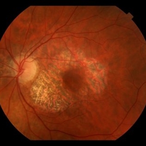
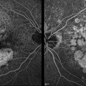
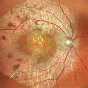
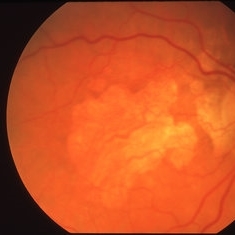
53 year old female with confirmed MIDD (genetic testing at Emory). Vision is stable with progressing GA but still central sparing OU. No evidence of choroidal neovascularization. Moderate myopia.
Photographer: Virginia Gebhart, Retina Consultants of Carolina
Imaging device: Topcon 50DX
Condition/keywords: geographic atrophy, Maternally inherited diabetes and deafness (MIDD), MIDD
-
 Pigmentary Degeneration of Retina (Secondary to Elmiron)
Nov 27 2024 by Virginia Gebhart
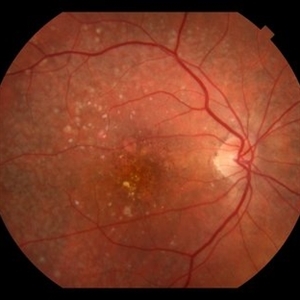
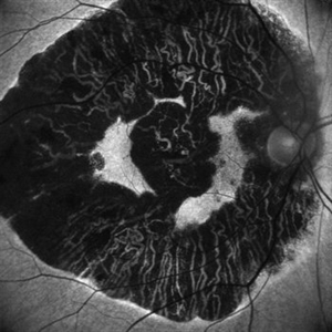
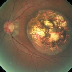
77 year old female with advanced geographic atrophy after years of Elmiron use (stopped in 2018). Serial exams show continued progression of GA. Central vision limited, vision remains stable and patient does not report noticing any changes.
Photographer: Virginia Gebhart, Retina Consultants of Carolina
Imaging device: Optos California
Condition/keywords: geographic atrophy, secondary pigmentary degeneration, toxic maculopathy
-
 Geographic Atrophy
Apr 22 2024 by Angela Rico
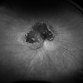
59 year-old female with MM1 Mitochondrial Genetic Defect. V/A- OD: 20/25, OS:20/40
Photographer: Angela Rico M.D.
Condition/keywords: Dystrophy of the Retinal Pigment Epithelium
-
 Familial Dominant Drusen
Mar 28 2024 by Houda Brarou
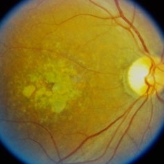
Familial Dominant Drusen is a genetically inherited retinal dystrophy and thought to represent an early-onset variant of age related macular degeneration. The gene responsible is EFEMP1 and inherited in autosomal dominant manner with variable expressivity. It is represented with multiple radially elongated small drusen in early stages and in later stages they become larger and more confluent. Geographic atrophy occurs in advanced stages.
Photographer: Houda Braou , Mohammed V military hospital of Rabat
Imaging device: TOPCON DRI OCT Triton Plus
Condition/keywords: FAMILIAL DOMINANT DRUSEN
-
 Remembrance Poppy Form Geographic Atrophy
Feb 15 2024 by HECTOR ARTURO MENDEZ PONCE
Autofluorescence that shows geographic atrophy in a 79 year old patient with dry age-related macular degeneration.
Photographer: Hector Arturo Mendez Ponce, MD
Imaging device: Heidelberg Spectralis
Condition/keywords: dry age-related macular degeneration (dry AMD), geographic atrophy
-
 Dry Age-related Macular Degeneration and Geographic Atrophy
Feb 1 2024 by Alejandro Cruz
88-year-old woman with dry age-related macular degeneration and geographic atrophy
Photographer: Alejandro Cruz, MD, Asociación para Evitar la Ceguera en México
Imaging device: Clarus 700
Condition/keywords: Drusen, dry age-related macular degeneration (dry AMD), geographic atrophy, macular degeneration, retina
-
 Dry AMD
Jan 25 2024 by Virginia Gebhart
79 year old female with intermediate dry AMD. Small area of geographic atrophy superior, large drusen and stippled RPE changes. BCVA 20/40
Photographer: Virginia Gebhart
Imaging device: Topcon
Condition/keywords: age-related macular degeneration (AMD), dry age-related macular degeneration (dry AMD), geographic atrophy
-
 Geographic Atrophy
Nov 16 2023 by Virginia Gebhart
67 year old female with Neovascular AMD with inactive CNV. Extensive geographic atrophy with minimal foveal sparing. Extensive ectopic CNV just superiorly to ON remains inactive. Discussed with pt treating with Syfovre to slow down GA progression
Photographer: Virginia Gebhart
Imaging device: Optos
Condition/keywords: advanced geographic atrophy, age-related macular degeneration (AMD), dry age-related macular degeneration (dry AMD), geographic atrophy
-
 Geographic Atrophy
Sep 21 2023 by Ben Serar
Fundus photograph of RE showing Geographic Atrophy in a case of Age- Related Macular Degeneration (ARMD).
Condition/keywords: Age- Related Macular Degeneration (ARMD), geographic atrophy
-
 MIDD
MIDD
May 26 2023 by Virginia Gebhart
51-year-old female with dry AMD, advanced atrophic without subfoveal involvement OU. Genetic testing confirmed MIDD (maternal inherited diabetes and deafness) which is a mitochondrial inherited dystrophy. Unaware of any family hx of macular degeneration.
Photographer: Virginia Gebhart, Retina Consultants of Carolina
Imaging device: Topcon TRC 50DX
Condition/keywords: advanced geographic atrophy, geographic atrophy
-
 Macular Degeneration with Extensive Geographic Atrophy
Jan 26 2022 by Olivia Rainey
Heidelberg Spectralis fluorescein angiography of a 94-year-old woman with Macular Degeneration affecting both eyes. The FA reveals transmission defects consistent with RPE changes and geographic atrophy of RPE of both eyes, as well as window defects consistent with peripheral scarring in the right eye. The patient's vision was Dcc20/70 in both eyes at the visit the images were taken.
Photographer: Olivia Rainey, OCT-C, COA
Imaging device: Heidelberg Spectralis
Condition/keywords: 30-degrees, choroidal neovascularization (CNV), dry age-related macular degeneration (dry AMD), early phase, fluorescein angiogram (FA), geographic atrophy, heidelberg spectralis, macular degeneration, neovascular age-related macular degeneration (AMD)
-
 Cuticular Drusen
Jun 13 2021 by Priya Rasipuram Chandrasekaran, MBBS, DO, DNB, FRCS
This is the fundus photo showing numerous yellow, small, hard drusen distributed throughout the retina. The corresponding OCT shows numerous elevated lesions underneath the RPE causing RPE elevations and arranged in a saw-tooth manner. Macular complications include acquired vitelliform lesion, choroidal neovascular membrane and geographic atrophy which are common after 60 years of age. It is usually associated with mutations in complement factor H. Basal laminar drusen, diffuse drusen and early adult onset grouped drusen are other alternative names. The differential diagnosis includes autosomal dominant drusen, pattern macular dystrophy, Sorsby macular drusen, mitochondrial macular dystrophy and so on.
Condition/keywords: cuticular drusen
-
 Geographic Atrophy
May 11 2020 by Gayathri Mohan
Fundus autofluorescence image of a patient with geographic atrophy.
Photographer: Gayathri Mohan, Retina Foundation
Imaging device: Mirante, Nidek
Condition/keywords: fundus autofluorescence (FAF), geographic atrophy
-
 Geographic Atrophy- Retro Mode
May 10 2020 by Gayathri Mohan
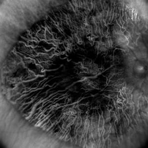
Image of a patient with geographic atrophy taken in retro mode. The deeper choroidal vessels are prominently seen.
Photographer: Gayathri Mohan, Retina Foundation
Imaging device: Mirante, Nidek
Condition/keywords: geographic atrophy, retro
-
 Geographic Atrophy
May 10 2020 by Gayathri Mohan
Color fundus photograph of a patient with geographic atrophy.
Photographer: Gayathri Mohan, Retina Foundation
Imaging device: Mirante, Nidek
Condition/keywords: fundus photograph, geographic atrophy
-
 Retinal Angiomatous Proliferation RAP
Mar 11 2020 by RAFAEL REIS PEREIRA, MD
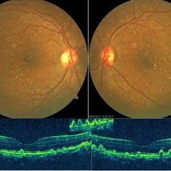
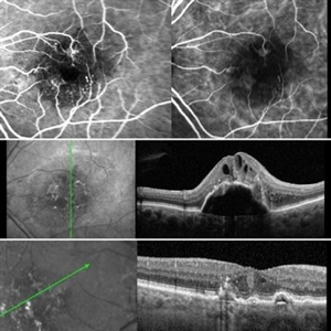
Retinal angiomatous proliferation (RAP) is a unique variant of neovascular age-related macular degeneration. Published studies have estimated that up to 15% of patients with neovascular age-related macular degeneration have RAP. Clinical features frequently associated with RAP include bilateral disease, presence of pigment epithelial detachments, and reticular pseudodrusen. RAP is more frequently associated with the development of retinal pigment epithelial tears and geographic atrophy that can lead to severe vision loss. We present a stereo fluorescein angiography and ICG (upper right and left image respectively) and OCT of left and right eye (middle and inferior image) of a RAP choroidal neovascularization in an 89-year-old patient.
Photographer: Rafael Reis Pereira
Imaging device: HRA Heildelberg Spectralis
Condition/keywords: retinal angiomatous proliferation (RAP)
-
 Macular Pattern Dystrophy Associated with MELAS
Dec 19 2019 by Olivia Rainey
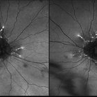
Bilateral wide field fundus autofluorescence images of a 54-year-old female with macular pattern dystrophy associated with MELAS. The patient is positive for m.3243A>G in MT-TL1. She had stroke in her 40s, hearing loss in her 30s, and has early onset diabetes. MyRetinaTracker shows VUS in RP1L1. Mutation in RP1L1 have been describe in other families with occult macular dystrophy. Farnsworth D15 is showing mild tritan abnormality, which is most commonly seen with acquired maculopathies. 12/17/19 patient's Optos and OCT show mild progression of atrophy.
Photographer: Olivia Rainey
Imaging device: Optos California
Condition/keywords: advanced geographic atrophy, bilateral, fundus autofluorescence (FAF), MELAS, Optos, pattern macular dystrophy, wide angle imaging
-
 Macular Pattern Dystrophy Associated with MELAS
Dec 19 2019 by Olivia Rainey
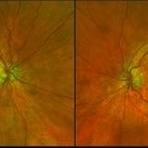
Bilateral wide field pseudocolor images of a 54-year-old female with macular pattern dystrophy associated with MELAS. The patient is positive for m.3243A>G in MT-TL1. She had stroke in her 40s, hearing loss in her 30s, and has early onset diabetes. MyRetinaTracker shows VUS in RP1L1. Mutation in RP1L1 have been describe in other families with occult macular dystrophy. Farnsworth D15 is showing mild tritan abnormality, which is most commonly seen with acquired maculopathies. 12/17/19 patient's Optos and OCT show mild progression of atrophy.
Photographer: Olivia Rainey
Imaging device: Optos California
Condition/keywords: advanced geographic atrophy, bilateral, fundus photograph, MELAS, Optos, pattern macular dystrophy, pseudocolor, wide angle imaging
-
 Geographic Atrophy in Dry AMD
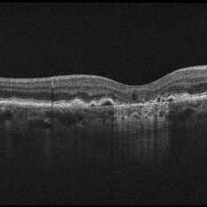
Dec 12 2019 by Darin R. Goldman, MD
This OCT B-scan shows geographic atrophy (GA) in dry age-related macular degeneration. There is focal atrophy of the RPE and outer retinal layers underneath the fovea, which is typical of GA. The loss of RPE in the affected area, relative to the surrounding macula, results in reverse shadowing within the underlying choroid. This effect is due to more penetration of the optical signal from the OCT illumination source owing to a relative absence of light being reflected as it normally would be from intact RPE. The result is a distinct border on each side of the affected area, where the underlying choroidal signal is more intense than the immediately adjacent areas. Additionally, adjacent to the area of GA are typical drusen, which are nodule-like diffusely hyperreflective accumulations within and under the RPE/Bruch complex, and pigment epithelial detachments (PEDs), which are nodule-like elevations of the RPE with underlying hyporeflective spaces.
Condition/keywords: dry age-related macular degeneration (dry AMD), geographic atrophy, optical coherence tomography (OCT)
-
 Geographic Atrophy Secondary to Central Areolar Choroidal Dystrophy
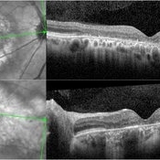
Dec 8 2019 by Anfisa Ayalon, MD
OCT pictures of a 37-year-old male with CACD. Note atrophic changes in the outer retinal layer.
Photographer: Anfisa Ayalon,MD., Meir Medical Center, Kfar Saba, Israel.
Condition/keywords: central areolar choroidal dystrophy (CACD), geographic atrophy, optical coherence tomography (OCT)
-
 Central Areolar Choroidal Dystrophy
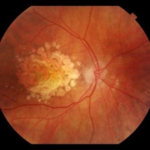
Dec 8 2019 by Anfisa Ayalon, MD
Fundus photograph of a 37-year-old male with CACD. The patient has visual acuity of 1/18 in the right eye and 6/30 in the left eye. Full-field ERG was normal under photopic and scotopic conditions.
Photographer: Anfisa Ayalon,MD., Meir Medical Center, Kfar Saba, Israel.
Condition/keywords: central areolar choroidal dystrophy (CACD), geographic atrophy, hereditary retinal degeneration
-
 Dry AMD with Central Geographic Atrophy, a Few Intraretinal Crystals, and Prominent Choroidal Vessels
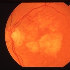
Sep 18 2019 by John S. King, MD
93-year-old white female routine AMD visit with VA of CF in OD due to central geographic atrophy.
Photographer: Gretchen Harper
Imaging device: Topcon 50
Condition/keywords: crystalline retinopathy, dry, geographic atrophy
-
 SMD/Geographic Atrophy
Apr 2 2019 by Gary R. Cook, MD, FACS
76-year-old white female with the geographic atrophy form of AMD OS; V.A. = 20/200
Imaging device: Topcon VT-50
Condition/keywords: age-related macular degeneration (AMD), geographic atrophy
-
 SMD/Geographic Atrophy
Apr 2 2019 by Gary R. Cook, MD, FACS
76-year-old white female with the geographic atrophy form of AMD OD; V.A. = 20/200
Imaging device: Topcon VT-50
Condition/keywords: age-related macular degeneration (AMD), geographic atrophy
-
 Geographic Atrophy
May 16 2018 by Bhavani Kulantayan, BA - Senior Retina Grader
The fundus photograph of a patient with geographic atrophy observed in the left eye. The geographic atrophy is one of the clinical signs of late stage in AMD.
Condition/keywords: geographic atrophy
 Loading…