Search results (38 results)
-
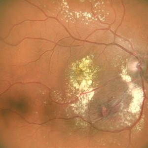
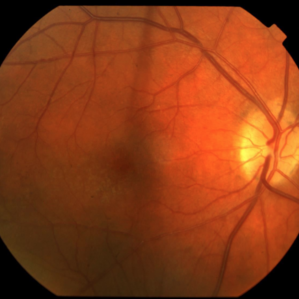
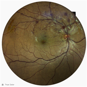 Superior Temporal Venous Branch Occlusion
Oct 23 2025 by Vicente Nicanor Mancilla Guerrero
Confocal laser retinography of the right eye of a 45-year-old female patient with hypertension (+). Venous dilation is evidenced together with the presence of flame hemorrhages and cottony spots in the upper temporal arch.
Photographer: Vicente Mancilla G, Medical Technologist in Ophthalmology
Imaging device: Compass CenterVue
Condition/keywords: branch retinal vein occlusion (BRVO)
-
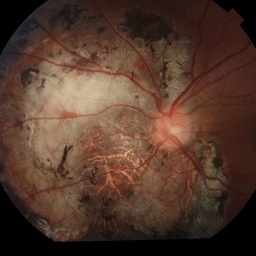
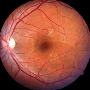
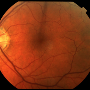
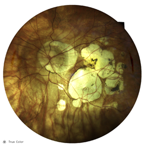 Myopic Degeneration
Oct 23 2025 by Vicente Nicanor Mancilla Guerrero
Confocal laser retinography of a female patient with -20.00Dp myopia. Typical findings of myopic retinopathy can be seen.
Photographer: Vicente mancilla G, Ophthalmic Medical Technologist
Imaging device: Compass Centervue
Condition/keywords: high myopia
-
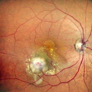 Lasered Retinal Artery Macroaneurysm
Sep 22 2025 by Tejaswita Verma
Fundus image of a 73 year old hypertensive female status post focal laser for exudative RAM. There was associated macular edema on OCT. Vision was 6/18.Patient was also planned for intravitreal anti VEGF injection on the same day.
Photographer: DR. TEJASWITA VERMA
Imaging device: MIRANTE
Condition/keywords: focal laser, RAM, retinal artery macroaneurysm
-
 Retinal Artery Macroaneurysm With Macular Edema
Sep 12 2025 by Tejaswita Verma
Fundus photo of a 73 year old hypertensive female with 6/18 vision, presenting with RAM ,with surrounding hard exudates and macular edema. She was advised focal laser, anti VEGF injection.
Photographer: DR. TEJASWITA VERMA
Imaging device: MIRANTE
Condition/keywords: RAM, retinal arterial macroaneurysm
-
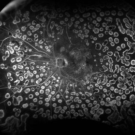
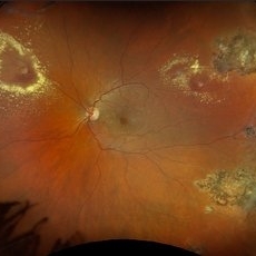 Retinal Aneurysms
Aug 6 2025 by Korey Starkey
54 year-old patient presents with scattered peripheral aneurysms with exudates. FA was performed showing peripheral nonperfusion and aneurysms. Treated patient with PRP and focal laser to aneurysms and continued observation.
Photographer: Kore Starkey
Imaging device: Optos
Condition/keywords: aneurysm, branch retinal vein occlusion (BRVO), chorioretinal scar, circinate ring, exudates, fundus photography, lesion, Optos, retinal aneurysms
-
 Leber's Miliary Aneurysm
Jan 23 2025 by Tejaswita Verma
A 41 year old male presented with 6/9 vision in the RE and fundus picture revealed miliary aneurysm with exudates and hemorrhages surrounded by old focal and sectoral laser marks. OCT revealed altered foveal contour with cystic spaces and IRHRM. He was advised RE injection antiVEGF with focal laser.
Photographer: DR. TEJASWITA VERMA
Imaging device: MIRANTE
Condition/keywords: Leber's miliary aneurysm
-
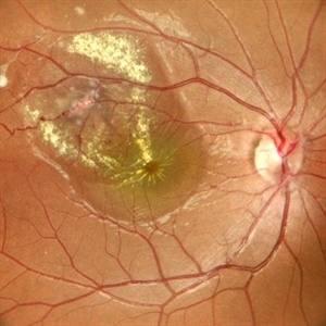
 Retinal Artery Macroaneurysm
Jul 13 2024 by Tejaswita Verma
A 53 year old female presented with blurred vision in RE since a month ,with borderline DM and HTN not on medications .H/o highest BP recording was 160/90 mm Hg.Vision 6/60 .FFA revealed leakages. She was advised RE focal laser with intravitreal anti-VEGF injections
Photographer: DR. TEJASWITA VERMA
Imaging device: MIRANTE
Condition/keywords: RETINAL ARTERY MACROANEURYSM
-
 Choroidal Osteoma
Jun 13 2024 by Virginia Gebhart
20 year old female with choroidal osteoma. Stable s/p PDT x3 and focal laser x 2, no obvious progression on last exam. Monitoring closely. Vision 20/30.
Photographer: Virginia Gebhart
Imaging device: Topcon 50 DX
Condition/keywords: barrier laser, choroidal osteoma, PDT
-
 FAF of Barricade Laser on Choroidal Osteoma
Jun 12 2024 by Virginia Gebhart
20 year old female with stable choroidal osteoma s/p PDT x 3 and focal laser x 2. No obvious progression on last exam, vision 20/30. Monitoring closely.
Photographer: Virginia Gebhart
Imaging device: Topcon 50 DX
Condition/keywords: autofluorescence imaging, barrier laser, choroidal osteoma, focal laser, fundus autofluorescence (FAF)
-
 Choroidal Osteoma
May 30 2024 by Virginia Gebhart
33 year old female with regressed osteoma OS s/p focal laser, TTT and PDT (first treatment in 2013). Vision 20/20, pt remains asymptomatic.
Photographer: Virginia Gebhart
Imaging device: Topcon 50DX
Condition/keywords: choroidal osteoma
-
 Diabetic Macular Edema
Feb 7 2024 by Virginia Gebhart
FA of 70 year old male with diabetic macular edema. FA shows early hyper-fluorescence with late leakage and capillary dropout in the temporal macula. Focal laser performed.
Photographer: Virginia Gebhart
Imaging device: Optos California
Condition/keywords: capillary dropouts, fluorescein angiogram (FA), macular edema
-
 Macroaneurysms
Jan 28 2024 by Anjana Mirajkar, MS Ophthalmology
Fundus image of post focal laser in a 20 year old female in a case of retinal arterial macroaneurysm
Photographer: Dr. Anjana Mirajkar -Retina Foundation, Ahmedabad
Imaging device: Mirante-Nidek
Condition/keywords: macroarterial aneurysm
-
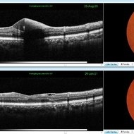
 CSR Treated with Focal Laser: Fundus, FFA, OCT Images
Dec 6 2021 by Nizamuddin HM Shaik, MD, FRCS
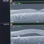
Fundus photograph , FFA and OCT ( Pre and Post ) of a 35-year-old lady with CSR treated with focal laser.
Photographer: Mahmoud , Ophthalmology Technician, International Medical Center
Imaging device: OCT
Condition/keywords: central serous chorioretinopathy (CSCR), laser photocoagulation
-
 CSR Treated with Focal Laser: FFA
Dec 6 2021 by Nizamuddin HM Shaik, MD, FRCS
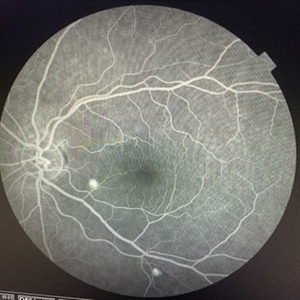
FFA of 35-year-old lady with CSR treated with focal laser.
Photographer: Mahmoud , Ophthalmology Technecian, International Medical Center
Imaging device: OCT
Condition/keywords: central serous chorioretinopathy (CSCR), FFA, focal laser, laser photocoagulation
-
 Focal Laser Treatment for Central Serous Retinopathy: FFA
Dec 6 2021 by Nizamuddin HM Shaik, MD, FRCS
FFA of a 35-year-old lady with CSR treated with focal laser.
Photographer: Mahmoud , Ophthalmology Technecian, International Medical Center
Imaging device: OCT
Condition/keywords: central serous chorioretinopathy (CSCR), laser photocoagulation
-
 CSR Treated with Focal Laser: Fundus Photo
Dec 6 2021 by Nizamuddin HM Shaik, MD, FRCS
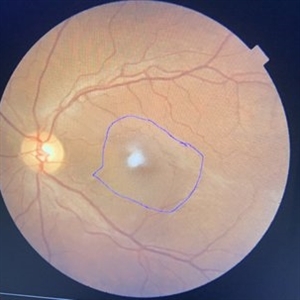
Fundus photograph of 35-year old lady with CSR treated with focal laser.
Photographer: Mahmoud , Ophthalmology Technician, International Medical Center
Imaging device: OCT
Condition/keywords: central serous chorioretinopathy (CSCR), focal laser, laser photocoagulation
-
 Posterior Ophthalmomyiasis Interna
Sep 20 2021 by Haley Tamanosky
Fundus photograph of 36-year-old woman after focal laser of a fly larva.
Photographer: Haley Tamanosky
Condition/keywords: focal laser, Posterior Ophthalmomyiasis Interna
-
 Sub-Internal Limiting Membrane Hemorrhage - Pre and Post YAG Laser
May 21 2021 by Anmol Naik
A 36-year-old male complained of central scotoma in the right eye after observing intense laser lights at a local festival celebration. His BCVA was 6/60. On examination, he had a sub-internal limiting membrane (ILM) hemorrhage, which was treated with focal frequency-doubled Nd:YAG laser. Two weeks later, the hemorrhage resolved completely with BCVA of 6/6.
Photographer: Anmol Naik, MS, Insight Institute of Ophthalmology, Pune, India.
Imaging device: Topcon 3D Maestro 1, integrated Fundus camera and OCT
Condition/keywords: focal laser, laser photocoagulation, sub-inner limiting membrane hemorrhage
-
 CSR Post Laser
May 15 2021 by Deepak Bhojwani, MS
Post focal laser fundus image of 38-year-old gentlemen with chronic CSR showing regression of entire subretinal fluid.
Photographer: Deepak Bhojwani
Condition/keywords: central serous retinopathy (CSR)
-
 Idiopathic Juxtafoveal Telangectasia Type 1
Nov 4 2019 by Thomas A. Ciulla, MD, MBA, FASRS
The telangiectasis occurs unilaterally in the temporal half of the macula in an area of 1–2 disc diameters. OCT originally showed significant macular edema temporally, mostly in the inner retina. He underwent a series of antiVEGF injections, as well as focal laser. The macular edema resolved and the visual acuity improved from 20/200 to 20/20.
Condition/keywords: idiopathic macular telangiectasia, juxtafoveal telangiectasis, parafoveal telangiectasia
-
 Idiopathic Juxtafoveal Telangectasia Type 1
Nov 4 2019 by Thomas A. Ciulla, MD, MBA, FASRS
The telangiectasis occurs unilaterally in the temporal half of the macula in an area of 1–2 disc diameters. OCT originally showed significant macular edema temporally, mostly in the inner retina. He underwent a series of antiVEGF injections, as well as focal laser. The macular edema resolved and the visual acuity improved from 20/200 to 20/20.
Condition/keywords: juxtafoveal telangiectasis, parafoveal telangiectasia
-
 BRVO
BRVO
Aug 28 2019 by Megan Fanelli
CASE: A 50-year-old male with past medical history significant for hypertension and a branch retinal vein occlusion. He complained of flashing lights and floaters for the past month. The floaters were consistent with red blood cells in the anterior vitreous. His visual acuity was 20/25 -1+2 in the left eye and 20/20 -1 in the right eye. The patient has been followed for BRVO since 2011 and received focal laser treatment and anti-VEGF injections. His last injection was 19 months prior to the vitreous hemorrhage. The plan is to treat the patient with sector pan-retinal photocoagulation. Image Description: Late phase wide field fluorescein angiogram of the left eye shows peripheral non-perfusion with neovascularization elsewhere with a pre-retinal hemorrhage. The image also displays leakage within the macula and previous focal laser treatment.
Condition/keywords: branch retinal vein occlusion (BRVO)
-
 Coats' Disease
Aug 24 2018 by Kim Barrett
Montage fluorescein angiography of 14-year-old male with Coats' Disease of the left eye. Multiple focal laser treatments. Current uncorrected visual acuity is 20/15-1 OU.
Photographer: Kim Barrett, C.O.A. Retina Specialist of Michigan
Imaging device: Heidelberg Spectralis
Condition/keywords: adolescent, Coats' disease, fluorescein angiogram (FA), Heidelburg Spectralis, laser photocoagulation, left eye, macroaneurysm, montage
-
 Heavy Focal Laser FAF Photograph - OS
Jul 20 2018 by Hosam Attia, MD
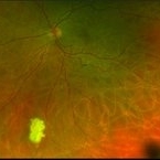
65-year-old, African American, woman with inactive PDR, S/P multiple PRP/ heavy focal OU, now receiving simultaneous Ozurdex/ Eylea injection OS, on regular basis w/ long standing poor vision 20/200-20/400 OS, since 2016 - patient was and currently being treated by another physician.
Imaging device: Optos California
Condition/keywords: focal laser, fundus autofluorescence (FAF), proliferative diabetic retinopathy (PDR)
 Loading…