Search results (37 results)
-
 The Retinal Tempest: Toxocara's Trail
Aug 31 2025 by Giriraj Vibhute
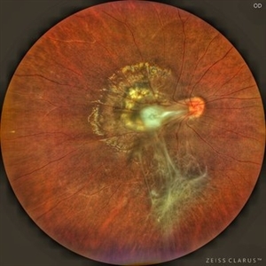
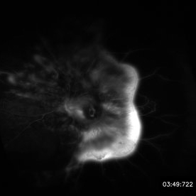
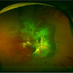
In this striking image, a central white granuloma spirals outward from the optic disc, surrounded by fibrous traction bands and scarring—the telltale markings of intraocular toxocara lesion. The retina is ravaged with proliferative vitreoretinal membranes and peripheral pigmentary changes, starkly illustrating the chronic inflammation and vision-threatening complications caused by Toxocara canis.
Photographer: Dr Giriraj Vibhute, MM Joshi eye institute, Hubli, India.
Condition/keywords: dragged disc, fibrous proliferation, Toxocara, toxocara canis
-
 Hamartoma of the Retina and Retinal Pigment Epithelium
Jan 5 2025 by César Adrián Gómez Valdivia, MD
Hamartoma of the retina and retinal pigment epithelium found in a 10 year-old male patient with type 2 neurofibromatosis history. Overlaying fibrous proliferation can be appreciated. Findings were unilateral.
Photographer: @eyemissu2
Imaging device: TOPCON TRC-50DX
Condition/keywords: hamartoma, retinal pigment epithelium (RPE) hamartoma
-
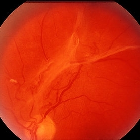
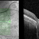 Proliferative diabetic retinopathy with fibrous proliferation over disc
Nov 4 2022 by T. P . VIGNESH, MBBS,MS
SD-OCT of a 60 year old man with proliferative diabetic retinopathy post PRP laser, revealing regressed fibrous proliferation attached to the disc .
Photographer: Shivanath
Imaging device: Heidelberg Spectralis
Condition/keywords: proliferative diabetic retinopathy (PDR)
-
 Ultra-Widefield Image of Tractional-Rhegmatogenous Retinal Detachment Sparing Fovea
Jul 16 2021 by Kushal S Delhiwala, MBBS, MS, FMRF,FICO, FAICO
Ultra-widefield fundus photograph of an 45-year-old phakic male with superior tractional-rhegmatogenous retinal detachment sparing fovea. Retinal break was observed at the base of fibrous proliferation. Scattered whitish outer retinal spots were noted in area of retinal detachment.
Photographer: Kushal Delhiwala, Netralaya superspeciality eye hospital, Ahmedabad, Gujarat,India
Imaging device: Optos Daytona
Condition/keywords: fibrovascular proliferation, fibrovascular tissue, outer retinal white spots, tractional retinal detachment, ultra-wide field imaging
-
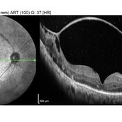 Fibrovascular Membrane
Apr 5 2018 by Mohamed Tawfik, MD
16 mm wide field OCT scan of a case of fiber-vascular membrane demonstrate the point of attachment of membrane.
Photographer: Mohamed A,Tawfik MD,FRCSed
Condition/keywords: fibrotic neovascularization, fibrous proliferation, fibrovascular change
-
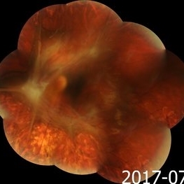 Advanced Proliferative Diabetic Retinopathy
Nov 4 2017 by Hamid Ahmadieh, MD
Merged color fundus photograph of the left eye of a 30-year-old woman with type1 diabetes since childhood. Note laser scars, severe fibrous proliferation, traction RD and macular dragging.
Photographer: Shabnam Poureh, Negah Eye Center, Tehran, Iran
Condition/keywords: color fundus photograph, diabetes, fibrous proliferation, proliferative diabetic retinopathy (PDR), severe traction
-
 Severe Fibrovascular Proliferative Mass With Tractional RD in PDR
Aug 1 2017 by Eitae Kim, MD
Severe fibrovascular proliferation with tractional retinal detachment is seen on UWF FAG.
Photographer: Eitae Kim, BOIM retinal center, Pureun eye hospital
Condition/keywords: fibrous proliferation, tractional retinal detachment
-
 Severe Fibrovascular Proliferative Mass With Tractional RD in PDR
Aug 1 2017 by Eitae Kim, MD
Severe fibrovascular proliferation with tractional retinal detachment is seen on UWF fundus photograph.
Photographer: Eitae Kim, BOIM retinal center, Pureun eye hospital
Condition/keywords: fibrous proliferation, tractional retinal detachment
-
---thumb.jpg/image-square;max$300,300.ImageHandler) Reduced Vision
Reduced Vision
Feb 4 2014 by Maurice F. Rabb
69-year-old female with a detachment of the pigment epithelium in the right eye with hemorrhagic and early fibrous proliferative changes. The left eye contained a large, turbid detachment of the pigment epithelium with patchy atrophy, a very shallow evident overlying sensory retinal detachment, but no subretinal hemorrhage. The visual acuity in each eye was 20/400.
Condition/keywords: fibrous proliferation, patchy atrophy, pigment epithelial detachment (PED), reduced vision
-
 Fibrotic Tractional Membrane in ROP Stage 5
Nov 7 2013 by Maria Ana Martinez-Castellanos, MD
Stage 5 retinopathy of prematurity in a 6 month old baby.
Photographer: Maria A. Martinez-Castellanos. Asociacion para Evitar la Ceguera en Mexico
Imaging device: RetCam II
Condition/keywords: fibrous proliferation, fibrovascular proliferation, retinopathy of prematurity (ROP)
-
 PDR
PDR
Mar 29 2013 by Henry J. Kaplan, MD
Fibrous proliferation (FPE) in a patient with PDR.
Condition/keywords: fibrous proliferation, FPE
 Loading…