Search results (44 results)
-
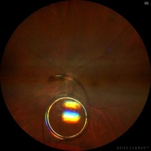
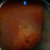
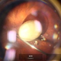
 Dropped Nucleus
Dec 16 2025 by Diana Elena Ornelas Rodríguez
Intraoperative photo of a dropped nucleus due to blunt trauma prior to fragging it.
Photographer: Diana Elena Ornelas Rodríguez, México.
Condition/keywords: dislocated crystalline lens, dislocated lens, Lens Luxation, Posterior Dislocated Nucleus
-
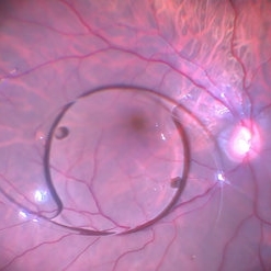
The B-scan Ballet
Dec 16 2025 by Malvika Singh
Dynamic B-scan showing post-traumatic dislocated lens in the vitreous, freely moving in the cavity with eye movements.
Condition/keywords: dislocated lens
-
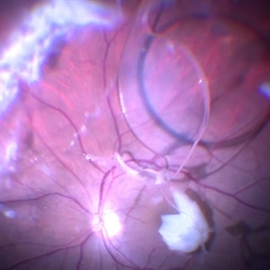
Unanchored
Dec 16 2025 by Malvika Singh
Dynamic B-scan showing free movements of the dislocated lens in the vitreous cavity post trauma.
Condition/keywords: dislocated lens
-
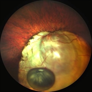
 Dislocated Lens
Dec 8 2025 by Parnian Arjmand, MD, MSc, FRCSC, DABO
A high myope patient presented 12 years after PPV a Cataract extraction for a retinal detachment repair with a new onset of vision loss. A dislocated IOL was noted on clinical examination.
Condition/keywords: Aphakia, dropped IOL, myopia, zonular dehiscence
-
 Dislocated IOL
Sep 20 2025 by JORGE SOBERANES
Fundus photograph of a 65-year-old man with a history of cataract surgery one year ago and bad vision since that.
Photographer: Dr. Jorge Soberanes, APEC, Universidad Nacional Autónoma México
Condition/keywords: dislocated lens, intraocular lens dislocation
-
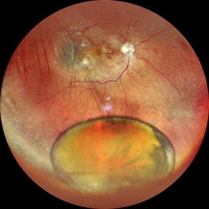 Dislocated Nucleus
Sep 12 2025 by Tejaswita Verma
Fundus photo of a middle aged male with 6/36 vision, spontaneously dislocated nucleus posteriorly with focal retinal detachment. Right eye Pars plana Vitectomy + nucleus removal + intravitreal C3F8 (12%) gas was performed for this patient.
Photographer: DR. TEJASWITA VERMA
Imaging device: MIRANTE
Condition/keywords: dislocated lens, retinal detachment
-
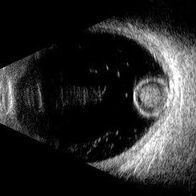 Morgagnian Ghost in the Deep
Jul 3 2025 by Gustavo Uriel Fonseca Aguirre
This B-mode para-axial ultrasound scan shows a posteriorly dislocated lens with cortical liquefaction, a dense nucleus, and an intact capsular bag. Vitreous bands are visible extending from the anterior to posterior segments. These findings were bilateral and not associated with trauma or prior surgery.
Photographer: Gustavo U. Fonseca Aguirre, Hospital Conde de Valenciana, Ciudad de México
Condition/keywords: ectopia lentis, morgagnian cataract
-
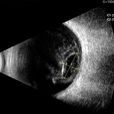 Ocular B-scan Ultrasound (Longitudinal Scan M6, gain 100 dB)
Jun 26 2025 by Hector Gabriel Moreno Solano, MD, MHA
B-scan ultrasound was performed in longitudinal section M6 with a gain of 100 dB. A hyperechoic structure with posterior acoustic shadowing is observed, consistent with lens luxation and condensed vitreous bands adjacent to the lens. The dislocated lens measures approximately 9.54 mm x 4.62 mm. The study was conducted following blunt ocular trauma caused by a golf ball. The remaining vitreous cavity appears anechoic, with no evidence of retinal detachment or other structural abnormalities in this section.
Photographer: Hector Gabriel Moreno Solano, Instituto Mexicano de Oftalmología “IMO I.A.P”
Imaging device: Quantel Medical
Condition/keywords: B scan ultrasound, lens luxation, ocular trauma
-
 Dislocated Lens
Jan 30 2025 by Kimberly Wakester
Fundus photograph of a 37-year-old man with an anteriorly dislocated lens in the left eye. The natural lens has displaced anteriorly in the AC secondary to trauma to the eye. There is also a Macular hole present with vitreous hemorrhage. Patient was recommended to proceed with lensectomy, iris repair and MH repair in the left eye.
Photographer: Kimberly Wakester, COA
Imaging device: Topcon TRC-50DX
Condition/keywords: dislocated lens, iridodialysis
-
 Macular Hole
Jan 30 2025 by Kimberly Wakester
Fundus photograph of a 37-year-old man with an anteriorly dislocated lens in the left eye. The natural lens has displaced anteriorly in the AC secondary to trauma to the eye. There is also a Macular hole present with vitreous hemorrhage. Patient was recommended to proceed with lensectomy, iris repair and MH repair in the left eye.
Photographer: Kimberly Wakester, COA
Imaging device: Optos California
Condition/keywords: dislocated lens, macular hole, vitreous hemorrhage
-
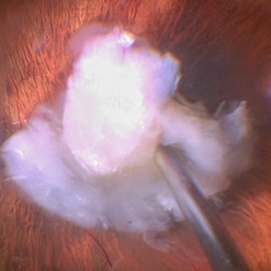
 IOL on Retinal Surface
Sep 8 2024 by Cesar Augusto Rocha Rojas, MD
Dislocated lens into the vitreous cavity during cataract surgery.
Photographer: Cesar Augusto Rocha Rojas, Hospital General de Zona #20, Instituto Mexicano del Seguro Social (IMSS)
Imaging device: Surgical microscope and smartphone
Condition/keywords: IOL drop
-
 Dislocated Lens
Jul 3 2024 by Anjana Mirajkar, MS Ophthalmology
An intra operative image showing us the dislocated cataractous lens piece eaten up by the cutter.
Photographer: Dr. Anjana Mirajkar -Retina Foundation, Ahmedabad.
Condition/keywords: Dislocated lens piece eaten up by the cutter
-
 Dislocated Iol With Hypotony Maculopathy and Hemorrhagic Choroidal
Feb 9 2024 by Sandra R Montezuma, MD
28 year old year-old male with history of congenital cataract of the right eye, s/p cataract extraction in 1999, s/p lens implant in 2011, presented with a dislocated IOL, hypotony, retina folds, hypotony maculopathy and hemorrhagic nasal choroidal after unsuccessful surgery to attempt remove the dislocated lens.
Photographer: Scott Baker, University of Minnesota
Condition/keywords: choroidals, dislocated posterior chamber intraocular lens (PCIOL), hypotony maculopathy, retina folds
-
 Dislocated Lens, Posterior OD
Jan 26 2024 by Corey Grant
OPTOS California photo presents a 71 year old male patient with a dislocated lens, posterior in the right eye. Presented on 1/26/24 with posteriorly dislocated SN60WF with a Soemmerring ring. Associated retinal hemorrhage within retinoschisis as well. This will result in a PPV/IOL exchange/SFIOL/STK for the right eye.
Photographer: Corey Grant, Ophthalmic Imager, Retina Specialist of Michigan
Imaging device: OPTOS California
Condition/keywords: color photo, IOL, OD, Optos, OPTOS CALIFORNIA, pars plana vitrectomy (PPV), retina
-
 Dislocated IOL
Oct 12 2023 by Virginia Gebhart
Fundus photo of an 83-year-old man with a 3 piece dislocated IOL. Surgery performed, PPV/removal of nonmagnetic FB/secondary Akreos. Eye is stable, vision limited due to grade 3 VH
Photographer: Virginia Gebhart, Retina Consultants of Carolina
Imaging device: Optos
Condition/keywords: dislocated intraocular lens (IOL), dislocated lens
-
 Dislocated Lens
Apr 26 2023 by Chloe Hanifan
Ultra wide field fundus photograph of a 41-year-old male with a dislocated lens affecting his right eye. IOL noted inferior vitreous base and vitrectomy surgery for removal of IOL was recommended. Patient has history of retinitis pigmentosa as well. Patient's vision at the time of presentation was counting fingers at 2 feet.
Photographer: Chloe Hanifan
Imaging device: Optos California
Condition/keywords: dislocated lens, fundus photography, Optos, pseudocolor, retinitis pigmentosa, ULTRA WIDE FIELD
-
 The Effects of Blunt Trauma
Feb 27 2022 by Jesus Lozano, MD
Axial Head Ct. 60 year old man with a history of blunt trauma and lost of vision after the event. VA HM. Iop 25mmhg. Cornea clear. Complete hyphema. BMode US: diffuse Vitreous Hemorrhage with a Dislocated Lens. Retina attached.
Photographer: Dr. Jesus Lozano. Retina Specialist. Hillel Yaffe Medical Center,Israel.
Imaging device: Axial Head CT
Condition/keywords: blunt trauma, hyphema, lens dislocation
-
 Dislocated Lens
Feb 18 2022 by Anthony Maida
Fundus photograph of 77 year old male with a dislocated lens secondary to contuse trauma
Photographer: Anthony Christopher Maida Medina
Imaging device: Artevo 800 microscope
Condition/keywords: dislocated lens, trauma
-
 Dislocated IOL Over Macula
Jan 11 2022 by Manish Nagpal, MD, FRCS (UK), FASRS
Intraoperative photo of a dislocated IOL sitting over the macular area.
Photographer: Manish Nagpal, Director, Retina Foundation, Ahmedabad
Imaging device: Sony PMW -10 MD surgical camera
Condition/keywords: dislocated intraocular lens (IOL), dislocated lens, dislocated posterior chamber intraocular lens (PCIOL)
-
 Dislocated IOL and Lens Matter
Jan 11 2022 by Manish Nagpal, MD, FRCS (UK), FASRS
Intraoperative photo of dislocated IOL and lens matter in the vitreous.
Photographer: Manish Nagpal, Retina Foundation, Ahmedabad, india
Imaging device: Sony PMW -10 MD surgical camera
Condition/keywords: dislocated crystalline lens, dislocated intraocular lens (IOL), dislocated lens, dislocated posterior chamber intraocular lens (PCIOL)
-
 Dislocated Brown Cataract with Chorioretinal Coloboma
Sep 8 2021 by Ram Sudarshan
A 44 year-old male with dislocated brown cataract resting within a chorioretinal coloboma.
Photographer: Mrs.Bharati
Imaging device: Clarus
Condition/keywords: Brown cataract, chorioretinal coloboma, coloboma, dislocated lens
-
 Dislocated Brown Cataract with a Chorioretinal Coloboma
Sep 8 2021 by Ram Sudarshan
A 44 year-old male with dislocated brown cataract along with a chorioretinal coloboma.
Photographer: Dr.Sivadarshan
Condition/keywords: Brown cataract, chorioretinal coloboma, d, dislocated lens
-
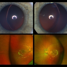 Ectopia Lentis
Jan 21 2021 by Jamin S. Brown, MD
This image serial demonstrates a patient with simple ectopia lentis. Anterior segment photographs in the upper panel demonstrate nasally subluxated crystalline lenses. Widefield fundus photography shows a "pseudo-buckle" which is the result of an optical effect due to the lens subluxation (artifactual image enlargement). Also note the juvenile macular reflex in this young patient. Ectopia lentis can present isolated ("simple") or in combination with various systemic defects (Marfan's syndrome, Weil-Marchesani syndrome or Ehlers-Danlos syndrome to name a few). Isolated ectopia lentis can be hereditary and causative genes have been identified as ADAMTSL4 located on chromosome 4 and FBN1 gene located on chromosome 15. Defects in the genes cause weakness in the zonular fibers which can lead to lens dislocation. Lastly, various ocular disorders such as Aniridia, Axenfeld-Rieger, Pseudoexfoliation or Trauma may also result in lens dislocation or subluxation.
Photographer: Stefanie Palmer CRA, Retina Vitreous Surgeons of CNY
Condition/keywords: dislocated lens, ectopia lentis
-
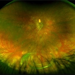
 Ultra-Widefield Montage of Traumatically Dislocated Posterior Polar Cataract in Vitreous
Dec 29 2020 by Kushal S Delhiwala, MBBS, MS, FMRF,FICO, FAICO
Fundus photograph of 50-year-old male with right eye posterior traumatic dislocation of lens having posterior polar cataract.
Photographer: Kushal Delhiwala, Netralaya superspeciality eye hospital, Ahmedabad, Gujarat,India
Imaging device: Optos Daytona
Condition/keywords: blunt trauma, dislocated lens, posterior subcapsular polar cataract, trauma, ultra-wide field imaging
-
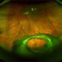
 Slide 7-77
Feb 25 2019 by Lancaster Course in Ophthalmology
Lens dislocated into the anterior chamber.
Condition/keywords: dislocated lens, lens
 Loading…