Search results (50 results)
-
 Cracking the Angioid Streaks Mystery: Multimodal Mayhem
Nov 26 2025 by SHRADDHA RAJ SHRIVASTAVA
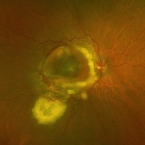
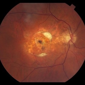
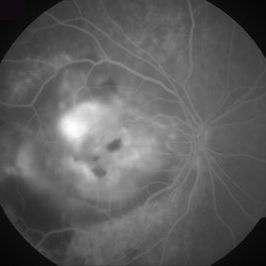
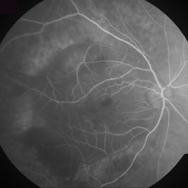
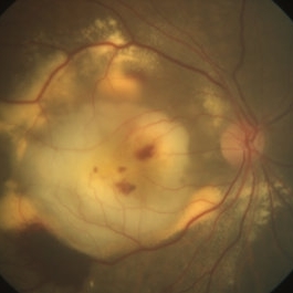
Multimodal imaging of right eye fundus showing Angioid Streaks with scarred CNVM. Color fundus photo shows hyperpigmented irregular lines emanating from the disc in a radiating fashion. Surrounding the angioid streaks and at the posterior pole, we can see numerous dot-like hypopigmented deposits and a disciform scar at macula. G-FAF images better reveal more extensive hypoautofluorescent streaks than are apparent on standard fundus photo. Characteristic “Para-streak phenomenon” of focal hyperautofluorescent spots are seen along the margins of the dark angioid streaks, corresponding to the areas of pigment clumping seen clinically. The para-streak pigment clumps are better delineated on the novel Retro-imaging method, appearing as raised bumps surrounding the angioid streaks.
Photographer: Dr. Shraddha Raj Shrivastava
Imaging device: Nidek Mirante SLO/OCT (Confocal scanning/Spectral domain OCT)
Condition/keywords: Angioid Streaks, Bruch's membrane, disciform scar, fundus autofluorescence (FAF), multimodal imaging, retro mode
-
 Macular Telangiectasia Type 2
Mar 29 2024 by Lucy V Cobbs, M.D.
Color fundus photograph of the left eye of a 70-year-old male with a disciform scar resulting from a neovascular membrane. A minority of MacTel type 2 patients develop neovascular disease, and the gold standard treatment is anti-VEGF intravitreal therapy. Without treatment, membranes may progress to severe central macular scarring. Late stages of proliferative MacTel type 2 may be confused with AMD, and a differentiating aspect is that MacTel type 2 typically lacks pigment epithelial detachments and drusen.
Condition/keywords: Mac Tel type 2
-
 DISCIFORM SCAR AND RETINAL PIGMENT EPITHELIUM (RPE) DETACHMENT IN A CASE OF IDIOPATHIC POLYPOIDAL CHOROIDAL VASCULOPATHY (IPCV)
Oct 21 2023 by Aditya S Kelkar, MS, FRCS, FASRS,FRCOphth
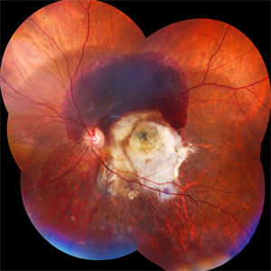
Right eye fundus photograph of a 83 year old female demonstrating Disciform Scar And Retinal Pigment Epithelium (RPE) Detachment In A Case Of Idiopathic Polypoidal Choroidal Vasculopathy (IPCV).
Photographer: DR APURVA MUKADAM
Imaging device: OPTOS DAYTONA
Condition/keywords: disciform scar
-
 Angiod Streaks and Disciform Scar
Jun 28 2022 by Julian Garcia-Sanchez
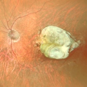
Fundus photograph of a 56-year-old man with angiod streaks and disciform scars due to CNV.
Photographer: Julian Garcia-Sanchez, MD, Asociación para Evitar la Ceguera en México
Imaging device: Zeiss Clarus 700
Condition/keywords: Angiod streaks, choroidal neovascularization (CNV), disciform scar
-
 Subretinal Hemorrhage and Disciform Scar
Feb 1 2022 by Lucas Zago Ribeiro, MD
Fundus photo of a 70-year-old man with acute subretinal hemorrhage, and previous disciform scar due to Wet AMD.
Photographer: Lucas Zago Ribeiro, UNIFESP, Brazil
Condition/keywords: disciform scar, subretinal hemorrhage, wet age-related macular degeneration (wet AMD)
-
 Disciform Scar
Aug 18 2020 by Aditya S Kelkar, MS, FRCS, FASRS,FRCOphth
Left eye fundus photograph of 75-year-old male, showing large disciform scar post subretinal bleeding secondary to idiopathic polypoidal choroidal vasculopathy
Photographer: Dr.Mounika Bolisetty
Imaging device: CLARUS 500
Condition/keywords: disciform scar, idiopathic polypoidal choroidal vasculopathy
-
 Disciform Scar
Jun 10 2020 by Manish Nagpal, MD, FRCS (UK), FASRS
Disciform scar from wet ARMD.
Photographer: gayathri mohan
Imaging device: nidek slo mirante
Condition/keywords: age-related macular degeneration (AMD), disciform scar
-
 Histoplasmosis and Old Disciform Macular Scar
Mar 27 2019 by Gary R. Cook, MD, FACS
Left eye of a 59-year-old white male with an old, inactive, disciform macular scar secondary to presumed ocular histoplasmosis (POHS); V.A.= counting fingers at 3 feet.
Imaging device: Topcon VT-50
Condition/keywords: central disciform scar, disciform scar, peripapillary atrophy, presumed ocular histoplasmosis syndrome (POHS)
-
 Slide 9-84
Feb 26 2019 by Lancaster Course in Ophthalmology
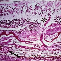
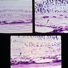
Senile macular degeneration with disciform scar. A retinal arteriole (asterisk) extends into the subretinal component of the scar, through a break in the thickened and detached inner layer of Bruch's membrane, and then into the vascularized intra-Bruch's-membrane component of the scar. Study of serial sections disclosed this retinal vessel to anastomose with the choroidal vessel (arrow) which extends through a branch in Bruch's membrane.
Condition/keywords: Bruch's membrane, disciform scar, macular degeneration, retinal arteriole
-
 Slide 9-83
Feb 26 2019 by Lancaster Course in Ophthalmology
Senile macular degeneration. A disciform scar is shown in a typical two component configuration. Arrows indicate the thickened and detached inner aspect of Bruch's membrane. There is one component of the disciform lesion that is located between the retina and the detached inner aspect of Bruch's membrane. RPE hyperplasia is evident in this component. The second component is located between the two layers of Bruch's membrane, and this component has choroidal neovascular tissue in the lower view.
Condition/keywords: Bruch's membrane, choroidal neovascular tissue, disciform scar, macular degeneration, retinal pigment epithelium
-
 Disciform Scar Due to Exudative Macular Degeneration
Feb 2 2018 by Olivia Rainey
Color fundus photograph of a 74-year-old male with a central disciform scar due to exudative macular degeneration.
Photographer: Olivia Rainey
Imaging device: Topcon 50dx
Condition/keywords: 50 degrees, age-related macular degeneration (AMD), central disciform scar, color fundus photograph
-
 Neovascular AMD
Jan 3 2017 by Jason Griffith
75-year-old male with neovascular AMD with disciform scar OS
Photographer: Jason Griffith
Imaging device: Topcon TRC-50EX
Condition/keywords: disciform scar, neovascular age-related macular degeneration (AMD)
-
 Macular Disciform Scar
Jun 8 2015 by ARIEL WANG
Fundus photograph and OCT scan of an 86-year-old man with long-standing type I diabetic proliferative retinopathy.
Photographer: Suber Huang, Retina Center of Ohio
Imaging device: Heidelberg Spectralis
Condition/keywords: central disciform scar
-
 Active Neovascular AMD With Disciform Scar
Apr 30 2015 by Mitzy E Torres Soriano, MD
Active neovascular AMD with disciform scar.
Photographer: Mitzy E. Torres Soriano, MD; Centro medico Cagua-Estado Aragua. Venezuela
Imaging device: TOPCON
Condition/keywords: disciform scar, disciform with hemorrhage, neovascular age-related macular degeneration (AMD), wet age-related macular degeneration (wet AMD)
-
 Chronical Submacular Hemorrhage in the Setting of Neovascular AMD
Mar 23 2015 by Rita Couceiro, MD, MS
An 80-year-old male, with a history of hypertension and high cholesterol, complained of acute and painless vision loss in his left eye (OS) in the previous 5 months. On observation best corrected visual acuity in OS was hand motion. A dense vitreous opacity in OS precluded fundus examination. Ocular ultrasound revealed vitreous hemorrhage and thickening of the macular area. The patient was submitted to pars plana vitrectomy, which disclosed a large submacular hemorrhage with chronical features and disciform scarring in the setting of neovascular AMD.
Imaging device: Intraoperative fundus photograph
Condition/keywords: neovascular age-related macular degeneration (AMD), submacular hemorrhage, wet age-related macular degeneration (wet AMD)
-
 Disciform Scar - Fluorescein Angiography
Oct 3 2013 by Gerardo Garcia-Aguirre, MD
Disciform scar - fluorescein angiogram.
Condition/keywords: disciform scar
-
 Disciform Scar - Fluorescein Angiography
Oct 3 2013 by Gerardo Garcia-Aguirre, MD
Disciform scar - fluorescein angiogram.
Condition/keywords: disciform scar
-
 Disciform Scar - Fluorescein Angiography
Oct 3 2013 by Gerardo Garcia-Aguirre, MD
Disciform scar - fluorescein angiogram.
Condition/keywords: disciform scar
-
 Disciform Scar - Fluorescein Angiography
Oct 3 2013 by Gerardo Garcia-Aguirre, MD
Disciform scar - fluorescein angiogram.
Condition/keywords: disciform scar
-
 Disciform scar - Red Free Image
Oct 3 2013 by Gerardo Garcia-Aguirre, MD
Disciform scar - red free image.
Condition/keywords: disciform scar, red-free
-
 Disciform Scar - Fundus Photograph
Oct 3 2013 by Gerardo Garcia-Aguirre, MD
Disciform scar - fundus photograph.
Condition/keywords: disciform scar, fundus photograph
-
---thumb.jpg/image-square;max$300,300.ImageHandler) Congenital Toxoplasmosis
Congenital Toxoplasmosis
Aug 14 2013 by From the Collections of Thomas M. Aaberg, MD and Thomas M. Aaberg Jr., MD
Disciform scar.
Condition/keywords: congenital toxoplasmosis, disciform scar
-
---thumb.jpg/image-square;max$300,300.ImageHandler) Congenital Toxoplasmosis
Congenital Toxoplasmosis
Aug 14 2013 by From the Collections of Thomas M. Aaberg, MD and Thomas M. Aaberg Jr., MD
Disciform scar.
Condition/keywords: congenital toxoplasmosis, disciform scar
-
---thumb.jpg/image-square;max$300,300.ImageHandler) Congenital Toxoplasmosis
Congenital Toxoplasmosis
Aug 14 2013 by From the Collections of Thomas M. Aaberg, MD and Thomas M. Aaberg Jr., MD
Disciform scar.
Condition/keywords: congenital toxoplasmosis, disciform scar
-
---thumb.jpg/image-square;max$300,300.ImageHandler) Congenital Toxoplasmosis
Congenital Toxoplasmosis
Aug 14 2013 by From the Collections of Thomas M. Aaberg, MD and Thomas M. Aaberg Jr., MD
Disciform scar.
Condition/keywords: congenital toxoplasmosis, disciform scar
 Loading…