Search results (43 results)
-
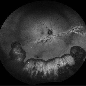
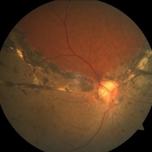
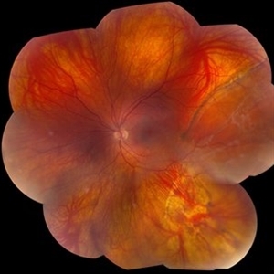
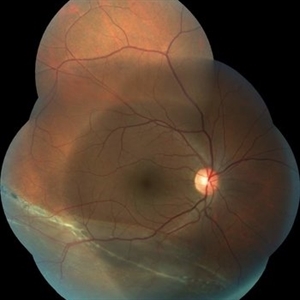
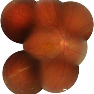
 Choroidal Melanoma with Exudative Detachment
Apr 7 2025 by Virginia Gebhart
Autofluorescence image of 36 year old female showing demarcation line of fluid/detachment from new choroidal melanoma. Pt will be scheduled for brachytherapy pending CT scan results.
Photographer: Virginia Gebhart, Retina Consultants of Carolina
Imaging device: Optos California
Condition/keywords: Autoflourescence, autofluorescence imaging, choroidal melanoma, melanoma, retinal detachment
-
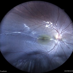
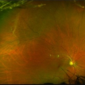
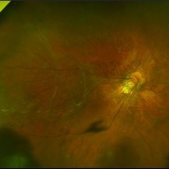
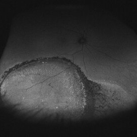
 Guardian Angel
Dec 11 2024 by Virginia Gebhart
48 year old female 3 months s/p brachytherapy for choroidal melanoma. Persistent subretinal and increased subfoveal fluid. Will observe for now, will consider Ozurdex if no improvement. BCVA 20/80
Photographer: Virginia Gebhart, Retina Consultants of Carolina
Imaging device: Optos California
Condition/keywords: brachytherapy, demarcation line, fundus autofluorescence (FAF), serous detachment, subretinal fluid
-
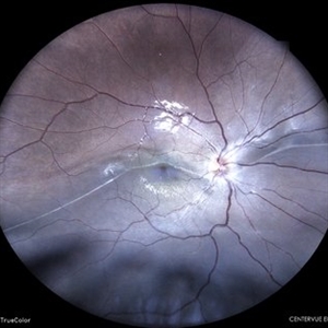
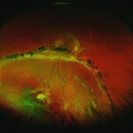
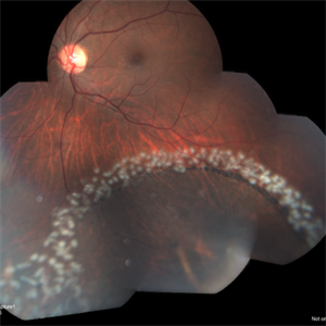
 Repaired Retinal Detachment with Multiple Breaks
Dec 9 2024 by Virginia Gebhart
FAF in 25 year old female of repaired retinal detachment 1.5 year s/p scleral buckle/cryo. Pt had been having symptoms for over a year, inferior demarcation line from retinal fluid that was present. Retina remains flat and attached under buckle. Treated lattice inferiorly, no new holes or tears. VA 20/20
Photographer: Virginia Gebhart, Retina Consultants of Carolina
Imaging device: Optos California
Condition/keywords: autofluorescence imaging, cryotherapy, demarcation line, lattice degeneration, scleral buckle
-
 Old Retinal Detachment
Jun 17 2024 by Akansha Sharma
Color fundus photograph of a 10 year old male with old retinal detachment with subretinal band.
Photographer: Dr. Akansha Sharma, Bharati Eye Hospital
Condition/keywords: demarcation line, OLD RD, subretinal bands
-
 Old Retinal Detachment
Apr 17 2024 by Akansha Sharma
Color fundus photograph of a 13 year old female with old retinal detachment at presentation as is demonstrated by a demarcation line.
Photographer: Dr. Akansha Sharma, Bharati Eye Hospital
Condition/keywords: delimited old retinal detachment, demarcation line, RD, Retinal Detachment
-
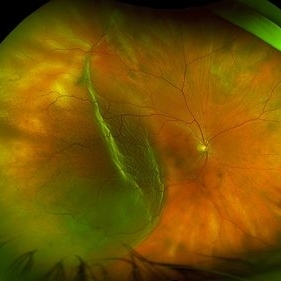 Retinal Detachment
Mar 28 2024 by Virginia Gebhart
68 year male with chronic appearing retinal detachment with subretinal bands and subretinal fibrosis. Demarcation line present, SRF splits the fovea on OCT.
Photographer: Virginia Gebhart
Imaging device: Optos California
Condition/keywords: chronic retinal detachment, Retinal Detachment
-
 Retinal detachment
Apr 12 2023 by Ahmed Abbas Hashmi, OD
Color fundus photograph of the left eye of a 30-year-old man with asymptomatic inferior retinal detachment with pigmented demarcation line. Macula and Disc healthy.
Photographer: Ahmed Abbas Hashmi
Imaging device: Topcon TRC-NW8F
Condition/keywords: Pigmentary demarcation line, Retinal Detachment
-
 Spontaneous reattachment of retina
Aug 30 2022 by Ruchir Mehta, DO, DNB, FRCS
Fundus photograph of a 22 year old male with spontaneously reattached retina with pigmented demarcation line involving the macula as seen in the picture
Photographer: Ruchir Mehta, Mehta Superspeciality Eye Hospital, Jamnagar, Gujarat, India
Imaging device: Zeiss Visucam 500
Condition/keywords: spontaneous retinal reattachment
-
 Chronic retinal detachment changes
Apr 29 2022 by Otakar Dušek, M.D. Ph.D.
Colour fundus photo of 22-year-old woman with bulous retinal detachment number 5-9, old demarcation lines and inferotemporal periheral secondary retinal cyst.
Photographer: Otakar Dušek, Charles University, Prague
Imaging device: Zeiss Clarus
Condition/keywords: chronic retinal detachment, demarcation line, peripheral retinal cyst
-
 Senile Retinoschisis
Jul 27 2021 by Dhaivat Shah
Senile retinoschisis is an acquired, idiopathic, degenerative condition of the neurosensory layer of the retina. It is characterized by separation at the outer plexiform layer or less commonly at the neurosensory layer of the retina. A 71-year-old male underwent cataract surgery in the right eye 1 week before his presentation to retina clinic. His chief complaint was minimal visual improvement after the surgery. His visual acuity in the right eye was 5/60 before cataract surgery and 6/60 after the surgery and no improvement with pinhole. On fundus examination of the right eye, an immobile, transparent subtle bullous elevation of the retina with minimal pigmentary changes was noted at the macula. The absence of a retinal tear , corrugations and demarcation lines differentiate it from rhegmatogenous retinal detachment. Optical Coherence Tomography is a confirmatory tool to diagnose senile retinoschisis. The OCT of this patient showed epiretinal membrane and coalescence of microcystic degenerations with splitting of the outer plexiform layer from rest of the outer retinal layers. Guarded visual prognosis was explained.
Photographer: Choithram Netralaya
Condition/keywords: optical coherence tomography (OCT), retinoschisis
-
 Retinal Holes, Demarcation Line
Jul 19 2021 by RUSHIK PATEL
Utlrawide pseudo-color fundus photograph of 28-year-old boy with 2 retinal hole surrounded by subretinal fluid less than 1 disc diameter and demarcation line.
Photographer: Rushik Patel, Netralaya Super Speciality Eye Hospital
Condition/keywords: retinal hole
-
 CHRPE
CHRPE
Jan 15 2021 by Priya Rasipuram Chandrasekaran, MBBS, DO, DNB, FRCS
This is the fundus photo and fundus photo montage of the left eye of a 25-year-old male showing flat, solitary, round, greyish pigmented lesion situated AT THE equator with a scalloped margin. Vessels overlying the lesion are normal and there is a clear demarcation line between this and normal retina. The margins are hypopigmented with few hypopigmented lacunae inside.
Condition/keywords: congenital hypertrophy of the retinal pigment epithelium (CHRPE)
-
 Bullous Retinoschisis with Outer Retinal Holes
Jun 15 2020 by Olivia Rainey
Ultra-widefield pseudocolor fundus photograph of a 56-year-old female with bullous retinoschisis with outer retinal holes affecting her right eye. The physician noted superotemporal retinoschisis in her monoculcar functioning eye. There was no demarcation line and no inner or outer layer breaks at her first appointment in February of 2020. On 6/15/20 she had a new onset outer holes and SRF tracking inferiorly. The physician recommended observation, however if this continues to progress we have discussed indications for barrier laser.
Photographer: Olivia Rainey, OCT-C, COA
Imaging device: Optos California
Condition/keywords: bullous retinoschisis, Optos, outer layer breaks, outer layer hole, pseudocolor, subretinal fluid, superior retina, ultra-wide field imaging
-
 Chronic Retinal Detachment in a Young Myopic Patient
Nov 6 2019 by Kamal Kishore, MD, MBBS
Chronic retinal detachment in a 27-year-old myopic female showing spontaneous reattachment in inferotemporal quadrant, and demarcation line and subretinal gliosis in superotemporal quadrant.
Photographer: Stephanie Shaver
Imaging device: Topcon 50 EX with OIS Winstation
Condition/keywords: chronic retinal detachment, high myopia
-
 Retinal Detachment with Demarcation Line
Apr 8 2019 by Gary R. Cook, MD, FACS
Pigmented demarcation line from a shallow, chronic retinal detachment OD
Imaging device: Topcon VT-50
Condition/keywords: demarcation line
-
 Self-Demarcated Retinal Tear
Apr 8 2019 by Gary R. Cook, MD, FACS
66-year-old white female with partially pigmented demarcation line surrounding an asymptomatic horseshoe retinal tear; no laser or cryo treatment ever performed; V.A. = 20/20.
Imaging device: Topcon VT-50
Condition/keywords: demarcation line, retinal tear
-
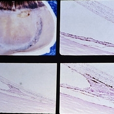 Slide 9-73
Feb 26 2019 by Lancaster Course in Ophthalmology
Inferior retinal dialysis with a localized area of long-standing retinal detachment and demarcation line (upper left). There is total atrophy of the photoreceptor cell layer (upper right). The demarcation line (lower views) is an area of RPE hypertrophy and hyperplasia with nodular basement membrane production and retinal adhesion.
Condition/keywords: photoreceptor cell, retinal dialysis, retinal pigment epithelium (RPE) hypertrophy
-
 Sub Total RD
Apr 5 2018 by Mohamed Tawfik, MD
Fundus Photo Of a case of Sub-total RD macular ON BCVA 1.0; demonstrated demarcation line denote old RD.
Photographer: Moahmed A,Tawfik MD , FRCSed
-
 Macula Sparring Tractional Retinal Detachment
Feb 9 2018 by Olivia Rainey
Ultra-wide field pseudocolor image of a 22-year-old male with a macula sparring tractional retinal detachment relating to retinopathy of prematuritiy affecting his right eye.
Photographer: Olivia Rainey
Imaging device: Optos
Condition/keywords: color fundus photograph, demarcation line, macula sparring, Optos, retinopathy of prematurity (ROP), tractional retinal detachment, ultra-wide field imaging
-
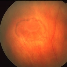
 Chronic Inferior Retinal Detachment
Mar 1 2017 by Philip J. Polkinghorne, MD
Color photograph of chronic retinal detachment with pigment demarcation line and atrophic holes visible. The vision was recorded at 20/20, and follow up is 3 years.
Photographer: Alex Fraser
Condition/keywords: atrophic retinal hole, demarcation line
-
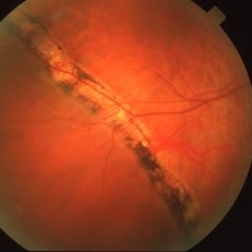
 Longstanding Retinal Detachment Due to a Larg Retinal Tear
Dec 27 2016 by Hamid Ahmadieh, MD
Wide-field color fundus photograph of the right eye of a patient with longstanding retinal detachment. Demarcation lines are visible.
Photographer: Shabnam Poureh, Negah Eye Center, Tehran, Iran
Condition/keywords: color fundus photograph
-
 Longstanding Retinal Detachment Secondary to a Larg Retinal Tear
Dec 27 2016 by Hamid Ahmadieh, MD
Montaged color fundus photograph of the right eye of a patient with longstanding retinal detachment. Demarcation lines are visible.
Photographer: Shabnam Poureh, Negah Eye Center, Tehran, Iran
Condition/keywords: color fundus photograph, demarcation line
-
 Asymptomatic Chronic Retinal Detachment With Demarcation Line
Jun 11 2016 by Philip J. Polkinghorne, MD
A 65-year-old emmetrope with asymptomatic chronic retinal detachment with demarcation line.
Photographer: Alex Fraser, Greenlane Clinical Center, Auckland, New Zealand
Condition/keywords: chronic retinal detachment, fundus autofluorescence (FAF)
-
 Retinal Detachment
May 13 2016 by Nichole Lewis
Inferior Retinal Detachment with some demarcation line s/p barrier laser.
Photographer: Nichole Lewis
Condition/keywords: barrier laser
-
 Retinal Detachment
May 9 2016 by Nichole Lewis
Retinal detachment with partial demarcation line and same day barrier laser treatment.
Photographer: Nichole Lewis
 Loading…