Search results (73 results)
-
 Desert Rose
Aug 4 2025 by KANWALJEET HARJOT MADAN, M.S. (Ophthalmology); FAICO (Vitreous - Retina)
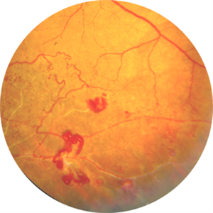
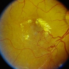
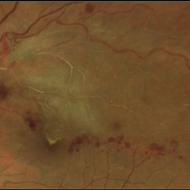
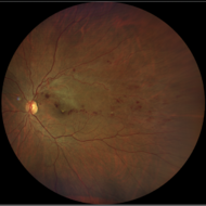
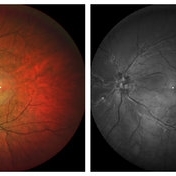
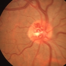
This is fundus picture of left eye of a 54 years male depicting Supero Temporal Branch Retinal Vein Occlusion with collaterals and neo vascularization elsewhere.
Photographer: Dr. Kanwaljeet Harjot Madan, Thind Eye Hospital, Jalandhar City (Punjab). INDIA.
Imaging device: Zeiss Fundus Camera
Condition/keywords: Neo Vascularization, Supero Temporal Branch Retinal Vein Occlusion
-
 Neovascularization of the Disc (NVD) - Red Free
Apr 28 2025 by Vishal Agrawal, MD, FRCS,FACS,FASRS
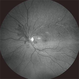
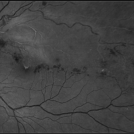
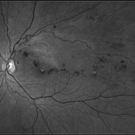
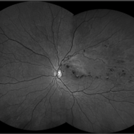
The red-free image enhances the visualization of the NVD, showing the fine neovascular fronds sprouting from the optic disc. Collateral vessels and vascular anastomosis are better appreciated.
Photographer: Dr Ayushi Gupta
Imaging device: Clarus 700
Condition/keywords: branch retinal vein occlusion (BRVO), collaterals
-
 Proliferative Diabetic Retinopathy
May 2 2024 by Aditya S Kelkar, MS, FRCS, FASRS,FRCOphth
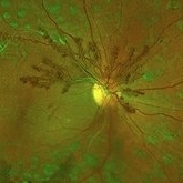
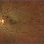
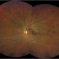
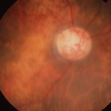
This fundus photo captures an intricate web of new vessels at optic disc.
Photographer: Dr Yash Garg , National Institute of Ophthalmology , Pune
Imaging device: OPTOS DAYTONA
Condition/keywords: web of collaterals
-
 Collaterals
Sep 21 2023 by Ben Serar
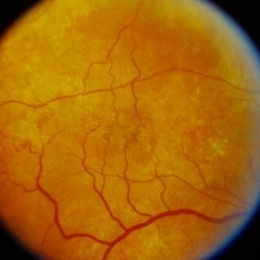
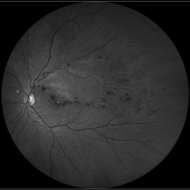
Fundus photograph showing arteriovenous shunts indicative of collaterals.
Condition/keywords: collaterals
-
 Branch Retinal Vein Occlusion (BRVO)
Sep 12 2023 by Ben Serar
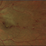
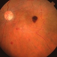
Fundus Photograph of RE showing exudates at the macula with macular edema, with collaterals and peri-venous sheathing along the inferotemporal vessel arcade, as a sequelae of branch retinal vein occlusion.
Condition/keywords: branch retinal vein occlusion (BRVO), macular edema
-
 Branch retinal vein occlusion - Colour & Red free image - ring shaped collaterals
Jul 18 2023 by Harsh Vardhan Singh, MS
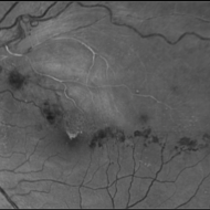
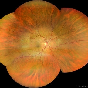
43-year-old woman presented with left eye old STBRVO with chronic CME of duration 6month showing ring shaped collaterals more evident on red free image
Photographer: Harsh Vardhan Singh, AIIMS, Guwahati
Imaging device: Zeiss Clarus 700
Condition/keywords: branch retinal vein occlusion (BRVO), BRVO, non-perfused branch retinal vein occlusion (BRVO)
-
 Branch retinal vein occlusion - Colour & Red free image - ring shaped collaterals
Jul 18 2023 by Harsh Vardhan Singh, MS
43-year-old woman presented with left eye old STBRVO with chronic CME of duration 6month showing ring shaped collaterals more evident on red free image
Photographer: Harsh Vardhan Singh, AIIMS, Guwahati
Imaging device: Zeiss Clarus 700
Condition/keywords: branch retinal vein occlusion (BRVO), BRVO, non-perfused branch retinal vein occlusion (BRVO)
-
 BRVO with Arteriovenous Malformation
Aug 4 2022 by KRISHNENDU NANDI, MS
An asymptomatic 55 years old male, hypertensive patient came for routine check up. On examination, his right eye was having BRVO with arteriovenous malformation with collaterals. Fundus photo of the right eye showed venous compression at the level of optic disc at 1 o'clock region with arteriovenous malformation around the disc and formation of collaterals extending up to foveal area. There was also presence of hard exudates and CME.
Photographer: Krishnendu Nandi, Netralayam Eye Care Centre, Kolkata, India
Condition/keywords: arteriovenous malformation, branch retinal vein occlusion (BRVO), collaterals
-
 Impending-CRVO
Feb 20 2022 by Vishal Gupta, MBBS, MS
Impending CRVO in a 54 year old hypertensive female patient with Collaterals and Grade 2 Chronic Hypertensive retinopathy.
Photographer: Dr Shobhit Chawla, Prakash Netra Kendr, Lucknow, UP, INDIA
Imaging device: Zeiss Clarus 500
Condition/keywords: central retinal vein occlusion (CRVO), collaterals, dilated tortuous vessels, hypertensive retinopathy
-
 Neovascular Membrane
Mar 14 2021 by Luiz A Zago, PhD
New and old neovascular membrane secondary to chorioretinal scar and a "drainage" vein.
Photographer: Luiz Zago PhD
Imaging device: Topcon 50IX
Condition/keywords: chorioretinal scar, collaterals, neovascular membrane, retina vessels
-
 Regressed CRVO
Jun 27 2020 by Aayesha Khanum
Regressed CRVO with severe collateral at disc
Photographer: Ravikrishna, Puttaswamy
Imaging device: Heidelberg Spectralis
Condition/keywords: central retinal vein occlusion (CRVO), collaterals
-
 Nonperfused BRVO with Collateral Vessels
Apr 8 2019 by Gary R. Cook, MD, FACS
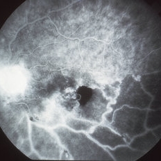
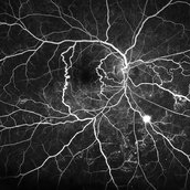
Late-phase fluorescein angiogram image of the left eye of a 73-year-old African-American female with a nonperfused BRVO showing flow through the collateral vessels, marked loss of the capillary bed, disc leakage from some NVD, and ischemic staining of the retinal veins; V.A. = 20/70-1
Imaging device: Topcon VT-50
Condition/keywords: branch retinal vein occlusion (BRVO), capillary nonperfusion, collaterals, disc leakage, FA late phase, fluorescein angiogram (FA)
-
 Nonperfused BRVO with Collateral Vessels
Apr 8 2019 by Gary R. Cook, MD, FACS
73-year-old African-American female with a remote, nonperfused inferotemporal branch retinal vein occlusion with collateral vessels visible across the horizontal raphae, mild NVD, and some macular hemorrhage and edema; V.A. = 20/70-1
Imaging device: Topcon VT-50
Condition/keywords: branch retinal vein occlusion (BRVO), collaterals, macular edema, macular hemorrhage, retinal hemorrhage
-
 CRVO with Disc Collaterals
Mar 27 2019 by Gary R. Cook, MD, FACS
81-year-old white female with remote CRVO with disc collaterals OD; V.A.= counting fingers 1 ft.
Imaging device: Topcon VT-50
Condition/keywords: central retinal vein occlusion (CRVO), collaterals, disc
-
 Disc Collaterals in CRVO
Mar 27 2019 by Gary R. Cook, MD, FACS
Elderly white male with remote, compensated CRVO showing persistent dilation of the venous vasculature and optic disc collaterals; V.A.= 20/20
Imaging device: Topcon VT-50
Condition/keywords: central retinal vein occlusion (CRVO), collaterals, disc
-
 Collateral Vessels in Resolved HRVO
Feb 12 2018 by John S. King, MD
Initial presentation: 25-year-old AAM; Hx DM, Sarcoidosis, Renal Disease, HTN with recent hypertensive crisis. Collateral vessels likely due to old HRVO with one front of NVE inferiorly; veins attenuated proximal to the collateral vessels.
Condition/keywords: collaterals, hemicentral retinal vein occlusion, retinal neovascularization
-
 Old Macular BRVO
Oct 10 2015 by Hamid Ahmadieh, MD
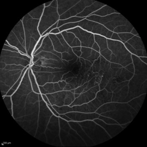
Mid -venous phase FA of the left eye of a 58-year-old woman with the history of macular BRVO. Note microaneurysms, collaterals , tortuosity of venules and an enlarged irregular FAZ compatible with macular ischemia.
Photographer: Nayereh Hadipour, Negah Eye Center, Tehran, Iran
Condition/keywords: collaterals, macular branch retinal vein occlusion (BRVO), macular ischemia, microaneurysms
 Loading…