 Initializing download.
-
 By KRISHNENDU NANDI, MS
Netralayam Superspeciality Eye Care Centre, Kolkata, India
Co-author(s): Ramesh Venkatesh, Narayana Nethralaya, Bangalore, India; Rupak Biswas, Netralayam Eye Care Centre, Kolkata, India - Uploaded on Aug 4, 2022.
- Last modified by Joshua Friedman on Aug 8, 2022.
- Rating
- Appears in
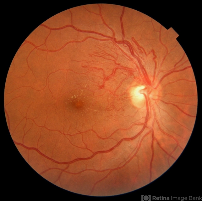
- BRVO with Arteriovenous Malformation
- Condition/keywords
- branch retinal vein occlusion (BRVO), arteriovenous malformation, collaterals
- Photographer
- Krishnendu Nandi, Netralayam Eye Care Centre, Kolkata, India
- Imaging device
- Fundus camera
- Description
- An asymptomatic 55 years old male, hypertensive patient came for routine check up. On examination, his right eye was having BRVO with arteriovenous malformation with collaterals. Fundus photo of the right eye showed venous compression at the level of optic disc at 1 o'clock region with arteriovenous malformation around the disc and formation of collaterals extending up to foveal area. There was also presence of hard exudates and CME.


 Caused due Branch Retinal Vein Occlusion (BRVO)")
 after Anti VEGF Treatment")
")


