Search results (34 results)
-
 Ciliary Body Melanoma
Feb 12 2025 by Virginia Gebhart
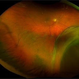
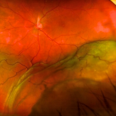
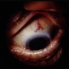
91 year old female with large collar button tumor emanating from the ciliary body with resolving vitreous hemorrhage. Melanoma cells in the AV as well as studded on the entire retina surface. Pt scheduled for enucleation. CT scans of chest and abdomen showed no evidence of metastatic disease.
Photographer: Virginia Gebhart, Retina Consultants of Carolina
Imaging device: Optos California
Condition/keywords: asteroid hyalosis, ciliary body mass, ciliary body melanoma, vitreous hemorrhage
-
 Ciliary Body Melanoma
Nov 2 2024 by Virginia Gebhart
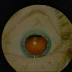
53 year old male with a large mass behind the lens as well as prominent scleral vessels. Clinical exam and ultrasound findings consistent with melanoma. Pt will be scheduled for enucleation pending CT scan results. Edit: Sadly patient has canceled all appointments and has requested no further contact
Photographer: Virginia Gebhart, Retina Consultants of Carolina
Imaging device: Optos California
Condition/keywords: ciliary body mass, ciliary body melanoma, ciliary body tumor
-
 New Choroidal Melanoma
Jan 4 2024 by Virginia Gebhart
77 year old male with a bilobed pigmented mass with exudative RD, and trace inflammation present in AV consistent with choroidal melanoma. Mass extends into ciliary body. Pt scheduled for MRI prior to plaque radiation to rule out metastasis.
Photographer: Virginia Gebhart
Imaging device: Optos California
Condition/keywords: ciliary body melanoma, exudative retinal detachment
-
 Ciliary Body Melanoma
Jul 4 2021 by Gerardo Rivera Arroyo
Clinical image taken in a slit lamp with a gonioscopy of a 39-year-old female patient with ciliary body melanoma before enucleation and pathological study.
Condition/keywords: ciliary body melanoma, gonioscopy
-
 Ciliary body melanoma
Jul 4 2021 by Gerardo Rivera Arroyo
Clinical image taken in a slit lamp with a gonioscopy of a 39-year-old female patient with ciliary body melanoma before enucleation and pathological study.
Condition/keywords: ciliary body melanoma, gonioscopy
-
 Ciliary Body Melanoma
May 18 2020 by McGill University Health Centre
Large tumors displace the lens. Of the 3 locations in the uveal tract, tumors of the ciliary body have the worst prognosis. This enucleation specimen shows a pigmented, bilobed, dome-shaped tumor arising from the ciliary body (arrow). The lens has been removed, and a diffuse, flat retinal detachment artifact is present.
Condition/keywords: melanoma
-
 Ciliary Body Melanoma
May 18 2020 by McGill University Health Centre
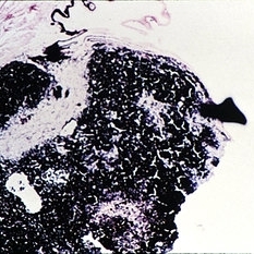
Uveal melanoma is the most common primary eye malignancy in adulthood, occurring mainly after age 60. The uveal tract — composed of the iris, ciliary body, and choroid — can be affected by uveal melanoma. Despite advances in treatment of the primary tumor, metastatic disease occurs in almost half of patients, generally affecting the liver and lungs via hematogenous dissemination of the primary tumor. Tumors have different levels of pigmentation, and some are amelanocytic (nonpigmented). The differential diagnosis for amelanotic choroidal melanoma is metastatic disease. Large tumors displace the lens. Of the 3 locations in the uveal tract, tumors of the ciliary body have the worst prognosis. This enucleation specimen shows a pigmented, nodular-shaped ciliary body melanoma (arrow) with extensive necrosis (*). A retinal detachment is present with subretinal fluid (arrowhead), and the retina is folded (•).
Condition/keywords: enucleation, melanoma
-
 Ciliary Body Melanoma
May 18 2020 by McGill University Health Centre
Uveal melanoma is the most common primary eye malignancy in adulthood, occurring mainly after age 60. The uveal tract — composed of the iris, ciliary body, and choroid — can be affected by uveal melanoma. Despite advances in treatment of the primary tumor, metastatic disease occurs in almost half of patients, generally affecting the liver and lungs via hematogenous dissemination of the primary tumor. Tumors have different levels of pigmentation, and some are amelanocytic (nonpigmented). The differential diagnosis for amelanotic choroidal melanoma is metastatic disease. Large tumors displace the lens. Of the 3 locations in the uveal tract, tumors of the ciliary body have the worst prognosis The enucleation specimen in (B) shows a large, dome-shaped, mixed melanotic and amelanotic choroidal melanoma. The anterior chamber is closed, and the angle is infiltrated (arrow). Total secondary retinal detachment with subretinal serous fluid and some subretinal hemorrhages are present (arrowhead). The lens is cataractous.
Condition/keywords: enucleation, melanoma
-
 Ciliary Body Melanoma
May 18 2020 by McGill University Health Centre
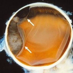
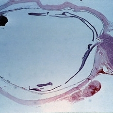
Uveal melanoma is the most common primary eye malignancy in adulthood, occurring mainly after age 60. The uveal tract — composed of the iris, ciliary body, and choroid — can be affected by uveal melanoma. Despite advances in treatment of the primary tumor, metastatic disease occurs in almost half of patients, generally affecting the liver and lungs via hematogenous dissemination of the primary tumor. Tumors have different levels of pigmentation, and some are amelanocytic (nonpigmented). The differential diagnosis for amelanotic choroidal melanoma is metastatic disease. Large tumors displace the lens. Of the 3 locations in the uveal tract, tumors of the ciliary body have the worst prognosis. The enucleation specimen in (A) shows a firm, dome-shaped, deeply pigmented tumor arising from the ciliary body (arrow). The lens has been removed, and a diffuse retinal detachment artifact is present.
Condition/keywords: enucleation, melanoma
-
 Ciliochoroidal Melanoma Photograph with Gonioscopy Lens
May 14 2020 by Anfisa Ayalon, MD
Gonioscopy photograph of a 71-year-old woman with ciliochoroidal melanoma. Note a melanoma-associated exudative retinal detachment and feeding vessels.
Photographer: Anfisa Ayalon, MD., Meir Medical Center, Kfar Saba, Israel.
Condition/keywords: ciliary body melanoma, exudative retinal detachment, gonioscopy
-
 Fundus Autofluorescence in Ciliochoroidal Melanoma
May 14 2020 by Anfisa Ayalon, MD
Fundus autofluorescence photograph of a 71-year-old woman with ciliochoroidal melanoma. Note a melanoma-associated massive exudative retinal detachment.
Photographer: Anfisa Ayalon, MD., Meir Medical Center, Kfar Saba, Israel.
Imaging device: California, Optos 200 DTX
Condition/keywords: ciliary body melanoma, exudative retinal detachment
-
 Ciliochoroidal Melanoma
May 14 2020 by Anfisa Ayalon, MD
Fundus photograph of a 71-year-old woman with ciliochoroidal melanoma. Note a melanoma-associated massive exudative retinal detachment.
Photographer: Anfisa Ayalon, MD., Meir Medical Center, Kfar Saba, Israel.
Imaging device: California, Optos 200 DTX
Condition/keywords: ciliary body melanoma, exudative retinal detachment, melanoma
-
 Superior Ciliochoroidal Melanoma with Diffuse Vitreous Seeding
Jan 21 2020 by Sophia El Hamichi, MD
Montage photograph of a fundus of a superior ciliochoroidal melanoma with diffuse pigmentary vitreous seeding.
Condition/keywords: ciliary body melanoma, melanoma, montage, vitreous seeding
-
 Ciliary Body Melanoma
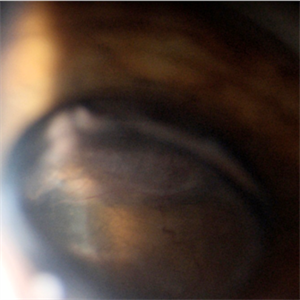
Apr 1 2019 by Gary R. Cook, MD, FACS
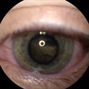
White male with a ciliary body melanoma OD seen as a dark, dome-shaped mass through a dilated pupil; VA=20/30-2.
Imaging device: Topcon VT-50
Condition/keywords: ciliary body mass, melanocytic lesion, melanoma
-
 Sentinel Vessel in Ciliary Body Melanoma
Apr 1 2019 by Gary R. Cook, MD, FACS
White male with a sentinel vessel OD due to the presence of a ciliary body melanoma; VA = 20/30-2.
Imaging device: Topcon VT-50
Condition/keywords: ciliary, melanocytic lesion, melanoma, sentinel vessel
-
 Slide 14-20
Mar 4 2019 by Lancaster Course in Ophthalmology
Ciliary body melanomas may appear early as a discrete mass or may show a diffuse growth pattern. As they enlarge, they may grow anteriorly onto the iris or extend posteriorly to the choroid.
Condition/keywords: ciliary body melanoma, melanoma
-
 Slide 14-19
Mar 4 2019 by Lancaster Course in Ophthalmology
Ciliary body melanomas may appear early as a discrete mass or may show a diffuse growth pattern. As they enlarge, they may grow anteriorly onto the iris or extend posteriorly to the choroid.
Condition/keywords: ciliary body melanoma, melanoma
-
 Ciliary Body Melanoma (Class 2 PRAME Positive)
Jan 17 2019 by Olivia Rainey
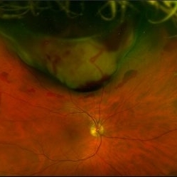
Pseudocolor optos fundus image of an 61-year-old male with ciliary body melanoma affecting his right eye, which tested PRAME positive placing him in class 2. The melanoma had caused a hemorrhagic retinal detachment. Patient was referred for a retinal detachment and was experiencing cloudy vision affecting his inferior field. He had been seeing flashes inferiorly 1-2x a daily for about 6-8 months.
Photographer: Olivia Rainey
Imaging device: Optos
Condition/keywords: ciliary body melanoma, class 2, Optos, PRAME positive
-
 Choroidal and Ciliary Body Melanoma
Apr 11 2018 by Jason Griffith
15-year-old male patient referred for suspicious mass.
Photographer: Jason Griffith, Tennessee Retina, Nashville, TN
Imaging device: Topcon TRC 50EX
Condition/keywords: ciliary body melanoma
-
 Ciliary Body Ocular Melanoma
May 9 2016 by Nichole Lewis
Ciliary body ocular melanoma.
Photographer: Nichole Lewis
Condition/keywords: ciliary body melanoma
-
 Ciliary Body Choroidal Melanoma
Jan 29 2015 by H. Michael Lambert, MD
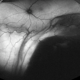
Large mass hanging posterior to the lens superiorly in a dilated eye.
Condition/keywords: ciliary body melanoma
-
 Ciliary Body Choroidal Melanoma
Jan 29 2015 by H. Michael Lambert, MD
Invivo presentation of ciliary body melanoma.
Condition/keywords: ciliary body melanoma
-
 Ciliary Body Melanoma-UBM
Feb 27 2014 by Susanna S. Park, MD, PhD
UBM of a large ciliary body melanoma in a 57-year-old man. Histopathology after eye removal showed a diffuse ring component that also involved the anterior choroid.
Photographer: Ellen Redenbo, University of California Davis Eye Center
Condition/keywords: ciliary body melanoma, ultrasound
-
 Ciliary Body Melanoma B-Scan Ultrasound
Feb 27 2014 by Susanna S. Park, MD, PhD
Large ciliary body melanoma in a 57-year-old man.
Photographer: Ellen Redenbo, University of California Davis Eye Center
Condition/keywords: B scan ultrasound, ciliary body melanoma
-
 Ciliary Body Melanoma With Partial Ring Configuration and Diffuse Sentinel Vessels
Feb 26 2014 by Susanna S. Park, MD, PhD
Slit lamp photo of a 57-year-old man with new vision loss from cataract formation. Large ciliary body mass with diffuse sentinel vessels is noted. The eye was removed and the tumor was noted to have a partial ring configuration with predominantly epithelioid cells and early vitreous seeding.
Photographer: Ellen Redenbo, University of California Davis Eye Center
Condition/keywords: ciliary body melanoma, melanoma
 Loading…