Search results (94 results)
-
 CHRPE with Lacunae
Dec 22 2025 by Kimberly Wakester
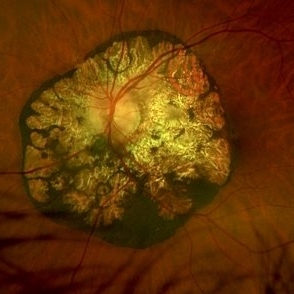
Optomap RGB image of an 48-year-old man with a CHRPE with lacunae in the right eye. Recommended yearly observation.
Photographer: Kimberly Wakester, COA, OCT-C
Imaging device: Optos California
Condition/keywords: congenital hypertrophy of the retinal pigment epithelium (CHRPE)
-
 CHRPE
CHRPE
Dec 11 2025 by Virginia Gebhart
48 year old female referred for pigmented lesion. Exam and photos consistent with well circumscribed CHRPE with lacunae. Patient previously unaware. Observation recommended.
Photographer: Virginia Gebhart, Retina Consultants of Carolina
Imaging device: Optos California
Condition/keywords: CHRPE, congenital hypertrophy of the retinal pigment epithelium (CHRPE)
-
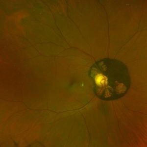
 Peripapillary CHRPE
Aug 29 2025 by Parnian Arjmand, MD, MSc, FRCSC, DABO
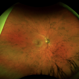
Optos color photo of a 64-year old male with an incidental finding of peri papillary congenital hypertrophy of the retinal pigment epithelium.
Condition/keywords: Peripapillary CHRPE
-
 Bear Tracks CHRPE - Red Channel
Jul 29 2025 by Drew Mitchell
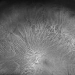
Green Free UWF image of extensive bear track patterned CHRPE.
Photographer: Drew Mitchell, OCT-C
Imaging device: Optos California
Condition/keywords: bear tracks, CHRPE, congenital hypertrophy of the retinal pigment epithelium (CHRPE), Green Free, OPTOS CALIFORNIA
-
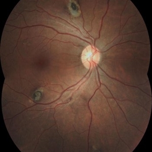
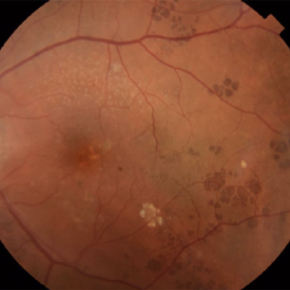
 CHRPE Spots
Jun 13 2025 by Brandon I Fram, MD
21 year-old, no past medical or family histories, with diffuse CHRPE spots suspicious for Gardner syndrome/FAP
Condition/keywords: bear tracks, CHRPE, congenital hypertrophy of the retinal pigment epithelium (CHRPE), familial adenomatous polyposis, Gardner Syndrome
-
 Bear Tracks (CHRPE)
Jun 4 2025 by Paulina Araujo
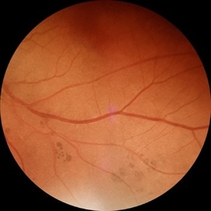
The 55-degree fundus photograph of the left eye shows bear tracks along the inferior temporal arcade.
Photographer: Paulina D.Araujo Martínez, Asociación para Evitar la Ceguera en México I.A.P., Hospital Dr Luis Sánchez Bulnes.
Condition/keywords: bear tracks, congenital hypertrophy of the retinal pigment epithelium (CHRPE)
-
 CHRPE
CHRPE
Mar 25 2025 by Toolie Winters
Ultra-wide field fundus photograph of a 78-year-old woman with extensive CHRPE lesions OS. Continued observation has been recommended at this time.
Photographer: Toolie Winters
Imaging device: Optos California
Condition/keywords: CHRPE, congenital hypertrophy of the retinal pigment epithelium (CHRPE), fundus photography, Optos, Optos California, pseudocolor, ultra-wide field imaging
-
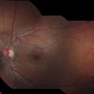
 Multimodal Imaging in CHRPE
Mar 6 2025 by Gerardo - Montante Montelongo, MD
Fundus photograph of an 83-year-old male with a history of Diabetes, smoking, cataract surgery on the right eye in 2022, and open-angle glaucoma. Asymptomatic. Indirect ophthalmoscopy revealed 80% excavation, peripapillary atrophy, and a hyperpigmented perifoveal lesion with 35% atrophy, 10% drusen, and 5.1 mm diameter, corresponding to a CHRPE. At multimodal imaging, FFA shows hypoautofluorescence of the lesion, OCT shows preservation of internal retinal layers, atrophy of external retinal layer, with an RPE disruption, and posterior shadowing. USG shows a flat hyperechoic lesion 5.1 mm in diameter and 1.32 mm in thickness, solid and with high internal reflectance.
Photographer: Gerardo Montante-Montelongo, MD, Mexican Institute of Ophthalmology
Imaging device: Clarus 700
Condition/keywords: congenital hypertrophy of the retinal pigment epithelium (CHRPE), multimodal imaging
-
 Lattice Degeneration With Atrophic Retinal Holes
Jan 30 2025 by Kimberly Wakester
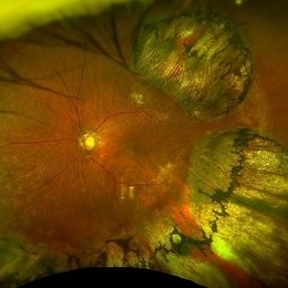
Ultra-wide field montage fundus photograph of a 56-year-old woman with lattice degeneration with atrophic holes statues post laser. Patient also has a small CHRPE temporal to macula and trace ERM that is not visually significant. Will continue follow up care to monitor and treat as needed.
Photographer: Kimberly Wakester, COA
Imaging device: Optos California
Condition/keywords: atrophic retinal hole, CHRPE, epiretinal membrane (ERM), lattice degeneration, montage photo
-
 CHRPE and Bear Tracks
Jan 7 2025 by Drew Mitchell
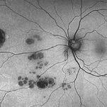
Fundus Autofluorescence of a CHRPE and Bear Tracks.
Photographer: Drew Mitchel, OCT-C
Imaging device: Optos Silverstone
Condition/keywords: bear tracks, CHRPE, congenital hypertrophy of the retinal pigment epithelium (CHRPE)
-
 CHRPE
CHRPE
Jan 6 2025 by Kavitha Duraipandi, MD DNB FICO FRCS
Bear tracks (animal tracks, grouped pigmentation spots) are simply many small CHRPEs located in isolated small area of the retina. These have not been reported to have the potential to transform into adenocarcinoma but yearly evaluations may be prudent.
Condition/keywords: CHRPE
-
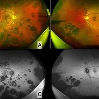 Bear Track Lesions in a Case of Congenital Hypertrophy of RPE
Sep 6 2024 by Giriraj Vibhute
Fundus photo and autofluorescence image in a 58-year-old woman A&B: Right and left eye ultrawidefield pseudocolor imaging in case of congenital hypertrophy of RPE. C&D: Fundus autofluorescence of right and left eye of the same patient. The patient/ family members did not have any history of colon cancer. Patient was advised colonoscopy and family members were screened.
Photographer: Giriraj Vibhute, dept of retina, M M Joshi eye institute, Hubli. India
Imaging device: Optos daytona
Condition/keywords: bear tracks, congenital hypertrophy of the retinal pigment epithelium (CHRPE)
-
 CHRPE and CHRRPE
May 26 2024 by shama sharief
Funds photo with CHRPE and CHRRPE.
Condition/keywords: CHRRPE, congenital hypertrophy of the retinal pigment epithelium (CHRPE)
-
 Benign Lobular Inner Nuclear Layer Proliferations (BLIP)
Apr 15 2024 by Virginia Gebhart
29 year old male with multiple flat CHRPE lesions, genetic testing negative for ACP genes associated with Gardner syndrome. Multiple intraretinal amelanotic lesions consistent with Benign Lobular Inner Nuclear Layer Proliferations (BLIP) of the retina
Photographer: Virginia Gebhart
Imaging device: Topcon
Condition/keywords: BLIP Benign Lobular Inner Nuclear Layer Proliferations, CHRPE, congenital hypertrophy of the retinal pigment epithelium (CHRPE)
-
 Gardner's Syndrome
Nov 10 2023 by Virginia Gebhart
17-year-old male with multiple pigmented spots most likely related to Gardner's Syndrome. Pt has not been diagnosed with FAP at this time, however pt receives regular screenings. Extensive maternal family hx of FAP syndrome and colon cancer. Pt's mother has FAP, who had colon resection. Pt's 2 aunts, 1 uncle, grandmother and great grandmother all passed from colon cancer. Pt has multiple maternal cousins diagnosed with FAP
Photographer: Virginia Gebhart
Imaging device: Topcon
Condition/keywords: CHRPE, congenital hypertrophy of the retinal pigment epithelium (CHRPE), familial adenomatous polyposis, Gardner Syndrome
-
 Gardner's Syndrome (FAP)
Nov 3 2023 by Virginia Gebhart
43 year-old female with multiple CHRPE lesions consistent with Gardner's Syndrome. Patient had colectomy at age 21. Patient is one of 7 children, 6 had FAP, 3 had colon removal and are alive and well, 3 have passed away from colon cancer. Maternal mother and grandmother also passed away from colon cancer. Patient has 2 children, one son (17) also has Gardner's Syndrome (FAP)
Photographer: Virginia Gebhart
Imaging device: Topcon
Condition/keywords: CHRPE, Gardner Syndrome
-
 Solitary large Congenital Hypertrophy of Retinal Pigment Epithelium (CHRPE)
Jul 1 2023 by Aditya S Kelkar, MS, FRCS, FASRS,FRCOphth
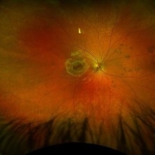
Right eye fundus photograph of a 42 year old asymptomatic male demonstrating a superotemporal solitary large Congenital Hypertrophy of Retinal Pigment Epithelium (CHRPE) lesion.
Photographer: Optom Komal Jangam
Imaging device: OPTOS DAYTONA
Condition/keywords: CHRPE
-
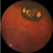
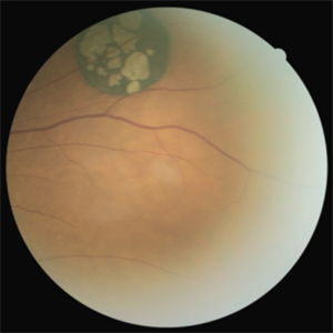 Solitary Congenital Hypertrophy of the Retinal Pigment Epithelium with Lacunae
Jun 11 2023 by Ethan K Sobol, MD
Fundus photograph of a solitary CHRPE in the superotemporal quadrant with central hypopigmented lacunae.
Condition/keywords: CHRPE, congenital hypertrophy of the retinal pigment epithelium (CHRPE), lacunae
-
 CHRPE
CHRPE
Jan 12 2023 by Christopher R. Adam, M.D.
Optos color photograph of a 51 y/o F with a 9x9mm CHRPE in the nasal quadrant. The lesion is flat with a bordering halo and lacunae.
Condition/keywords: congenital hypertrophy of the retinal pigment epithelium (CHRPE)
-
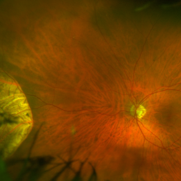
 Peripapillary Congenital Hypertrophy of the Retinal Pigment Epithelium
Dec 15 2022 by Jason Hsu, MD
Optos image of 62 year-old woman with incidental peripapillary congenital hypertrophy of the retinal pigment epithelium.
Photographer: Donnamarie Nielsen, COA
Imaging device: Optos California
Condition/keywords: congenital hypertrophy of the retinal pigment epithelium (CHRPE)
-
 Peripapillary Congenital Hypertrophy of the Retinal Pigment Epithelium Autofluorescence
Dec 15 2022 by Jason Hsu, MD
Optos fundus autofluorescence image of 62 year-old woman with incidental peripapillary congenital hypertrophy of the retinal pigment epithelium.
Photographer: Donnamarie Nielsen, COA
Imaging device: Optos California
Condition/keywords: congenital hypertrophy of the retinal pigment epithelium (CHRPE), fundus autofluorescence (FAF)
-
 Depigmented CHRPE
Nov 9 2022 by Maxwell J Wingelaar, MD
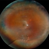
67 year-old female with a depigmented CHRPE
Condition/keywords: congenital hypertrophy of the retinal pigment epithelium (CHRPE)
-
 CHRPE - "BEAR TRACKS" PATTERN
Nov 8 2022 by Heitor Nogueira
BEAR TRACKS
Photographer: Heitor Nogueira
Condition/keywords: bear tracks, CHRPE, congenital hypertrophy of the retinal pigment epithelium (CHRPE)
-
 Congenital hypertrophy of the retinal pigment epithelium (CHRPE)
Jun 22 2022 by Dawson Winter
Ultrawide field fundus autofluorescence optos image of the left eye of a 28 year old female. Patient admits to floaters that come and go, but has no other ocular symptoms at this time. At the time of the appointment the patient was seeing 20/30+1 OS. Patient underwent MRI testing of the ocular orbit and results were found to be normal.
Photographer: Dawson Winters
Imaging device: Optos California
Condition/keywords: autofluorescence imaging, choroidal lesions, congenital hypertrophy of the retinal pigment epithelium (CHRPE), fundus autofluorescence (FAF), left eye, Optos, temporal retina, ultra-wide field imaging
-
 CHRPE
CHRPE
 Loading…