Search results (65 results)
-
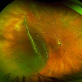
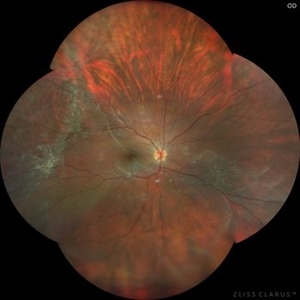
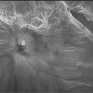
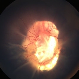
 Chronic RD with Retinal Dialysis
Jul 23 2025 by Virginia Gebhart
64 year old female with chronic retinal detachment from head trauma 41 years ago. Peripheral scarring from 6:00 to 11:00 with area of subretinal fluid inferotemporally, well demarcated with subretinal bands. Retinal dialysis inferotemporal from 7:00 to 9:00. No surgical repair needed or recommended at this time.
Photographer: Virginia Gebhart, Retina Consultants of Carolina
Imaging device: Optos California
Condition/keywords: chronic retinal detachment, demarcation, RD, Retinal Detachment, retinal dialysis, subretinal bands
-
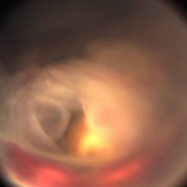
 Open Funnel
Apr 10 2025 by Gustavo Uriel Fonseca Aguirre
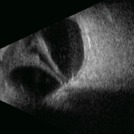
This B-mode longitudinal ultrasound scan demonstrates a long-standing rhegmatogenous retinal detachment, showing a characteristic open funnel configuration. The findings are consistent with chronic retinal detachment.
Photographer: Gustavo U. Fonseca Aguirre, Hospital Conde de Valenciana, Ciudad de México
Imaging device: Funnel
Condition/keywords: open funnel RD, Retina detachment
-
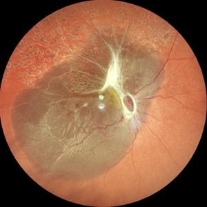
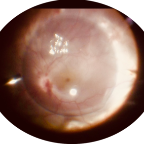
 Star Folds in a Chronic Retinal Detachment
Jul 3 2024 by Anjana Mirajkar, MS Ophthalmology
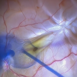
Intra-operative still RE showing a star fold at the parafoveal area causing traction at the macula. Brilliant blue dye being injected to the stain the ILM.
Photographer: Dr. Anjana Mirajkar -Retina Foundation, Ahmedabad
Condition/keywords: brilliant blue staining, proliferative vitreoretinopathy (PVR), star folds
-
 Fundus Autofluorescence of Closed Funnel Retinal Detachment
Apr 10 2024 by Max D Schlesinger, MD
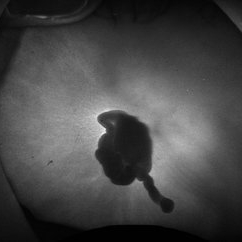
Fundus autofluorescence of a closed funnel retinal detachment; patient had previously undergone 360 degree retinectomy in attempt to re-attach retina for a chronic retinal detachment, which was unsuccessful.
Condition/keywords: Autoflourescence, Closed funnel RD, detachment
-
 Fundus Photo of Closed Funnel Retinal Detachment
Apr 10 2024 by Max D Schlesinger, MD
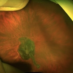
Wide-field funds photography of a closed funnel retinal detachment; patient had previously undergone 360 degree retinectomy in attempt to re-attach retina for a chronic retinal detachment, which was unsuccessful.
Condition/keywords: Closed funnel RD, detachment, Optos
-
 Retinal Detachment
Mar 28 2024 by Virginia Gebhart
68 year male with chronic appearing retinal detachment with subretinal bands and subretinal fibrosis. Demarcation line present, SRF splits the fovea on OCT.
Photographer: Virginia Gebhart
Imaging device: Optos California
Condition/keywords: chronic retinal detachment, Retinal Detachment
-
 The Bullet Ridden Retina
Feb 17 2024 by SHISHIR VERGHESE, MS, FVRS, FAICO (Retina)
Fundus image obtained of a case of lasered branch retinal vein occlusion (BRVO) with fibrovascular proliferation (FVP) where the laser marks have given way to multiple small retinal holes due to traction from the same.
Photographer: DIVYA SHAJI
Imaging device: NIDEK MIRANTE
Condition/keywords: BRVO, chronic retinal detachment
-
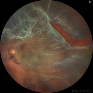
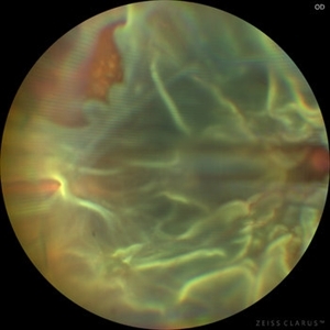 Chronic Open Funnel Retinal Detachment With Horse Shoe Tear
Feb 7 2024 by Harsh Vardhan Singh, MS
67 year old male with history of cataract surgery 1 year presented with old chronic retinal detachment with open funnel configuration with multiple breaks.
Photographer: Harsh Vardhan Singh
Imaging device: Clarus 700
Condition/keywords: chronic retinal detachment, Retinal Detachment, Retinal Detachment with multiple breaks
-
 Chronic Retinal Detachment with Proliferative Vitreoretinopathy
Jan 25 2024 by Isaac Agranoff
Widefield fundus photography of a 24 year old male presenting with subtotal retinal detachment with circumferential anterior proliferative vitreoretinopathy. The detachment is bullous inferiorly with atrophic retina and subretinal bands. There are also scattered patches of lattice with atrophic holes and associated detachment in the periphery. Patient presented with flashes for 2 years with worsening vision over the past 6-8 months, measured at 20/100 ph 20/60 OS.
Photographer: Isaac Agranoff, Ashley Rigdon
Imaging device: Optos California
Condition/keywords: atrophic hole, chronic retinal detachment, lattice degeneration, proliferative vitreoretinopathy (PVR), subretinal bands
-
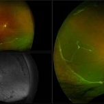
 Chronic retinal detachment in a young myope
Jan 5 2024 by Kamal Kishore, MD, MBBS
Fundus photograph of an 18-year-old -5.50 D myope patient presenting with loss of vision in the right eye
Photographer: Tanya Huston, COA, Illinois Retina and Eye Associates, Peoria, IL
Imaging device: Zeiss Clarus
-
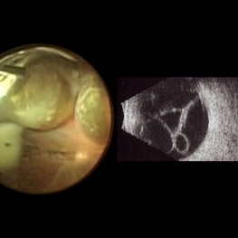 Macrocysts in Kickboxer
Nov 17 2023 by Bradley T. Smith, MD, FASRS
Intraoperative photo and preoperative b scan of chronic retinal detachment with macrocysts in a kickboxer
Condition/keywords: B scan ultrasound, chronic retinal detachment, ocular trauma, pars plana vitrectomy (PPV), retinal macrocyst
-
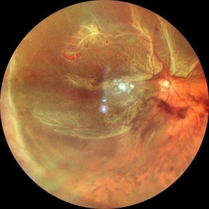 Superior Retinal Detachment
Aug 13 2023 by Anjana Mirajkar, MS Ophthalmology
A color photo of RE of a 55 year old male in a case of Superior Retinal Detachment with macula off with a superior horse shoe tear with vitreous Haze
Photographer: Dr. Anjana Mirajkar -Retina Foundation, Ahmedabad
Condition/keywords: chronic retinal detachment
-
 Total Rhegmatogenous retinal detachment with lattice degeneration & Vitreous haemorrhage
Jul 31 2023 by Harsh Vardhan Singh, MS
72-year male presented PVD induced total retinal detachment with vitreous hemorrhage
Photographer: Dr Harsh Vardhan Singh, AIIMS, Guwahati
Imaging device: Zeiss Clarus 700
Condition/keywords: chronic retinal detachment, hemorrhage, rrd
-
 Total Rhegmatogenous retinal detachment with opened posterior margin of lattice degeneration
Jul 18 2023 by Harsh Vardhan Singh, MS
78-year-old man with history of defective following cataract surgery showed total retinal detachment on examination
Photographer: Harsh Vardhan Singh, AIIMS, Guwahati
Imaging device: Zeiss Clarus 700
Condition/keywords: chronic retinal detachment, peripheral lattice degeneration, rrd
-
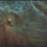
 Chronic retinal detachment changes
Apr 29 2022 by Otakar Dušek, M.D. Ph.D.
Colour fundus photo of 22-year-old woman with bulous retinal detachment number 5-9, old demarcation lines and inferotemporal periheral secondary retinal cyst.
Photographer: Otakar Dušek, Charles University, Prague
Imaging device: Zeiss Clarus
Condition/keywords: chronic retinal detachment, demarcation line, peripheral retinal cyst
-
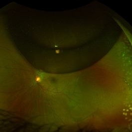 Chronic Retinal Detachment after Pneumatic Retinopexy
Jan 8 2022 by Parnian Arjmand, MD, MSc, FRCSC, DABO
This is a fundus photo in the eye of a young phakic patent who presented with a 6 month history of "difficulty seeing at night" and subjective nasal "blurriness" in the left eye. There was a chronic temporal RD, OS, extending to the arcades (Mac on). This photo is week 1 s/p Pneumatic retinopexy with SF6 gas and laser retinopexy to temporal breaks (6 holes, lattice); no PVD. As you can see, there is a "bleb" of viscous schlieren given the chronic nature of this RD that persist posterior to the breaks and temporal to the macula. This type of sub retinal fluid may take months to years to resorb.
Condition/keywords: chronic retinal detachment, pneumatic retinopexy
-
 Intraretinal cysts
Nov 15 2021 by Marcelo Zas, MD PhD
Left eye from a young patient with a chronic rhegmatogenous retinal detachment presenting intraretinal cysts.
Photographer: Zas Marcelo MD PhD
Condition/keywords: chronic retinal detachment, intraretinal cyst
-
 Intraretinal cysts
Nov 15 2021 by Marcelo Zas, MD PhD
Left eye from a young patient with a chronic rhegmatogenous retinal detachment presenting intraretinal cysts.
Photographer: Zas Marcelo MD PhD
Condition/keywords: chronic retinal detachment, intraretinal cyst
-
 Macrocyst in the Fovea
Feb 2 2021 by Peter J Belin, MD
36-year-old male with a white cataract and a chronic total retinal detachment for 1 year presented with a recurrent PVR detachment after primary repair 2 weeks prior. This OCT- EDI demonstrates a large retinal cyst through the fovea.
Photographer: Holly Cheshier, CRA, OCT-C, COT
Imaging device: Heidelberg Spectralis
Condition/keywords: chronic retinal detachment, proliferative vitreoretinopathy (PVR), retinal cyst, retinal macrocyst
-
 Intraretinal Cysts in Chronic Retinal Detachment
Dec 8 2020 by Alice Kim
B-scan ultrasound showing multiple intraretinal cysts in the setting of chronic retinal detachment and proliferative vitreoretinopathy.
Condition/keywords: chronic retinal detachment, intraretinal cyst, proliferative vitreoretinopathy (PVR)
-
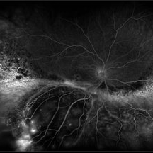 Exudative Retinal Detachment and Branch Retinal Vein Occulsion
Oct 29 2020 by Olivia Rainey
Ultra-widefield fluorescein anigogram of a 51-year-old female with an exudative retinal detachment and branch retinal vein occlusion with retinal neovascularization affecting her right eye. The physician stated that the multiple aneurysmal dilations noted in the inferior periphery are responsible for the exudative RD seen on exam. He is considering Coat's vs FEVR given family history of aneurysms/congenital heart pathology per patient. He encouraged the patient to control their blood pressure, cholesterol, blood sugar, and co-morbidities which may have promoted the BRVO. He recommended antiVEGF injections to control the vascular leakage. Given the severe presentation and imminent threat to her vision, he recommended Eylea as first line therapy.
Photographer: Olivia Rainey, OCT-C, COA
Imaging device: Optos California
Condition/keywords: branch retinal vein occlusion (BRVO), chronic retinal detachment, fluorescein angiogram (FA), fluorescein leakage, inferior retina, inferior retinal detachment, Optos, ultra-wide field imaging
-
 Retinal Detachment Associated with Coloboma
Aug 23 2020 by Noy Ashkenazy, MD, MS
Fundus photograph of a 2-year-old boy with a history of CHARGE syndrome. The image nicely illustrates a retinal detachment associated with a congenital coloboma.
Photographer: Giselle DeOliveira
Imaging device: Retcam III
Condition/keywords: CHARGE syndrome, chronic retinal detachment, coloboma, pediatric retina
-
 Retinal Cyst
Aug 14 2020 by Noy Ashkenazy, MD, MS
Fundus photograph of a 13-year-old male with a chronic retinal detachment following a penetrating ocular trauma. There is a retinal cyst and proliferative vitreoretinopathy.
Photographer: Giselle DeOliveira
Imaging device: Retcam III
Condition/keywords: chronic retinal detachment, proliferative vitreoretinopathy (PVR), retinal cyst
-
 PFC- Thor's Mjolnir in RD cases.
Jul 25 2020 by SANDEEP KUMAR
PFC in chronic retinal detachment has a vital role. The image is of a 12-year-old girl with open funnel RD. Characteristically PFCLs have high specific gravity ranging from 1.76 to 2.03, low surface tension, and viscosity. These physical properties make perfluorocarbon liquids an ideal for intraoperative tool in vitreoretinal surgery. With high specific gravity they flatten the detcahed retina and push the SRF anteriorly giving surgeon room for maneuvers.
Photographer: Dr Daraius Shroff . Shroff Eye Centre New Delhi
Condition/keywords: perfluorooctane
 Loading…