Search results (18 results)
-
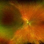
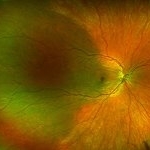
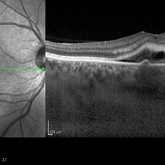
 MIDD (Maternally Inherited Diabetes and Deafness) - OCT OD
Nov 30 2024 by John S. King, MD
OCT shows mild RPE deposit inferiorly (corresponds to area of FA blockage and HyperAF) and a focal area of iRORA with loss of EZ more superiorly (possibly due to regression of RPE deposit). No choroidal thickening (like in pachychoroid pigment epitheliopathy or cscr) 57 yo WF referred for AMD vs Pattern Dystrophy that was diagnosed 10 years ago. Reported some slow progressive vision loss in both eyes for distance and near. Denies nyctalopia or hemeralopia. Background medical history includes HTN, CVD, and DM. No family history of eye problems. Denied pentosan use. Anterior segment showed moderate cataracts (OD>OS). Posterior segment exam showed macular changes and mild NPDR. The macular appearance showed a symmetrical, paramacular ring of fleck-like drusenoid material with some faint focal areas of RPE hyperplasia. Fundus Photos, AF, OCT were performed as well as a gene test. Further questioning showed revealed that her mother and maternal grandmother had both diabetes mellitus and sensorineural hearing loss. The patient developed diabetes in her teens, and some high frequency hearing loss in her early twenties. She had not had a previous genetic test or diagnosis of MIDD. Gene testing is pending for the mitochondrial component. Invitae's retinal panel, which does not include mitochondrial disorders, only showed a variant of uncertain significance, HMCN1. I discussed this case with Dr. Freund, and it is similar to a the case report : Inoue M, Kiss S, Freund KB. MACULAR PIGMENT RINGS AS THE PRESENTING FINDING OF MITOCHONDRIAL MYOPATHY, ENCEPHALOPATHY, LACTIC ACIDOSIS, AND STROKELIKE EPISODES. Retin Cases Brief Rep. 2015 Fall;9(4):260-4. doi: 10.1097/ICB.0000000000000182. PMID: 26200388.
Photographer: Grace Melton and Carley Gunn
Imaging device: Zeiss Cirrus
Condition/keywords: Macular Dystrophy, Maternally Inherited Diabetes and Deafness, MIDD, Mitochondrial Disorder
-
 Uveal Effusion Syndrome
Sep 19 2024 by Virginia Gebhart
61 year old female with idiopathic uveal effusion syndrome. 360 degrees of choroidal thickening, especially anterior with exudative fluid inferior. Mild vitritis present. Unable to gain venous access for FA, ultrasound and UBM performed which confirm choroidal and ciliary body thickening. Pt sent for inflammatory work up including MRI of brain and orbits. Treatment pending results.
Photographer: Virginia Gebhart, Retina Consultants of Carolina
Imaging device: Optos California
Condition/keywords: idiopathic uveal effusion syndrome, uveal effusion
-
 Isolated Choroidal Melanocytosis - Montage
Oct 27 2019 by John S. King, MD
23-year-old white female consulted for a large pigmented choroidal lesion in the right eye. Healthy, no history of glaucoma, 20/20 OU without scleral pigment changes; large, flat pigmented choroidal lesion that is sectoral (temporally) and pigment appears to spare the major choroidal vessels. On OCT (not shown) there is mildly increased choroidal thickening in the area of the lesion.
Photographer: Shelly Blair
Imaging device: Optos CA
Condition/keywords: choroidal melanocytosis, ocular melanocytosis
-
 Isolated Choroidal Melanocytosis
Oct 27 2019 by John S. King, MD
23-year-old white female consulted for a large pigmented choroidal lesion in the right eye. Healthy, no history of glaucoma, 20/20 OU without scleral pigment changes; large, flat pigmented choroidal lesion that is sectoral (temporally) and pigment appears to spare the major choroidal vessels. On OCT (not shown) there is mildly increased choroidal thickening in the area of the lesion.
Photographer: Shelly Blair
Imaging device: Optos CA
Condition/keywords: choroidal melanocytosis, ocular melanocytosis
-
 Posterior Microphthalmos
Nov 22 2015 by Mallika Goyal, MD
Bilateral posterior microphthalmos, high hypermetropia, crowded disc, papillomacular fold, retinitis punctata albescens (white retinal flecks over the retinal midperiphery and periphery) in a 22-year-old male patient. B-scan revealed sclera-choroidal thickening. Best corrected VA was 20/80 each eye.
Photographer: Mallika Goyal, MD, Apollo Health City, Jubilee Hills, Hyderabad, India
Condition/keywords: posterior microphthalmos
-
 Posterior Microphthalmos
Nov 16 2015 by Mallika Goyal, MD
Bilateral posterior microphthalmos, high hypermetropia, crowded disc, papillomacular fold, retinitis punctata albescens. B-scan revealed sclera-choroidal thickening.
Photographer: Mallika Goyal, MD
Condition/keywords: posterior microphthalmos
-
---thumb.jpg/image-square;max$300,300.ImageHandler) Sturge-Weber Diffuse Hemangioma and Retinal Detachment on B-scan
Sturge-Weber Diffuse Hemangioma and Retinal Detachment on B-scan
Apr 18 2014 by Susanna S. Park, MD, PhD
B-scan ultrasonogram of the right eye of an 8 year old Hispanic boy with Sturge -Weber Syndrome showing diffuse choroidal thickening from diffuse choroidal hemangioma and associated total exudative retinal detachment.
Photographer: Ellen Redenbo, University of California Davis Eye Center
Condition/keywords: B scan ultrasound, diffuse choroidal hemangioma, Sturge-Weber syndrome
-
---thumb.jpg/image-square;max$300,300.ImageHandler) Choroidal Metastasis B-Scan
Choroidal Metastasis B-Scan
Jan 10 2014 by Susanna S. Park, MD, PhD
B-scan ultrasound image showing choroidal thickening and exudative retinal detachment in a patient with diffuse choroidal metastasis from breast carcinoma.
Photographer: Ellen Redenbo, University of California Davis Eye Center
Condition/keywords: B scan ultrasound, choroidal metastasis
-
 Choroidal Thickening
Nov 29 2013 by Jason S. Calhoun
Patient comes in with blurred vision in the right eye. VA is CF. with pinhole at 20/400. Fundus photography shows moderate thickening of the choroid. IOP was 5 in the right eye.
Photographer: Jason S. Calhoun, Ophthalmic Photographer, Department of Ophthalmology, Mayo Clinic Jacksonville
Condition/keywords: choroid
-
 Diffuse Choroid hemangioma
Nov 7 2012 by Rajiv Anand, MD, FRCS, FASRS
B scan ultrasound shows the thickened choroid
Condition/keywords: choroidal thickening, Sturge-Weber syndrome
-
 Central Serous Retinopathy with Fibrin
Oct 13 2012 by Edwin H. Ryan, MD
EDI scan of choroid in patient with CSC
Condition/keywords: central serous chorioretinopathy (CSCR), choroidal thickening, fluorescein leakage
-
 Recurrent Central Serous Choroidopathy
Aug 21 2012 by Edwin H. Ryan, MD
EDI-OCT showing thickened choroid and subretinal fluid
Photographer: Edwin Ryan Jr. MD, VitreoRetinal Surgery, PA
Imaging device: Heidelberg Spectralis
Condition/keywords: central serous chorioretinopathy (CSCR), choroidal thickening, enhanced depth imaging
 Loading…