Search results (151 results)
-
 Choroidal Rupture
Dec 4 2025 by Vishal Agrawal, MD, FRCS,FACS,FASRS
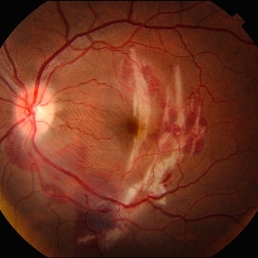
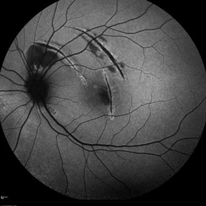
Color fundus pic of Left Eye showing curvilinear, crescent-shaped choroidal rupture, extending across the macular region, post blunt trauma. The rupture appears bright, yellow-white streak with well-delineated margins, consistent with exposed underlying sclera due to disruption of the choriocapillaris and Bruch's membrane.
Photographer: Dr Ayushi Gupta, Agrawal Hospital, Jaipur
Imaging device: Clarus 700
Condition/keywords: blunt trauma, choroidal rupture
-
 Multilayer Trauma
Nov 3 2025 by Malvika Singh
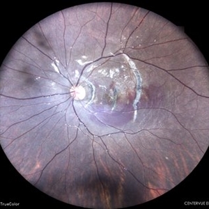
Fundus photograph of a 34 year old following trauma showing a choroidal rupture, a sub RPE and sub retinal bleed.
Photographer: Dr Malvika Singh, Retina Foundation, Ahmedabad, India
Imaging device: Mirante SLO/OCT
Condition/keywords: Choroidal Rupture, subretinal hemorrhage
-
 Choroidal Rupture
Sep 30 2025 by César Adrián Gomez Valdivia, MD
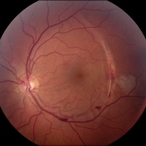
This fundus photograph shows curvilinear streaks of choroidal rupture radiating from the fovea, associated with subretinal hemorrhage. The rupture lines appear as crescent-shaped, whitish streaks representing a break in Bruch’s membrane, choriocapillaris, and retinal pigment epithelium.
Photographer: @eyemissu2
Imaging device: TOPCON TRX
Condition/keywords: Choroidal, Rupture
-
 Post Blunt Trauma Posterior Break
Aug 13 2025 by Debarun Sharma
Fundus photograph of a 21 year old male with history of blunt trauma with fist presenting with a large linear posterior break just adjacent to the infero-temporal arcade with choroidal rupture and subretinal bleed passing through fovea. Successful barrage laser of the break can be seen.
Photographer: Debarun Sharma, Sri Sankardeva Nethralaya, Guwahati
Imaging device: Optos
Condition/keywords: blunt trauma, full thickness retinal tear, laser photocoagulation
-
 Fluorescein Angiography in Choroidal Rupture
Jun 26 2025 by Hector Gabriel Moreno Solano, MD, MHA
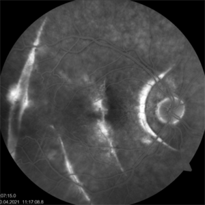
Fluorescein angiography of the right eye reveals three linear hypofluorescent lesions with progressive staining at the edges, consistent with choroidal ruptures. These lesions are temporally located in the posterior pole, with one of them situated near the fovea but without direct foveal involvement. The pattern is suggestive of previous blunt ocular trauma.
Photographer: Héctor Gabriel Moreno Solano, Instituto Mexicano de Oftalmología “IMO I.A.P”
Imaging device: CLARUS
Condition/keywords: Choroidal Rupture, fluorescein angiogram (FA)
-
 OCT Choroidal Rupture
Jun 26 2025 by Hector Gabriel Moreno Solano, MD, MHA
High-resolution OCT of the right eye shows a localized disruption of the retinal pigment epithelium (RPE)–Bruch’s membrane complex, consistent with a choroidal rupture. There is loss of the normal outer retinal architecture over the lesion, with focal elevation and irregularity of the underlying RPE. Hyperreflective material is noted at the level of the break, without associated subretinal fluid or signs of active choroidal neovascularization.
Photographer: Hector Gabriel Moreno Solano, Instituto Mexicano de Oftalmología “IMO I.A.P”
Imaging device: REVO
Condition/keywords: Choroidal Rupture, OCT
-
 Autofluorescence in Multiple Choroidal Ruptures
Jun 26 2025 by Hector Gabriel Moreno Solano, MD, MHA
Fundus autofluorescence imaging of the right eye shows three hypoautofluorescent linear lesions located temporally to the fovea, consistent with choroidal ruptures. The lesions demonstrate sharply demarcated borders with variable surrounding hyperautofluorescence, suggestive of retinal pigment epithelium (RPE) disruption and potential remodeling. One rupture is located near the foveal region, though the foveal center remains spared.
Photographer: Hector Gabriel Moreno Solano, Instituto Mexicano de Oftalmología “IMO I.A.P”
Imaging device: CLARUS
Condition/keywords: autofluorescence imaging, Choroidal Rupture
-
 Multiple Chorodial Ruptures
Jun 26 2025 by Hector Gabriel Moreno Solano, MD, MHA
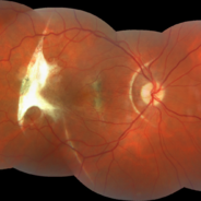
Color fundus photograph of the right eye reveals three well-defined, curvilinear choroidal ruptures located temporal to the fovea running parallel. The lesions appear as pale, crescent-shaped bands, with underlying retinal pigment epithelium disruption. One of the ruptures is situated near the foveal center, though without direct involvement.
Photographer: Hector Gabriel Moreno Solano, Instituto Mexicano de Oftalmología “IMO I.A.P”
Imaging device: CLARUS
Condition/keywords: Choroidal Rupture, color fundus photograph, color wide field
-
 Post-traumatic Choroidal Rupture
Jun 20 2025 by Alexander Babaev
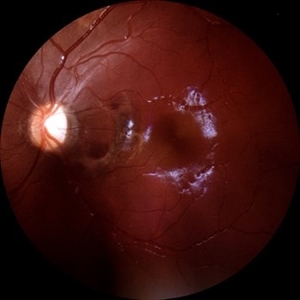
Fundus photograph of a 46-year-man with a choroidal rupture after blunt trauma, complicated CNV.
Photographer: Babaev Alexander, Saint-Petersburg, medical clinic "Vision"
Imaging device: Carl Zeiss Visucam 500
Condition/keywords: choroidal rupture
-
 Post-traumatic Choroida Rupture-Fluorescein Angiography
Jun 20 2025 by Alexander Babaev
Fluorescein angiography of an 46-year-man with a choroidal rupture after blunt trauma, complicated CNV. 07.15, Dye leakage is visible along the edges of the rupture
Photographer: Babaev Alexander, Saint-Petersburg, medical clinic "Vision"
Imaging device: Carl Zeiss Visucam 500
Condition/keywords: blunt trauma
-
 Post-traumatic Choroidal Rupture-Fluorescein Angiography
Jun 20 2025 by Alexander Babaev
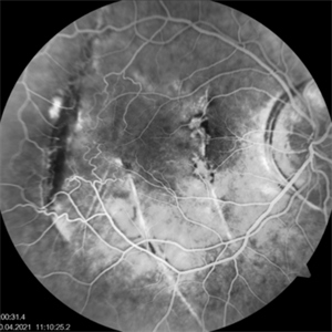
Fluorescein angiography of an 46-year-man with a choroidal rupture after blunt trauma, complicated CNV. 00.31s, Dye leakage is visible along the edges of the rupture
Photographer: Babaev Alexander, Saint-Petersburg, medical clinic "Vision"
Imaging device: Carl Zeiss Visucam 500
Condition/keywords: fluorescein angiogram (FA)
-
 Post-traumatic Choroidal Rupture-Fluorescein Angiography
Jun 20 2025 by Alexander Babaev
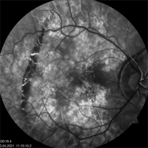
Fluorescein angiography of an 46-year-man with a choroidal rupture after blunt trauma, complicated CNV. 00.16s
Photographer: Babaev Alexander, Saint-Petersburg, medical clinic "Vision"
Imaging device: Carl Zeiss Visucam 500
Condition/keywords: blunt trauma
-
 Post-traumatic Choroidal Rupture
Jun 20 2025 by Alexander Babaev
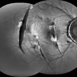
FAF
Photographer: Babaev Alexander, Saint-Petersburg, medical clinic "Vision"
Imaging device: Carl Zeiss Visucam 500
Condition/keywords: choroidal neovascularization (CNV), Choroidal rupture
-
 Choroidal Rupture
Jun 4 2025 by Paulina Araujo
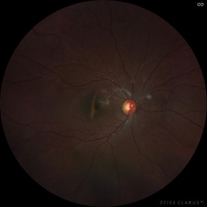
The 55-degree central fundus photograph of the left eye reveals a choroidal rupture in the nasal parafoveal area secondary to blunt ocular trauma.
Photographer: Paulina D.Araujo Martínez, Asociación para Evitar la Ceguera en México I.A.P., Hospital Dr Luis Sánchez Bulnes.
Condition/keywords: choroidal rupture
-
 Choroidal Rupture
Apr 7 2025 by Ramses Rosales-Diaz
Autofluorescence image of a 39-year-old female patient who sustained blunt ocular trauma resulting in three choroidal ruptures.
Photographer: Ramses Rosales-Diaz, Asociación Para Evitar la Ceguera en México I.A.P., Mexico City
Imaging device: Heidelberg Spectralis
Condition/keywords: blunt trauma, Choroidal Rupture
-
 Firework Injury
Feb 13 2025 by Virginia Gebhart
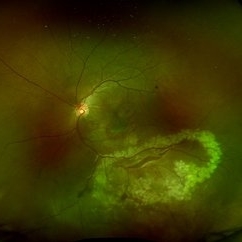
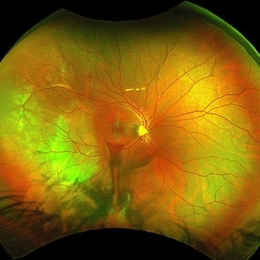
44 year old male presented New Year's Day for trauma after fireworks injury. Choroidal rupture temporal macula, inferior vitreous hemorrhage, and extensive RPE changes in the macula. Significant improvement since initial presentation. Limited central vision, guarded prognosis due to extensive blunt trauma.
Photographer: Virginia Gebhart, Retina Consultants of Carolina
Imaging device: Optos California
Condition/keywords: blunt trauma, choroidal rupture, commotio retinae, firework injury, secondary glaucoma, subretinal hemorrhage, VH, vitreous hemorrhage
-
 Choroidal Rupture With Traumatic Subretinal Hemorrhage
Apr 19 2024 by Akansha Sharma
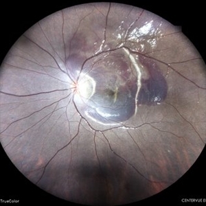
Color fundus photograph of a 11 year old female with subretinal bleed and choroidal rupture status post trauma.
Photographer: Dr. Akansha Sharma, Bharati Eye Hospital
Condition/keywords: Choroidal Rupture, subretinal hemorrhage, subretinal blood
-
 Choroidal Rupture With Traumatic Subretinal Hemorrhage
Apr 19 2024 by Akansha Sharma
Color fundus photograph of a 11 year old female with subretinal bleed and choroidal rupture status post trauma.
Photographer: Dr. Akansha Sharma, Bharati Eye Hospital
Condition/keywords: Choroidal Rupture, subretinal hemorrhage, subretinal blood
-
 Choroidal-rupture
Jan 2 2024 by Tahsin Khundkar, MD
37-year-old male with blunt ocular trauma presented with a choroidal rupture, pre -retinal and sub-retinal heme, and a heart shaped patch of commotio retinae.
Photographer: Jeffrey Zeigler, Concord Eye Center
Imaging device: Topcon
Condition/keywords: Choroidal Rupture, commotio retinae, Trauma
-
 Choroidal Rupture Autofluorescence
Nov 26 2023 by stephen oconnell
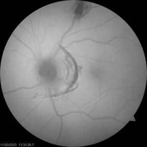
Autofluorescence better defines extent of choroidal rupture after blunt trauma - closed fist
Condition/keywords: Choroidal Rupture
-
 Choroidal Rupture Color
Nov 26 2023 by stephen oconnell
10 days after blunt trauma with closed fist
Condition/keywords: Choroidal Rupture
-
 Choroidal Rupture
Nov 5 2023 by Karen Flores Guevara
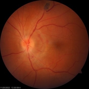
Fundus photograph of a 19-year-old man with a choroidal rupture.
Photographer: Hector Arturo Mendez-Ponce MD, Karen Flores-Guevara MD Asociación para Evitar la Ceguera en México
Condition/keywords: Choroidal, rupture, trauma
-
 Choroidal Rupture
Sep 30 2023 by Jacob D. Grodsky, MD
24 year old female who presented after being hit in the head with a metal softball bat after an altercation. The patient reported blurred vision as well as a zig-zag line described as a “lightning strike” across her vision. Examination was significant for a choroidal rupture OD as well as commotio retinae OU.
Condition/keywords: choroidal rupture, commotio retinae, trauma
-
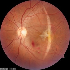 The sun, the star and the crescent moon
Jun 7 2023 by Prerana Shah
Fundus photograph of a 35 year old gentleman one week post blunt trauma.
Photographer: Swati Kadam and Disha Malusare, Shreeramkrishna Netralaya, Thane , Maharashtra ,India
Imaging device: Zeiss Visucam
Condition/keywords: choroidal rupture, ERM
-
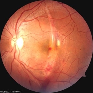 Choroidal rupture
Jun 7 2023 by Prerana Shah
Fundus photograph of a 35 year old gentleman on the day of blunt trauma.
Photographer: Swati Kadam and Disha Malusare, Shreeramkrishna Netralaya, Thane , Maharashtra ,India
Imaging device: Zeiss Visucam
Condition/keywords: choroidal rupture, ERM
 Loading…