Search results (143 results)
-
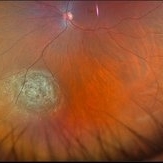
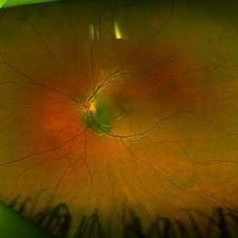
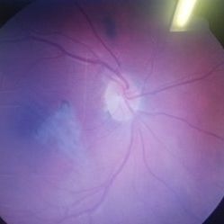
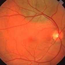
 Suspicious Nevus
Jan 15 2025 by Virginia Gebhart
14 year female with suspicious nevus located adjacent to the optic nerve. Questionable orange pigment present and worsening SRF compared to previous photos/OCT. RPE atrophy also present from previous fluid. No elevation. Will continue observation. BCVA 20/25
Photographer: Virginia Gebhart, Retina Consultants of Carolina
Imaging device: Topcon 50DX
Condition/keywords: choroidal nevus, nevus
-
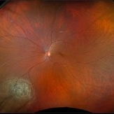
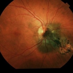
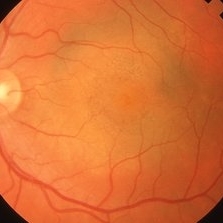
 Choroidal Nevus
Nov 7 2024 by Ashley Romero-Martinez
57 year old female presents with choroidal nevus in left eye. Has been present for 20 years, remains stable and will continue to observe.
Photographer: Ashley Romero-Martinez
Imaging device: Optos
Condition/keywords: choroidal nevus
-
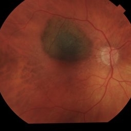
 Choroidal Nevus
Nov 7 2024 by Ashley Romero-Martinez
57 year old female presents with choroidal nevus in left eye. Has been present for 20 years, remains stable and will continue to observe.
Photographer: Ashley Romero-Martinez
Imaging device: Optos
Condition/keywords: choroidal nevus
-
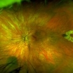
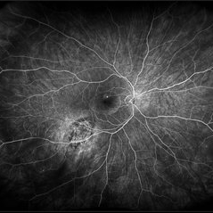 Suspicious Nevus / CSR
Aug 8 2024 by Virginia Gebhart
Fluorescein angiogram of 54 year old male with a suspicious appearing choroidal nevus and central serous retinopathy. Will monitor closely and follow up with serial exams.
Photographer: Virginia Gebhart
Imaging device: Optos California
Condition/keywords: central serous retinopathy (CSR), choroidal nevus, fluorescein angiogram (FA), FLUORESCEIN ANGIOGRAPHY, nevus
-
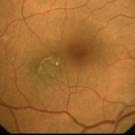
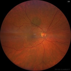 Choroidal Nevus
May 25 2024 by Gustavo Del Castillo-Marquez, MD
Fundus photograph of a 65 year old woman with a choroidal nevus.
Photographer: Gustavo Del Castillo-Márquez, Asociación Para Evitar la Ceguera en México, CDMX
Imaging device: Zeiss Clarus
Condition/keywords: choroidal nevus
-
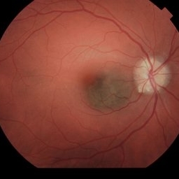
 Suspicious Choroidal Nevus
May 8 2024 by Virginia Gebhart
13 year old female with suspicious appearing choroidal nevus. High risk features present, adjacent to optic nerve, questionable orange pigment, SRF. No significant elevation on ultrasound. Will follow up with serial exams.
Photographer: Virginia Gebhart
Imaging device: Optos California
Condition/keywords: choroidal nevus, nevus
-
 Macula-threatening PEHCR causing a nasal visual-field defect in a patient with AMD
Apr 15 2024 by David A Reichstein, MD
(A) Ultra-widefield color fundus photograph demonstrates a PEHCR encroaching upon the temporal macula. Lipid exudation is apparent at the lesion’s anterior and inferior border. Subretinal hemorrhage is apparent at the lesion’s inferior border. Drusen are apparent in the macula. An unrelated, small choroidal nevus is apparent in the inferior fundus. (B) Ultra-widefield FA taken in early stage demonstrates hypofluorescence within the lesion consistent with blockage by possible sub-RPE or subretinal heme. (C) Ultra-widefield fundus photograph taken 6 months following the initiation of monthly anti-VEGF therapy demonstrates considerable reduction in the size of the lesion and resolution of the subretinal hemorrhage and lipid exudation. (D) Ultra-widefield fundus photograph taken 1 year after presentation where a treat-and-extend approach was performed for the most recent 6 months. The lesion had almost completely resolved.
Condition/keywords: peripheral exudative hemorrhagic chorioretinopathy (PEHCR)
-
 Suspicious Nevus
Feb 14 2024 by Virginia Gebhart
61 year old female with a suspicious choroidal nevus involving the optic nerve head. Patient asymptomatic, will continue to observe.
Photographer: Virginia Gebhart
Imaging device: Topcon TRC 50DX
Condition/keywords: choroidal nevus, nevus
-
 Suspicious Choroidal Nevus
Dec 6 2023 by Virginia Gebhart
55 year old female with suspicious pigmented choroidal nevus. 3 high risk features present. Ultrasound shows SRF and high internal reflectivity. Will observe closely
Photographer: Virginia Gebhart
Imaging device: Topcon
Condition/keywords: choroidal nevus, orange pigment
-
 Benign Nevus
Dec 6 2023 by Virginia Gebhart
71 year old female with benign choroidal nevus with fibrotic PED
Photographer: Virginia Gebhart
Imaging device: Topcon
Condition/keywords: choroidal nevus, fibrotic neovascularization, nevus
-
 Suspicious Choroidal Nevus
Dec 6 2023 by Virginia Gebhart
82 year old male with suspicious choroidal nevus and ERM. Remains unchanged since first visit 6 mos ago.
Photographer: Virginia Gebhart
Imaging device: Topcon
Condition/keywords: choroidal nevus
-
 Suspicious Choroidal Nevus / Optic Disc Drusen
Nov 1 2023 by Virginia Gebhart
29 year-old female with suspicious choroidal nevus adjacent to optic nerve and extending into fovea. Optic disc drusen OU. Pt is asymptomatic
Photographer: Virginia Gebhart
Imaging device: Topcon
Condition/keywords: choroidal nevus, disc drusen, drusen of optic disc, nevus
-
 Choroidal-Nevus
Feb 25 2023 by Aditya S Kelkar, MS, FRCS, FASRS,FRCOphth
Color fundus photograph of the left eye of a 55 year old male showing large choroidal nevus.
Photographer: Dr. Apoorva Jadhav, National Institute of Ophthalmology, Pune. India.
Imaging device: Zeiss Clarus 500
Condition/keywords: Choroidal nevus
-
 Choroidal nevus with halo
Aug 21 2022 by Niloofar Piri, MD
Fundus photograph of the right eye in a 43 yo patient demonstrating a flat choroidal nevsu with halo nasal to the optic nerve
Photographer: Andrew Polk, MD
Condition/keywords: Choroidal nevus, nevus with halo
-
 Nevus
Nevus
Jan 21 2021 by AGNES KIM
Fundus photograph of 30-year-old female of choroidal nevus. Another nevus was found in the same eye in the periphery. Macula has a lens artifact.
Condition/keywords: choroidal nevus
-
 Macular Nevus
Jan 20 2021 by Jamin S. Brown, MD
Macular Nevus as well as CSR.
Photographer: Stefanie Palmer CRA, Retina Vitreous Surgeons of CNY
Condition/keywords: choroidal nevus, macula lesion
-
 Choroidal Nevus Associated with Drusen
Jan 11 2021 by Gabriel Costa Andrade, PhD
Fundus photograph of an 53-year-old man with a macular melanotic nevus.
Photographer: Gabriel Andrade
Condition/keywords: choroidal nevus
-
 Polypoidal Choroidal Vasculopathy Complicating Benign Choroidal Nevus
Apr 23 2020 by Patrícia José Figueiredo Lopes
A 51-year-old female presented with a left pigmented choroidal lesion, involving the supero-temporal macula (A). Fundus autofluorescence (B) showed mottling and focal areas of hiperautofluorescence and the structural optical coherence tomography (OCT) (C) was compatible with choroidal nevus, with choriocapillaris compression and choroidal shadowing. In the nasal aspect of the nevus, there was sharp-peaked pigmented epithelium detachment (PED) with associated subretinal fluid, suspicious of a polypoidal lesion. Complementary testing with OCT-angiography (D) confirmed the vascularized nature of the sharp PED. Fluorescein angiography (E) showed masking from the pigmented choroidal lesion with hyperfluorescence from the PEDs and indocyanine green angiography (F) revealed two “hot spots” confirming the presence of choroidal polyps. Polypoidal choroidal vasculopathy complicating benign choroidal nevus is an unusual presentation but should be excluded and differentiated from malignant transformation.
Condition/keywords: choroidal nevus, polypoidal choroidal vasculopathy (PCV)
-
 Operculated Hole with Barrier Laser
Nov 5 2019 by Nichole Lewis
66-year-old female with an operculated retinal hole s/p barrier laser treatment. Choroidal Nevus Inferior.
Photographer: Nichole Lewis
Imaging device: Optos
Condition/keywords: barrier laser, choroidal nevus, operculated retinal hole, operculated tear
-
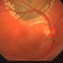 Suspicious Choroidal Nevus S/P Laser Photocoagulation
Apr 2 2019 by Gary R. Cook, MD, FACS
62-year-old white male with suspicious choroidal nevus OD with NSRD involving the macula; immediately S/P delimiting photocoagulation; V.A. = 20/200
Imaging device: Topcon VT-50
Condition/keywords: choroidal nevus, laser photocoagulation, neurosensory detachment of retina
-
 Choroidal Nevus with NSRD
Apr 2 2019 by Gary R. Cook, MD, FACS
62-year-old white male with suspicious choroidal nevus OD with neurosensory retinal detachment involving the macula; VA=20/200
Imaging device: Topcon VT-50
Condition/keywords: choroidal nevus, neurosensory detachment of retina
-
 Macular Choroidal Nevus
Apr 2 2019 by Gary R. Cook, MD, FACS
38-year-old patient with macular choroidal nevus OS; V.A.= 20/30.
Imaging device: Topcon VT-50
Condition/keywords: choroidal nevus, nevus
-
 Halo Nevus
Apr 2 2019 by Gary R. Cook, MD, FACS
67-year-old white male with a typical halo nevus OD with a couple of drusen on its surface; V.A. = 20/25.
Imaging device: Topcon VT-50
Condition/keywords: choroidal nevus, nevus
-
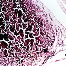 Slide 14-28
Mar 4 2019 by Lancaster Course in Ophthalmology
Other lesions mistaken for melanomas such as other tumors including hemangiomas, metastic tumors, melanocytoma of the disc, nevus of the choroid, hypertrophy of the pigment epithelium, adenoma and adenocarcinoma of the pigment epithelium, reactive proliferation of the pigment epithelium, and lymphoma and leukemia.
Condition/keywords: choroidal nevus, hemangioma, leukemia, lymphoma, melanoma, optic disc melanocytoma, retinal pigment epithelium (RPE) hypertrophy
-
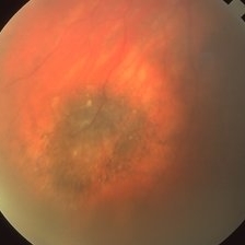
 Choroidal Melanoma
Jan 30 2019 by Karen Panzegrau
Ultra-wide field optos image of a 27-year-old male patient who presented with loss of vision for about 6-8 weeks. Previous choroidal nevus seen. Recommended annual monitoring. No exam for since 10/2014. Brachytherapy vs enucleation was discussed. Brachytherapy was decided as treatment. Full metastatic work up is being performed.
Photographer: Karen Panzegrau
Imaging device: Optos
Condition/keywords: choroidal nevus, exudative retinal detachment, malignant neoplasm of eye, Optos, ultra-wide field imaging
 Loading…