 Initializing download.
-
 By Patrícia José Figueiredo Lopes
Co-author(s): Filomena Pinto, Sara Vaz-Pereira - Uploaded on Apr 23, 2020.
- Last modified by Caroline Bozell on Apr 23, 2020.
- Rating
- Appears in
- 23-Apr-2020
- Condition/keywords
- polypoidal choroidal vasculopathy (PCV), choroidal nevus
- Description
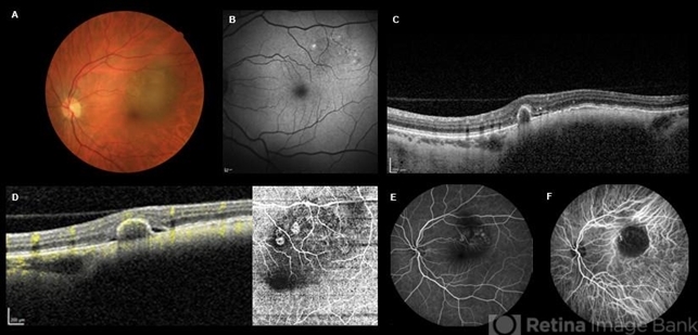
- A 51-year-old female presented with a left pigmented choroidal lesion, involving the supero-temporal macula (A). Fundus autofluorescence (B) showed mottling and focal areas of hiperautofluorescence and the structural optical coherence tomography (OCT) (C) was compatible with choroidal nevus, with choriocapillaris compression and choroidal shadowing. In the nasal aspect of the nevus, there was sharp-peaked pigmented epithelium detachment (PED) with associated subretinal fluid, suspicious of a polypoidal lesion. Complementary testing with OCT-angiography (D) confirmed the vascularized nature of the sharp PED. Fluorescein angiography (E) showed masking from the pigmented choroidal lesion with hyperfluorescence from the PEDs and indocyanine green angiography (F) revealed two “hot spots” confirming the presence of choroidal polyps. Polypoidal choroidal vasculopathy complicating benign choroidal nevus is an unusual presentation but should be excluded and differentiated from malignant transformation.


---thumb.jpg/image-square;max$79,0.ImageHandler "Polypoidal Choroidal Vasculopathy: Case 1 - Image 1 of 7")
---thumb.jpg/image-square;max$79,0.ImageHandler "Polypoidal Choroidal Vasculopathy: Case 1 - Image 3 of 7")

---thumb.jpg/image-square;max$79,0.ImageHandler "Polypoidal Choroidal Vasculopathy: Case 1 - Image 2 of 7")


---thumb.jpg/image-square;max$79,0.ImageHandler "Polypoidal Choroidal Vasculopathy - Case 1")