Search results (103 results)
-
 Ciliary Body Metastasis
Mar 26 2025 by Virginia Gebhart
54 year old female referred for iris mass. UBM shows large solid mass originating in the ciliary body and eroding into the anterior chamber under the iris epithelium. Recent CT scans revealed multiple bilateral pulmonary and hepatic nodules. Pt has been scheduled for PET scan and liver biopsy by radiation oncologist.
Photographer: Virginia Gebhart, Retina Consultants of Carolina
Imaging device: Samsung Galaxy
Condition/keywords: choroidal metastasis, ciliary body mass, metastatic cancer
-
 Choroidal Metastasis With Orange Pigment in a Patient With Endometrial Carcinoma
Aug 8 2024 by Guilherme Sturzeneker, MD, MSc
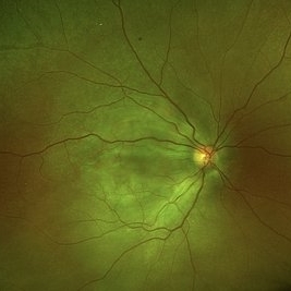
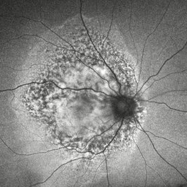
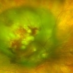
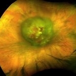
Ultra-widefield fundus photograph and autofluorescence of a 62-year-old woman with endometrial cancer, denoting choroidal metastasis with unusual orange pigment. This presentation is a reminder that the development of orange pigment is not pathognomonic for choroidal melanoma, as it may be seen in other lesions such as carcinoma metastasis.
Photographer: Andrea Almeida
Imaging device: Optos Silverstone
Condition/keywords: choroidal metastasis, metastatic cancer, orange pigment
-
 Choroidal Metastasis With Orange Pigment in a Patient With Endometrial Carcinoma
Aug 8 2024 by Guilherme Sturzeneker, MD, MSc
Ultra-widefield fundus photograph and autofluorescence of a 62-year-old woman with endometrial cancer, denoting choroidal metastasis with unusual orange pigment. This presentation is a reminder that the development of orange pigment is not pathognomonic for choroidal melanoma, as it may be seen in other lesions such as carcinoma metastasis.
Photographer: Andrea Almeida
Imaging device: Optos Silverstone
Condition/keywords: choroidal metastasis, metastatic cancer, orange pigment
-
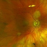
 Exudative Retinal Detachment With Choroidal Metastasis
May 1 2024 by Vishal Agrawal, MD, FRCS,FACS,FASRS
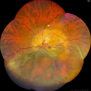
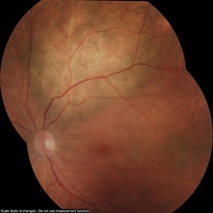
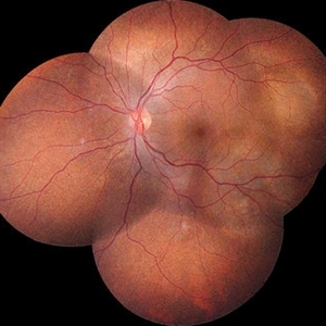
Left eye fundus picture of a 65-year-old female with choroidal metastases and exudative retinal detachment. The patient is under treatment for breast carcinoma.
Photographer: Dr Ayushi
Imaging device: Clarus 700
Condition/keywords: choroidal metastasis, exudative detachment
-
 Choroidal Metastasis
Apr 11 2024 by Corey Grant
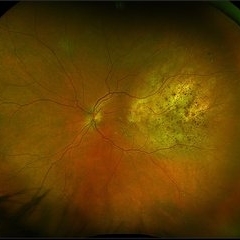
Ultra-Widefield fundus photography and fundus autofluorescence images of a 61 year old female with Choroidal Metastasis affecting both eyes. Patient presented with blurred vision and flashes for a few weeks. Patient visual acuity was cc20/100 PH20/60 in the right eye and cc20/200 in the left eye. Patient admits to history of smoking for many years bit no known history of cancer prior to the visit. Physician recommended going to the ER for full body PET CT and stated that the first line of treatment is usually systemic chemo therapy. Patient will be reassessed in one month.
Photographer: Corey Grant
Imaging device: OPTOS CALIFORNIA RGB
Condition/keywords: cancer, choroidal metastasis, fundus autofluorescence (FAF), fundus photography, hyperautofluorescence, hypoautofluorescence, Optos, OPTOS CALIFORNIA RGB, Retina, ULTRA WIDE FIELD
-
 Choroidal Metastasis
Dec 6 2023 by Virginia Gebhart
60 year old female with totally regressed tumor in temporal macula s/p external beam radiation and chemo. Pt diagnosed with stage IV metastatic lung cancer.
Photographer: Virginia Gebhart
Imaging device: Optos
Condition/keywords: choroidal metastasis, choroidal tumor
-
 Regressed Choroidal Metastasis
Dec 6 2023 by Virginia Gebhart
60 year old female with regressing metastasis along STA. Tumor has continued to regress s/p radiation in Feb 2023 and Tagrisso. VA 20/20. No elevation or SRF on most recent ultrasound
Photographer: Virginia Gebhart
Imaging device: Topcon
Condition/keywords: choroidal metastasis, choroidal tumor
-
 Choroidal Metastasis
Jun 12 2023 by Ethan K Sobol, MD
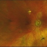
A 46-year-old male with a large choroidal metastatic lesion of the right eye, in the setting of stage IV lung cancer. Several atrophic holes are also seen in the superotemporal periphery.
Condition/keywords: choroidal metastasis, lung cancer metastasis
-
 Breast cancer metastatic to choroid
Jul 13 2021 by Odette M. Houghton, MD
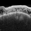
EDI-OCT image of a 59-year-old female with a choroidal tumor secondary to metastatic breast cancer.
Photographer: David Saiz COT, Mayo Clinic Arizona
Imaging device: Heidelberg Spectralis
Condition/keywords: breast cancer, choroidal metastasis, metastatic lesion
-
 Breast cancer metastatic to choroid
Jul 13 2021 by Odette M. Houghton, MD
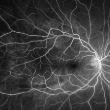
Arteriovenous phase fluorescein angiogram of a 59-year-old female with a choroidal tumor secondary to metastatic breast cancer.
Photographer: David Saiz COT, Mayo Clinic Arizona
Imaging device: Optos California
Condition/keywords: breast cancer, choroidal metastasis, metastatic lesion
-
 Rare Bilateral Choroidal Metastasis from Occult Primary Lung Cancer
May 5 2021 by Deependra Vikram Singh, MD FASRS
Fundus photographs and OCT scans of a 73-year-old non-smoker Indian male who presented to our retina clinic in 2013 with blurred vision in left eye for past 2 weeks. BCVA was 20/20 in right eye and 20/40 in left eye. Slit lamp exam was unremarkable for both eyes with no cells in aqueous or anterior vitreous. Fundus examination revealed creamy yellow choroidal lesions in both eyes. Lesion in right eye was one disc diameter (DD) in size and was located close to fovea (Fig-1a). Lesion in the left eye was bigger with a size of 2 DD located superior to fovea (Fig-1b). OCT scan for left eye revealed neurosensory detachment involving fovea (Fig-1c). Fundus fluorescein angiography was inconclusive for right eye and showed late hyper fluorescence the choroidal lesion in left eye. Patient underwent detailed systemic work up for malignancy that revealed primary lung non-small cell carcinoma. He had widespread metastasis affecting liver and brain. Palliative chemotherapy and radiotherapy were initiated 4 weeks after he presented to us. The choroidal lesions show progression on fundus picture and OCT scans done at 4 weeks follow up after initial presentation (Fig – 1d, e, f). The lesions in both eyes show regression at 4 weeks and 12 weeks follow up after initiation of therapy. Unfortunately, patient succumbed at 13 weeks follow up due to disease progression. The case demonstrates rare bilateral choroidal metastasis from primary lung cancer and also highlights that lesions can be asymptomatic till they develop neurosensory detachment as evident from asymptomatic lesion in right eye despite proximity to fovea and symptomatic lesion in left eye with NSD.
Photographer: Deependra Vikram Singh, Eye-Q Superspecialty Eye Hospitals, Gurugram
Imaging device: Topcon
Condition/keywords: choroidal mass, choroidal metastasis
-
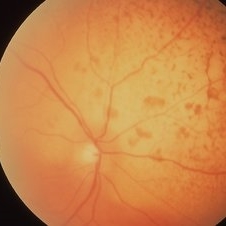
 Choroidal Metastasis from Pancreatic Carcinoma
Jan 10 2021 by Ivan G. Castillo Salazar, MD, FASRS
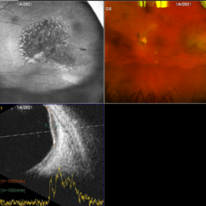
Fundus photograph of the left eye in a 72-year-old patient diagnosed with Stage I pancreatic carcinoma involving the head of the pancreas diagnosed three months ago. The patient was seen for a routine diabetic retinopathy evaluation. Incidentally, this mass was observed in the temporal periphery of the left eye. Visual acuity 20/25 OU. OCT normal OU.
Photographer: Amber Ozment
Imaging device: Optos California camera and Accutome 4 sight B scan ultrasonographer
Condition/keywords: choroidal metastasis
-
 Choroidal Metastases
Jan 18 2020 by Vishal Agrawal, MD, FRCS,FACS,FASRS
Left eye fundus montage of a 55-year-old female with choroidal metastases with the primary being breast carcinoma. The right eye had exudative retinal detachment.
Photographer: Dr Vishal Agrawal MD,FRCS
Imaging device: Zeiss
Condition/keywords: breast cancer, choroidal metastasis, metastatic lesion
-
 Choroidal Metastasis from Breast Cancer
Oct 1 2019 by John S. King, MD
60-year-old white female with four year history of breast cancer associated with metastases to many organs including the CNS, was sent her to r/o melanoma, found on routine exam. Visual acuity was HM; there was NSC/PSC; there was a unilateral, large choroidal lesion in the posterior pole that was yellow, well circumscribed, with plateau configuration associated with SRF adn heme.
Photographer: Kay Dalby
Imaging device: Optos CA
Condition/keywords: breast cancer, choroidal lesions, choroidal metastasis
-
 Choroidal Metastasis from Breast Cancer
Oct 1 2019 by John S. King, MD
60-year-old white female with four year history of breast cancer associated with metastasis to many organs including the CNS, was sent her to r/o melanoma, found on routine exam. Visual acuity was HM; there was NSC/PSC; there was a unilateral, large choroidal lesion in the posterior pole that was yellow, well circumscribed, with plateau configuration associated with SRF adn heme.
Photographer: Kay Dalby
Imaging device: Optos CA
Condition/keywords: breast cancer, choroidal lesions, choroidal metastasis
-
 Metastatic NSCLCA to the Choroid: lesions regressing while undergoing chemotherapy
May 27 2019 by John S. King, MD
Two small, yellow, choroidal lesions can be seen above the nerve and IT arcade can be seen that have regressed compared to the initial photos. Vision 20/20.
Photographer: Shelly Blair
Imaging device: Optos CA
Condition/keywords: choroidal metastasis, lung cancer metastasis
-
 Metastatic NSCLCA to the Choroid: Initial Appearance
May 27 2019 by John S. King, MD
60-year-old white male non-smoker presented to Dr. Zocchi with acute transient decreased vision in the right eye. Background history includes metastatic NSCLC (adenocarcinoma). Acuity OD 20/60, and posterior segment had two small, yellow, choroidal lesions, above the nerve and IT arcade (these had a fairly smooth and dome shaped appearance on the OCT, and top lesion had mild SRF) (see photo)
Photographer: Shelly Blair
Imaging device: Optos CA
Condition/keywords: choroidal metastasis, lung cancer metastasis
-
 Non-Rhegmatogenous Retinal Detachment
Mar 26 2019 by Gary R. Cook, MD, FACS
56-year-old white female with a non-rhegmatogenous retinal detachment secondary to metastatic breast carcinoma lesion OS; VA= counting fingers at 4 feet.
Imaging device: Topcon VT-50
Condition/keywords: breast carcinoma, choroidal metastasis, exudative detachment, metastatic lesion
-
 Non-Rhegmatogenous Retinal Detachment
Mar 26 2019 by Gary R. Cook, MD, FACS
56-year-old white female with non-rhegmatogenous retinal detachment secondary to metastatic breast lesion OS; VA= counting fingers at 4 feet.
Imaging device: Topcon VT-50
Condition/keywords: breast carcinoma, choroidal metastasis, exudative detachment
-
 Metastatic Cancer
Mar 26 2019 by Gary R. Cook, MD, FACS
64-year-old WF with metastatic breast carcinoma OD s/p radiation treatment; VA improved to 20/25.
Imaging device: Topcon VT-50
Condition/keywords: breast cancer, breast carcinoma, choroidal metastasis, metastatic lesion
-
 Metastatic Breast Carcinoma
Mar 26 2019 by Gary R. Cook, MD, FACS
64-year-old white female with metastatic breast carcinoma lesion superior to optic disc OD; VA= 20/70-1.
Imaging device: Topcon VT-50
Condition/keywords: breast cancer, breast carcinoma, choroidal metastasis, metastatic cancer, metastatic lesion
-
 Choroidal Metastasis
Mar 26 2019 by Gary R. Cook, MD, FACS
Fluorescein angiogram image of the choroidal metastasis secondary to breast carcinoma OD.
Imaging device: Topcon VT-50
Condition/keywords: breast cancer, breast carcinoma, choroidal metastasis, fluorescein angiogram (FA), metastatic lesion
-
 Choroidal Metastasis
Mar 26 2019 by Gary R. Cook, MD, FACS
Choroidal metastasis secondary to breast carcinoma.
Imaging device: Topcon VT-50
Condition/keywords: breast cancer, breast carcinoma, choroidal metastasis, metastatic lesion
-
 Metastatic Breast Carcinoma
Mar 26 2019 by Gary R. Cook, MD, FACS
55-year-old white female with metastatic breast carcinoma OD; V.A. = 20/60.
Imaging device: Topcon VT-50
Condition/keywords: breast cancer, breast carcinoma, choroidal metastasis, metastatic lesion
-
 Metastatic Cancer
Mar 26 2019 by Gary R. Cook, MD, FACS
75-year-old white male with metastatic prostate cancer of the right eye; VA= counting fingers at 1 foot.
Imaging device: Topcon VT-50
Condition/keywords: choroidal metastasis, metastatic lesion, retinal hemorrhage
 Loading…