 Initializing download.
-
 By Ivan G. Castillo Salazar, MD, FASRS
Texas Retina Associates
Co-author(s): Ivan Castillo MD, Texas Retina Associates, Waco TX - Uploaded on Jan 10, 2021.
- Last modified by Caroline Bozell on Jan 12, 2021.
- Rating
- Appears in
- 10-Jan-2021
- Condition/keywords
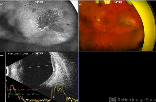
- choroidal metastasis
- Photographer
- Amber Ozment
- Imaging device
-
Fundus camera
Optos California camera and Accutome 4 sight B scan ultrasonographer - Description
- Fundus photograph of the left eye in a 72-year-old patient diagnosed with Stage I pancreatic carcinoma involving the head of the pancreas diagnosed three months ago. The patient was seen for a routine diabetic retinopathy evaluation. Incidentally, this mass was observed in the temporal periphery of the left eye. Visual acuity 20/25 OU. OCT normal OU.


---thumb.jpg/image-square;max$79,0.ImageHandler "Metastatic Breast Cancer")


