Search results (46 results)
-
 Uveal Melanoma
Apr 26 2025 by Vishal Agrawal, MD, FRCS,FACS,FASRS
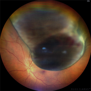
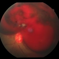
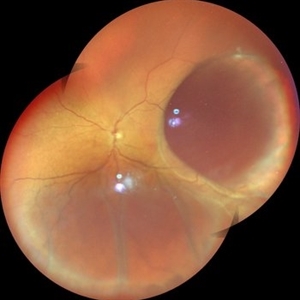
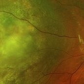
A 32 year-old male presented with complaints of perceiving a shadow in OS for 15-20 days. His BCVA was 20/20 OU. On Fundus examination, a large, elevated, well-defined, pigmented choroidal mass with few hemorrhages over the lesion was seen and a provisional diagnosis of uveal melanoma was made. urgent oncological consultation was recommended for further treatment.
Photographer: Dr Ayushi Gupta
Imaging device: Clarus 700
Condition/keywords: melanoma
-
 Choroidal Melanoma
Feb 6 2025 by Virginia Gebhart
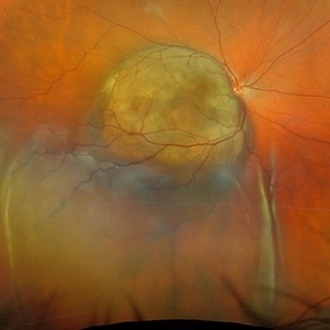
81 year old female with large pigmented collar button ciliochoroidal mass extending into the mid-vitreous cavity. Clinical exam and ultrasound finding consistent with melanoma. Due to size of tumor, pt scheduled for enucleation. CT scan of abdomen showed no evidence of metastatic disease.
Photographer: Virginia Gebhart, Retina Consultants of Carolina
Imaging device: Optos California
Condition/keywords: ciliochoroidal melanoma, collar button, melanoma
-
 New Choroidal Melanoma
Jan 3 2025 by Virginia Gebhart
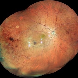
22 year old male referred for 2nd opinion on large choroidal mass with subretinal fluid. Clinical exam and ultrasound consistent with choroidal melanoma. CT scan of orbits showed possible inflammation involving orbital fat. Pt has been on oral prednisone for 1 week, inflammation has not responded. Referred to Emory for 2nd opinion on treatment
Photographer: Virginia Gebhart
Imaging device: Optos California
Condition/keywords: melanoma
-
 Choroidal Melanoma with Serous Retinal Detachment
Dec 20 2024 by Daniel Davis, OCT-C
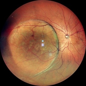
67 year old male presenting with large pigmented choroidal mass with serous retinal detachment.
Photographer: Daniel Davis, OCT-C, The Retina Institute
Imaging device: Optos California
Condition/keywords: Retina detachment
-
 Choroidal Detachment OS
Jul 5 2024 by Zach Seim
Optos Fundus Photograph of a Choroidal Detachment OS in a 75 year old male. VA at presentation was DCC HM.
Photographer: Zach Seim
Imaging device: Optos California
Condition/keywords: choroidal detachment, choroidal mass, left eye, optos, OPTOS CALIFORNIA
-
 Choroidal Mass
Mar 4 2024 by ANKIT JAIN
RE color photo montage of right eye of 48 year old with sub retinal hemorrhage with sub retinal fluid at level of fovea.
Photographer: Dr Ankit Jain
Imaging device: MIRANTE
Condition/keywords: macroaneurysm, retinal arterial macroaneurysm
-
 Choroidal Mass
Mar 4 2024 by ANKIT JAIN
Left eye color photo montage of 39 year old female with sub retinal mass in nasal quadrant with hemorrhages and subretinal fluid with inferior retinal detachment.
Photographer: Dr Ankit Jain
Imaging device: MIRANTE
Condition/keywords: choroidal mass
-
 Choroidal Mass
Sep 21 2023 by Vaidehi Sathaye
Widefield photograph of RE of a 68 year male with choroidal mass.
Photographer: Dr. Vaidehi Sathaye
Imaging device: Mirante
Condition/keywords: choroidal mass
-
 Choroidal Melanoma with Exudative Retinal Detachment
Mar 2 2023 by Aditya S Kelkar, MS, FRCS, FASRS,FRCOphth
Color fundus photograph of the left eye of a 45 year old male showing choroidal melanoma with exudative retinal detachment.
Photographer: Dr. Pranali Surawase, National Institute of Ophthalmology, Pune, India.
Imaging device: Zeiss Clarus 500
Condition/keywords: choroidal mass, exudative retinal detachment, Retinal detachment
-
 CLOUDS OF BLOOD-Sub Hyloid Haemorrhage secondary to choroidal mass
Oct 28 2022 by Magna Mary Kuruvila
Fundus photograph of a 50 year old male patient with sudden loss of vision showing sub hyloid hemorrhage secondary to choroidal mass visualised by bscan.
Photographer: Magna Mary Kuruvila
Condition/keywords: B scan ultrasound, choroidal tumor, subhyaloid hemorrhage
-
 Choroidal Osteoma
Jan 3 2022 by Thirumalesh Mochi Basavaraj, MD
Fundus photograph of a young female in her second decade with a choroidal mass lesion with calcification suggestive of choroidal osteomalacia.
Photographer: Putta Swamy, Narayana Nethralaya
Imaging device: Topcon DRI Triton
Condition/keywords: macular choroidal osteoma
-
 Choroidal-Mass with Exudative Retinal Detachment
Nov 23 2021 by VIRAL SHAH
48 year-old male patient has complaint of dimness of vision in left eye for 1-1/2 months. He has history of Radical Nephrectomy of left side due to clear cell renal cell carcinoma 4 months back.
Photographer: VIRAL SHAH, NETRALOK RETINA CLINIC, AHMEDABAD
Condition/keywords: choroidal mass, unilateral exudative retinal detachment
-
 Rare Bilateral Choroidal Metastasis from Occult Primary Lung Cancer
May 5 2021 by Deependra Vikram Singh, MD FASRS
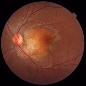
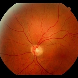
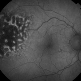
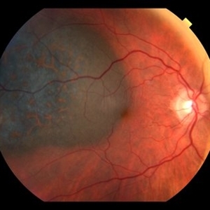
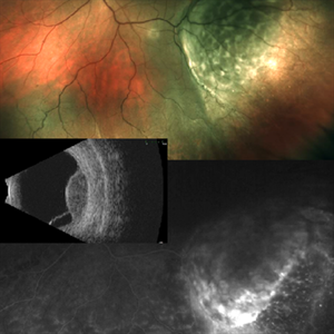
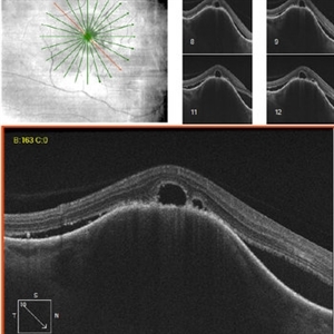
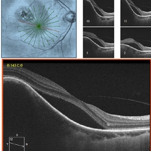
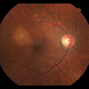
Fundus photographs and OCT scans of a 73-year-old non-smoker Indian male who presented to our retina clinic in 2013 with blurred vision in left eye for past 2 weeks. BCVA was 20/20 in right eye and 20/40 in left eye. Slit lamp exam was unremarkable for both eyes with no cells in aqueous or anterior vitreous. Fundus examination revealed creamy yellow choroidal lesions in both eyes. Lesion in right eye was one disc diameter (DD) in size and was located close to fovea (Fig-1a). Lesion in the left eye was bigger with a size of 2 DD located superior to fovea (Fig-1b). OCT scan for left eye revealed neurosensory detachment involving fovea (Fig-1c). Fundus fluorescein angiography was inconclusive for right eye and showed late hyper fluorescence the choroidal lesion in left eye. Patient underwent detailed systemic work up for malignancy that revealed primary lung non-small cell carcinoma. He had widespread metastasis affecting liver and brain. Palliative chemotherapy and radiotherapy were initiated 4 weeks after he presented to us. The choroidal lesions show progression on fundus picture and OCT scans done at 4 weeks follow up after initial presentation (Fig – 1d, e, f). The lesions in both eyes show regression at 4 weeks and 12 weeks follow up after initiation of therapy. Unfortunately, patient succumbed at 13 weeks follow up due to disease progression. The case demonstrates rare bilateral choroidal metastasis from primary lung cancer and also highlights that lesions can be asymptomatic till they develop neurosensory detachment as evident from asymptomatic lesion in right eye despite proximity to fovea and symptomatic lesion in left eye with NSD.
Photographer: Deependra Vikram Singh, Eye-Q Superspecialty Eye Hospitals, Gurugram
Imaging device: Topcon
Condition/keywords: choroidal mass, choroidal metastasis
-
 Choroidal Osteoma
Apr 10 2020 by Dipak Nag, MBBS, FCPS, MSc, FRF
The B scan showed slightly elevated, high reflective choroidal mass (red arrow) with acoustic shadowing of “pseudo-optic nerve” (yellow arrow). The mass persists even in lower gain. A scan showed a high intensity spike.
Photographer: SSN Nishi
Condition/keywords: B scan ultrasound, choroidal osteoma
-
 Circumscribed Choroidal Hemangioma
Oct 12 2019 by John S. King, MD
67-year-old white male with 6 days of decreased vision and known history of choroidal hemangioma, who had received PDT years ago for symptomatic SRF, had recurrence of SRF. PDT was applied to the lesion and one month later there is less subfoveal SRF, and vision has increased to 20/50 from 20/150. Will follow up in a month. Pictured is an orange-red choroidal mass with margins that blend with the surrounding choroid.
Photographer: Shelly Blair
Condition/keywords: choroidal hemangioma, photodynamic therapy
-
 Collar Button Appearance on B-Scan
Aug 28 2019 by Gayathri Mohan
B-scan showing an intraocular mass with collar button appearance. Suspected case of choroidal melanoma.
Photographer: Dr.Gayathri Mohan, Retina Foundation
Imaging device: Nidek Mirante SLO
Condition/keywords: choroidal mass, collar button
-
 Wide Field Fundus Montage of Intraocular Mass with Retinal Detachment
Aug 28 2019 by Gayathri Mohan
50 year old female came with diminution of vision in the LE. Wide field fundus photograph showing an intraocular mass temporally along with an exudative retinal detachment inferiorly. Ultrasonography showed an intraocular mass with collar button appearance suggestive of a Choroidal melanoma. She underwent enucleation and histopathology confirmed a spindle cell choroidal melanoma
Photographer: Dr. Gayathri Mohan, Retina Foundation
Imaging device: Nidek Mirante SLO
Condition/keywords: choroidal mass, collar button
-
 Intraocular Mass With Retinal Detachment
Aug 28 2019 by Gayathri Mohan
Wide field fundus image showing an intraocular mass temporally along with a retinal detachment.
Photographer: Dr. Gayathri Mohan, Retina Foundation
Imaging device: Nidek Mirante SLO
Condition/keywords: choroidal mass
-
 Hyper-autofluorescence of Orange Pigment Overlying a Lesion Suspicious for a Choroidal Melanoma
Jan 16 2019 by John S. King, MD
76-year-old white male saw his eye doctor with a three week complaint of photopsias and a shadow in his vision. Found to have a 10.5/12.5/2.5 (medium reflectivity) pigmented, choroidal mass associated with SRF and orange pigment (hyper-autofluorescence of lipofuscin, see image), and without drusen or halo.
Photographer: Stacey Coleman
Imaging device: Topcon 50
Condition/keywords: lipofuscin, orange pigment
-
 Orange Pigment Overlying a Lesion Suspicious for a Choroidal Melanoma
Jan 16 2019 by John S. King, MD
76-year-old white male saw his eye doctor with a three week complaint of photopsias and a shadow in his vision. Found to have a 10.5/12.5/2.5 (medium reflectivity) pigmented, choroidal mass associated with SRF and orange pigment (hyper-autofluorescence of lipofuscin), and without drusen or halo. See photo
Photographer: Stacey Coleman
Imaging device: Topcon 50
Condition/keywords: lipofuscin, orange pigment
-
 Choroidal Mass
Sep 21 2018 by Sarah Oelrich
Choroidal mass
Photographer: Sarah Oelrich CRA COT, Southeastern Retina Associates Knoxville Tn
Imaging device: OPTOS 200tx
Condition/keywords: choroidal mass
-
 Nasal Choroidal Mass
Jan 7 2018 by John S. King, MD
Mets vs Melanoma
Imaging device: Optos
Condition/keywords: choroidal mass
-
 Choroidal Mets - Renal Cell Carcinoma
Dec 2 2017 by John S. King, MD
54-year-old white female s/p nephrectomy 9 months ago for renal cell carcinoma; noticed blurry vision one month ago. SRF, ORTs, Choroidal Mass
Imaging device: Cirrus
Condition/keywords: choroidal metastasis, renal cell carcinoma
-
 Choroidal Mets - Renal Cell Carcinoma
Dec 2 2017 by John S. King, MD
54-year-old white female s/p nephrectomy 9 months ago for renal cell carcinoma; noticed blurry vision one month ago. Foveal SRF, choroidal mass.
Imaging device: Cirrus
Condition/keywords: choroidal metastasis, renal cell carcinoma
-
 Choroidal Mets - Renal Cell Carcinoma
Dec 2 2017 by John S. King, MD
54-year-old white female s/p nephrectomy 9 months ago for renal cell carcinoma; noticed blurry vision one month ago. Amelanotic choroidal mass in macula with overlying RPE pigmentary changes
Imaging device: Topcon
Condition/keywords: choroidal metastasis
 Loading…