Search results (32 results)
-
 Chorioretinal Coloboma
Oct 6 2025 by Seif Allah Anwar
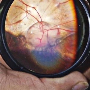
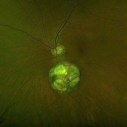
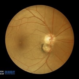
Fundus photograph of the patient left eye showing large, well-demarcated, excavated chorioretinal coloboma involving the inferior fundus, extending from the optic disc to the periphery. The lesion appears white due to bare sclera visibility, with absence of overlying choroid and retina. Retinal vessels course over the colobomatous area inferiorly.
Photographer: Dr. Seif Anwar, FRCSEd
Imaging device: Centervue Eidon
Condition/keywords: chorioretinal coloboma
-
 Unilateral Coloboma Involving Disc and Macula
Dec 27 2024 by Tejaswita Verma
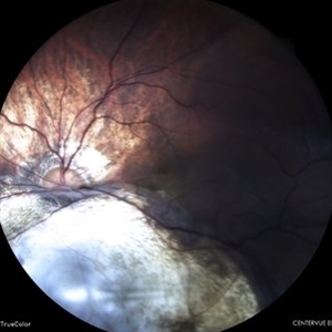
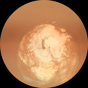
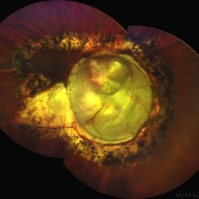
Fundus image of a 15 years old male presenting with unilaterally diminished vision since childhood in RE with CF3mt vision and inferior iris coloboma and retinochoroidal coloboma with nystagmus and cataract.
Photographer: DR. TEJASWITA VERMA
Imaging device: MIRANTE
Condition/keywords: chorioretinal coloboma, iridofundal coloboma
-
 Fundal Coloboma
Sep 25 2024 by DR Rohit Gupta
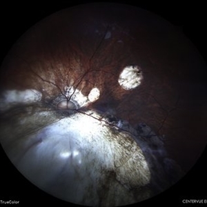
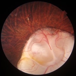
Fundus photograph of 16year old female patient with a fundal coloboma in left eye
Photographer: Dr Rohit gupta
Imaging device: Samsung S21
Condition/keywords: chorioretinal coloboma, coloboma of macula, coloboma of optic disc, congenital anomaly
-
 Retinal Detachment With Irido-fundal Coloboma
Feb 7 2024 by Akansha Sharma
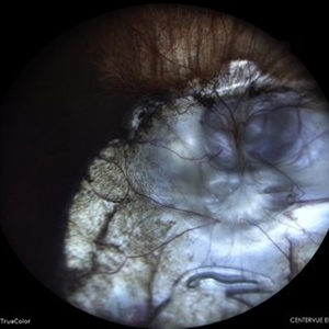
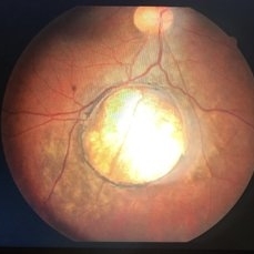
Color fundus photograph of a 43 year old male with retinal detachment in a case of iridofundal coloboma.
Photographer: Dr. Akansha Sharma, Bharati Eye Hospital
Condition/keywords: chorioretinal coloboma, RD
-
 Status Post Retinal Detachment Surgery in a Case of Iridofundal Coloboma
Feb 7 2024 by Akansha Sharma
Color fundus photograph of a 43 year old male post retinal detachment surgery in a case of iridofundal coloboma.
Photographer: Dr. Akansha Sharma, Bharati Eye Hospital
Condition/keywords: chorioretinal coloboma, RD
-
 IOL Drop in a Case of Iridofundal Coloboma
Feb 7 2024 by Akansha Sharma
Color fundus photograph of a 43 year old male with IOL drop in a case of iridofundal coloboma.
Photographer: Dr. Akansha Sharma, Bharati Eye Hospital
Condition/keywords: chorioretinal coloboma, IOL drop
-
 Chorioretinal Coloboma
Aug 7 2023 by Aditya S Kelkar, MS, FRCS, FASRS,FRCOphth
Fundus photograph of an 68-year-old woman with a chorioretinal coloboma observed.
Photographer: Optom Komal Jangam, National Institute of Ophthalmology, Pune, India.
Imaging device: OPTOS DAYTONA
Condition/keywords: chorioretinal coloboma
-
 Chorioretinal Coloboma
May 2 2023 by RAKESH SHAH, MS DNB FACS FRF FICO MBA
Young man with blurring of vision in both eyes
Photographer: Dr.Rakesh shah
Condition/keywords: chorioretinal coloboma
-
 Choroidal Coloboma
May 2 2023 by RAKESH SHAH, MS DNB FACS FRF FICO MBA
Young male patient came with blurring of vision in both eyes
Photographer: Dr Rakesh shah
Condition/keywords: chorioretinal coloboma
-
 Retino choroidal coloboma
Apr 27 2023 by Harshal Sahare, MBBS, MS, DNB, MNAMS, FICO,FVRS, FAICO
Fundus photograph of an 8 year old male child presented with a retino choroidal coloboma involving the disc.
Photographer: Dr Harshal Sahare, Sankara Eye Hospital, Shimoga, Karnataka
Imaging device: Topcon DRI SS OCT Triton
Condition/keywords: chorioretinal coloboma, coloboma of optic disc
-
 Retino choroidal coloboma
Apr 27 2023 by Harshal Sahare, MBBS, MS, DNB, MNAMS, FICO,FVRS, FAICO
Fundus photograph of an 8 year old male child presented with a retino choroidal coloboma involving the disc.
Photographer: Dr Harshal Sahare, Sankara Eye Hospital, Shimoga, Karnataka
Imaging device: Topcon DRI ss-OCT Triton
Condition/keywords: chorioretinal coloboma
-
 The Chorioretinal Coloboma
Oct 18 2022 by Magna Mary Kuruvila
Colour fundus photograph of an 32 year old male patient showing Chorioretinal Coloboma
Photographer: Dr Magna Mary Kuruvila,Blde University,Vijayapura
Imaging device: Canon
Condition/keywords: choroid, coloboma, retina
-
 Double disc sign
Oct 13 2022 by Vaibhavi Noticewala, M S Ophthalmology, FVRS
Double disc sign Doubling of the optic disc is rare and can manifest as true or pseudo doubling. Duke-Elder describes duplication of the optic disc as a rare anomaly wherein two discs, each provided with retinal vessels are seen in an otherwise normal eye. Rare cases of true duplication of optic discs with separation of optic nerve into two or more strands have been reported, based either on incidental necropsy findings, demonstration of two optic foramina in the same orbit on x ray, or angioscotomas as indirect evidence of the existence of double optic nerves. Pseudo doubling of the optic discs caused by lesions such as optic disc coloboma, peripapillary chorioretinal coloboma, or inflammatory foci are more common. Our case had Ipsilateral isolated ectatic peripapillary chorioretinal coloboma simulating double optic discs.
Photographer: Priyal Mistry
Condition/keywords: Pseudoduplication of optic disc
-
 Chorioretinal coloboma involving disc and macula
Mar 21 2022 by T. P . VIGNESH, MBBS,MS
Fundus photo of Right eye of a 55 year male patient revealing a fovea sparing well barraged chorioretinal coloboma involving the disc and the macula .
Photographer: Bharathi Singaravel
Imaging device: Zeiss Clarus
Condition/keywords: chorioretinal coloboma, coloboma of optic disc
-
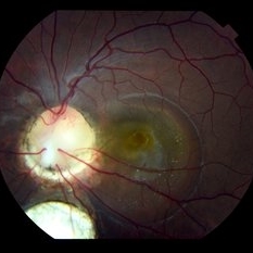
 Dislocated Brown Cataract with Chorioretinal Coloboma
Sep 8 2021 by Ram Sudarshan
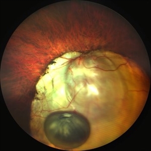
A 44 year-old male with dislocated brown cataract resting within a chorioretinal coloboma.
Photographer: Mrs.Bharati
Imaging device: Clarus
Condition/keywords: Brown cataract, chorioretinal coloboma, coloboma, dislocated lens
-
 Dislocated Brown Cataract with a Chorioretinal Coloboma
Sep 8 2021 by Ram Sudarshan
A 44 year-old male with dislocated brown cataract along with a chorioretinal coloboma.
Photographer: Dr.Sivadarshan
Condition/keywords: Brown cataract, chorioretinal coloboma, d, dislocated lens
-
 Retinal Blood Vessels in Retinochoroidal (RC) Coloboma
May 4 2021 by Priya Rasipuram Chandrasekaran, MBBS, DO, DNB, FRCS
This is the fundus photo of a 10-year-old girl showing RC coloboma along the infero nasal retina and involving the disc. This belongs to grade 4 of Ida Mann’s classification and grade 5 of Lingam Gopal’s classification of RC coloboma. The optic disc has no cup and BV for superior fundus emanates from superior part of optic disc and that for inferior fundus in the colobomatous area from multiple points. The blood vessels are discontinuous and are cork screw shaped.
Condition/keywords: chorioretinal coloboma
-
 Chorioretinal Coloboma with Retinal Detachment
Dec 5 2020 by Niloofar Piri, MD
14-year-old female with 1q21.1 microdeletion syndrome and behavioral, intellectual, and systemic abnormalities, including congenital microcornea, iris coloboma, and chorioretinal and optic nerve coloboma presented with decreased vision. Right eye fundus taken with RetCam shows coloboma with retinal detachment. (Left eye showed white cataract with funnel RD on B-scan).
Photographer: Niloofar Piri MD, Douglas Snyder MD
Condition/keywords: chorioretinal coloboma, optic nerve coloboma
-
 Surgery in MPPC Syndrome
Nov 17 2020 by Linda A Cernichiaro- Espinosa, MD
MPPC Syndrome: microcornea, posterior megalolenticonus, persistent fetal vasculature, chorioretinal coloboma. This surgery was performed with 3D printed spacers to shorten 25g trocars and small BIOM visualization system to repair a post traumatic rhegmatogenous retinal detachment.
Photographer: Rodrigo Gómez, PhD
Imaging device: iPhone X
Condition/keywords: chorioretinal coloboma, microcornea, pars plana vitrectomy (PPV), pediatric retina, persistent fetal vasculature (PFV)
-
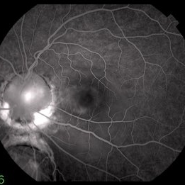
 Pseudo-Doubling of the Optic Disc
Aug 28 2020 by Catarina Almeida
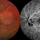
A 82-year-old woman presented for diabetic retinopathy screennig. In addition to a diabetic retinopathy and an epiretinal membrane, the left eye presented a well-defined round lesion in the inferonasal quadrant, adjacent to the optic disc, with identifiable bridging retinal vessels from the optic disc and no leakage on the fluorescein angiography, suggesting a pseudo-doubling of the optic disc. Pseudo-doubling of the optic disc is a rare condition, where a lesion resembling na optic disc is situated adjacent to the true optic disc, and may be caused by optic disc coloboma, peripapillary chorioretinal coloboma or inflammatory foci. As in this case, typical chorioretinal colobomas are located inferiorly and slightly nasally, resulting from failure of closure of the fetal fissure. Pseudo-doubling must be differentiated from the extremely rare true optic disc doubling by fluorescein angiography, head and orbits computerized tomography or magnetic resonance imaging.
Photographer: Catarina Almeida, Centro Hospitalar Tondela-Viseu
Imaging device: Retinography and fluorescein angiography (SPECTRALIS® Heidelberg Engineering, Germany)
Condition/keywords: coloboma, optic disc
-
 Colobomatous Optic Disc Maculopathy
Feb 13 2020 by Yoshihiro Yonekawa, MD, FASRS
EDI-OCT of a teenage girl with submacular fluid from a colobomatous optic disc. Note the subtle tracking of the subretinal fluid into the disc.
Photographer: Netanya Lerner, COA, Wills Eye Hospital/Mid Atlantic Retina
Imaging device: Topcon
Condition/keywords: chorioretinal coloboma, coloboma of optic disc, congenital optic nerve pit, subretinal fluid
-
 Colobomatous Optic Disc Maculopathy
Feb 13 2020 by Yoshihiro Yonekawa, MD, FASRS
Fluorescein angiography, late frame, of a teenage girl with submacular fluid from a colobomatous optic disc. The camera is focused is on the elevated macula, and the disc is subtly defocused.
Photographer: Netanya Lerner, COA, Wills Eye Hospital/Mid Atlantic Retina
Imaging device: Topcon
Condition/keywords: chorioretinal coloboma, coloboma of optic disc, congenital optic nerve pit, subretinal fluid
-
 Colobomatous Optic Disc Maculopathy
Feb 13 2020 by Yoshihiro Yonekawa, MD, FASRS
Beautifully focused fundus photograph of a teenage girl with submacular fluid from a colobomatous optic disc.
Photographer: Netanya Lerner, COA, Wills Eye Hospital/Mid Atlantic Retina
Imaging device: Topcon
Condition/keywords: chorioretinal coloboma, coloboma of optic disc, congenital optic nerve pit, subretinal fluid
-
 Cat Eye Syndrome
Feb 11 2020 by Sophia El Hamichi, MD
A 3-year-old female with cat eye syndrome including iris, chorioretinal and optic nerve colobomas. Note the CNV temporally to the optic nerve coloboma (blue arrows)
Photographer: Giselle De Oliveira, Bascom Palmer Eye Institute, Miami
Imaging device: RetCam
Condition/keywords: cat eye syndrome, chorioretinal coloboma, choroidal neovascularization (CNV), coloboma, coloboma of optic disc, optic nerve coloboma
-
 Coloboma
Oct 2 2019 by John S. King, MD
27-year-old white female with bilateral, isolated, inferior, chorioretinal colobomas; she has a history of retinal laser anterior to the edge of the coloboma OD secondary to a limited RD. This is the left eye.
Photographer: Shelly Blair
Imaging device: Optos CA
Condition/keywords: coloboma of choroid
 Loading…