Search results (24 results)
-
 Cat Scratch Disease
Mar 29 2021 by Gabriel Costa Andrade, PhD
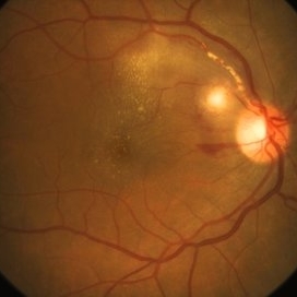
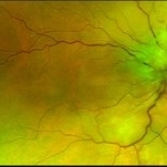
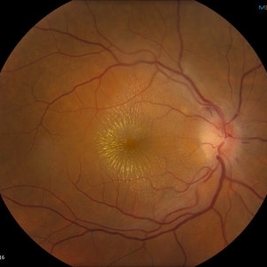
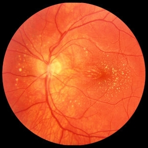
Fundus photograph of an 36-year-old woman with a macular vasculitis, pre retinal hemorrhage and exudation due to Bartonella henselae infection.
Photographer: Gabriel Andrade
Condition/keywords: cat scratch retinitis
-
 Serous Retinal Detachment and Retinal Infiltrate due to B. Hensele, Cat-Scratch Disease
Dec 21 2020 by John S. King, MD
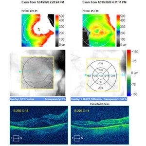
64-year-old female had at least a two week history of blurry vision in the right eye. She was being followed for a CRVO in the right eye, and as vision worsened, was referred to our clinic, and saw Dr. Zocchi. Vision in the right eye was CF; there was 1+ cell in the A/C; 1+ vitreous cell was present; disc edema with surrounding SRF as well as a small, white, retinal infiltrate just superior to the optic disc; vessel tortuosity was present as well as a few IRHs (left eye was u/r). There was sub-foveal and PP SRF on OCT. FA in the early to mid phase showed optic disc hyperfluorescence and early filling into the subretinal space. In the later frames there was disc leakage, staining/leakage of the retinal infiltrate, and filling into the subretinal space (See OCT - Left Image is initial visit). .... Multiple tests were done, she was started on doxycycline 100 mg BID, and Bartonella serology test came back positive. One week later vision improved to 20/100, a/c cell present, disc edema improved and the SRF was resolving (See OCT - Right Image is latest visit).
Imaging device: Zeiss Cirrus
Condition/keywords: Bartonella bacteria, cat scratch retinitis
-
 Serous Retinal Detachment and Retinal Infiltrate due to B. Hensele, Cat-Scratch Disease
Dec 19 2020 by John S. King, MD
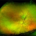
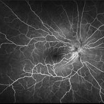
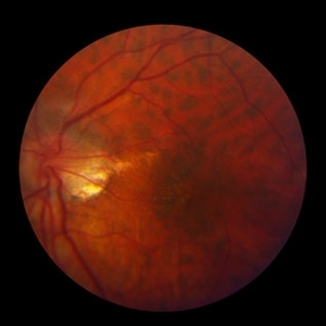
64-year-old female had at least a two week history of blurry vision in the right eye. She was being followed for a CRVO in the right eye, and as vision worsened, was referred to our clinic, and saw Dr. Zocchi. Vision in the right eye was CF; there was 1+ cell in the A/C; 1+ vitreous cell was present; disc edema with surrounding SRF as well as a small, white, retinal infiltrate just superior to the optic disc; vessel tortuosity was present as well as a few IRHs (left eye was u/r). There was sub-foveal and PP SRF on OCT. FA in the early to mid phase showed optic disc hyperfluorescence and early filling into the subretinal space. In the later frames there was disc leakage, staining/leakage of the retinal infiltrate, and filling into the subretinal space (See Image). Multiple tests were done, she was started on doxycycline 100 mg BID, and Bartonella serology test came back positive. One week later vision improved to 20/100, a/c cell present, disc edema improved and the SRF was resolving. (will add more photos next visit)
Photographer: Shelly Blair
Imaging device: Optos CA
Condition/keywords: cat scratch retinitis
-
 Serous Retinal Detachment and Retinal Infiltrate due to B. Hensele, Cat-Scratch Disease
Dec 19 2020 by John S. King, MD
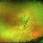
64-year-old female had at least a two week history of blurry vision in the right eye. She was being followed for a CRVO in the right eye, and as vision worsened, was referred to our clinic, and saw Dr. Zocchi. Vision in the right eye was CF; there was 1+ cell in the A/C; 1+ vitreous cell was present; disc edema with surrounding SRF as well as a small, white, retinal infiltrate just superior to the optic disc; vessel tortuosity was present as well as a few IRHs (See Image) (left eye was u/r). There was sub-foveal and PP SRF on OCT. FA in the early to mid phase showed optic disc hyperfluorescence and early filling into the subretinal space. In the later frames there was disc leakage, staining/leakage of the retinal infiltrate, and filling into the subretinal space. Multiple tests were done, she was started on doxycycline 100 mg BID, and Bartonella serology test came back positive..... One week later vision improved to 20/100, a/c cell present, disc edema improved and the SRF was resolving. (will add more photos next visit)
Photographer: Shelly Blair
Imaging device: Optos CA
Condition/keywords: cat scratch retinitis
-
 Serous Retinal Detachment and Retinal Infiltrate due to B. Hensele, Cat-Scratch Disease
Dec 19 2020 by John S. King, MD
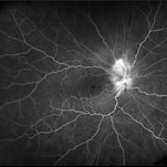
64-year-old female had at least a two week history of blurry vision in the right eye. She was being followed for a CRVO in the right eye, and as vision worsened, was referred to our clinic, and saw Dr. Zocchi. Vision in the right eye was CF; there was 1+ cell in the A/C; 1+ vitreous cell was present; disc edema with surrounding SRF as well as a small, white, retinal infiltrate just superior to the optic disc; vessel tortuosity was present as well as a few IRHs (left eye was u/r). There was sub-foveal and PP SRF on OCT. FA in the early to mid phase showed optic disc hyperfluorescence and early filling into the subretinal space (See Image). In the later frames there was disc leakage, staining/leakage of the retinal infiltrate, and filling into the subretinal space. Multiple tests were done, she was started on doxycycline 100 mg BID, and Bartonella serology test came back positive. One week later vision improved to 20/100, a/c cell present, disc edema improved and the SRF was resolving. (will add more photos next visit)
Photographer: Shelly Blair
Imaging device: Optos CA
Condition/keywords: cat scratch retinitis
-
 Serous Retinal Detachment and Retinal Infiltrate due to B. Hensele, Cat-Scratch Disease
Dec 19 2020 by John S. King, MD
64-year-old female had at least a two week history of blurry vision in the right eye. She was being followed for a CRVO in the right eye, and as vision worsened, was referred to our clinic, and saw Dr. Zocchi. Vision in the right eye was CF; there was 1+ cell in the A/C; 1+ vitreous cell was present; disc edema with surrounding SRF as well as a small, white, retinal infiltrate just superior to the optic disc; vessel tortuosity was present as well as a few IRHs (left eye was u/r). There was sub-foveal and PP SRF on OCT. FA in the early to mid phase showed optic disc hyperfluorescence and early filling into the subretinal space. In the later frames there was disc leakage, staining/leakage of the retinal infiltrate, and filling into the subretinal space (See Image). Multiple tests were done, she was started on doxycycline 100 mg BID, and Bartonella serology test came back positive. One week later vision improved to 20/100, a/c cell present, disc edema improved and the SRF was resolving. (will add more photos next visit)
Photographer: Shelly Blair
Imaging device: Optos CA
Condition/keywords: cat scratch retinitis
-
 Cat Scratch
Feb 15 2017 by Hua Gao, MD, PhD, FASRS
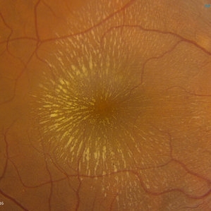
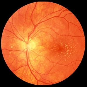
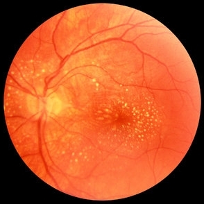
A female patient of 57-year-old presented with neuroretinitis due to cat-scratch disease with positive Bartonella henselae antibodies. Two weeks after symptom onset she developed exudates in a "macular star" pattern.
Condition/keywords: cat scratch retinitis
-
 Cat Scratch
Feb 15 2017 by Hua Gao, MD, PhD, FASRS
A female patient of 57-year-old presented with neuroretinitis due to cat-scratch disease with positive Bartonella henselae antibodies. Two weeks after symptom onset she developed exudates in a "macular star" pattern.
Condition/keywords: cat scratch retinitis
-
 Partial Macular Star
Jan 23 2017 by Chuck Terranova
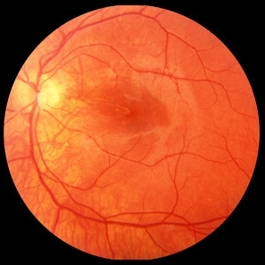
Partial macular star.
Photographer: Chuck Terranova, Ross Eye Institute, Buffalo, NY. USA
Imaging device: Zeiss FF-4
Condition/keywords: cat scratch retinitis
-
 Cat Scratch Disease
Dec 27 2016 by Elad Moisseiev, MD
A 31-year-old man with cat scratch disease (positive serology for Bartonella hensleae), which resulted in a 2 nasal branch retinal artery occlusions associated with a temporal visual field defect.
Photographer: Gailt Yair-Pur
Condition/keywords: branch retinal artery occlusion (BRAO), cat scratch retinitis
-
 BRAO d/t cat scratch disease - FA 5:23 min.
Jan 2 2013 by Roy Schwartz, MD
A 38-year-old male complained of a grey spot in visual field in his left eye. On clinical exam BRAO in LE, confirmed by FA, as seen in picture. The image was taken after late filling of artery. CWS blocks proximal part of artery. Serology for bartonella was positive.
Photographer: Galit Yair-Pur
Condition/keywords: branch retinal artery occlusion (BRAO), cat scratch retinitis
-
 BRAO d/t cat scratch disease - FA 00:18 min.
Jan 2 2013 by Roy Schwartz, MD
A 38-year-old male complained of a grey spot in visual field in his left eye. On clinical exam BRAO in LE, confirmed by FA, as seen in picture. Image shows delayed filling of artery. Serology for bartonella was positive.
Photographer: Galit Yair-Pur
Condition/keywords: branch retinal artery occlusion (BRAO), cat scratch retinitis
-
 BRAO d/t cat scratch disease - LE fundus photograph
Jan 2 2013 by Roy Schwartz, MD
A 38 year-old-male complained of a grey spot in visual field in his left eye. On clinical exam BRAO in LE, as seen in picture. Serology for bartonella was positive.
Photographer: Galit Yair-Pur
Condition/keywords: branch retinal artery occlusion (BRAO), cat scratch retinitis
-
 BRAO d/t cat scratch disease - RE fundus photograph
Jan 2 2013 by Roy Schwartz, MD
A 38-year-old male complained of a grey spot in visual field in his left eye. On clinical exam BRAO in LE, and a CWS in RE, as seen in this photograph. Serology for bartonella was positive.
Photographer: Galit Yair-Pur
Condition/keywords: branch retinal artery occlusion (BRAO), cat scratch retinitis
-
 Cat Scratch Disease
Oct 18 2012 by Raj K. Maturi, MD
Photographer: Tom Steele, CRA
Imaging device: Topcon 50dx
Condition/keywords: Bartonella bacteria, cat scratch retinitis
-
 Cat Scratch Disease
Oct 18 2012 by Raj K. Maturi, MD
Photographer: Tom Steele, CRA
Imaging device: Topcon 50dx
Condition/keywords: Bartonella bacteria, cat scratch retinitis
-
 Cat Scratch Disease
Oct 18 2012 by Raj K. Maturi, MD
Photographer: Tom Steele, CRA
Imaging device: Topcon 50dx
Condition/keywords: Bartonella bacteria, cat scratch retinitis, red-free
-
 Cat Scratch Disease
Oct 18 2012 by Raj K. Maturi, MD
Photographer: Tom Steele, CRA
Imaging device: Topcon 50dx
Condition/keywords: Bartonella bacteria, cat scratch retinitis
-
 Cat Scratch Disease
Oct 18 2012 by Raj K. Maturi, MD
Photographer: Tom Steele, CRA
Imaging device: Topcon 50dx
Condition/keywords: Bartonella bacteria, cat scratch retinitis
-
 Cat Scratch Retinitis with Macular Lipid Resolved
Oct 9 2012 by Jeffrey G. Gross, MD, FASRS
Cat scratch retinitis with macular lipid, resolved, s/p treatment, 20/70.
Condition/keywords: 20/70, cat scratch retinitis, macular lipid
-
 Cat Scratch Retinitis with Macular Lipid Resolving
Oct 9 2012 by Jeffrey G. Gross, MD, FASRS
Cat scratch retinitis with macular lipid, resolving.
Condition/keywords: cat scratch retinitis, macular lipid
-
 Cat Scratch Retinitis with Macular Lipid Resolving
Oct 9 2012 by Jeffrey G. Gross, MD, FASRS
Cat scratch retinitis with macular lipid, resolving, 20/100.
Condition/keywords: 20/100, cat scratch retinitis, macular lipid
-
 Cat Scratch Retinitis with Macular Lipid
Oct 9 2012 by Jeffrey G. Gross, MD, FASRS
Cat scratch retinitis, with macular lipid.
Condition/keywords: cat scratch retinitis, macular lipid
 Loading…