Search results (71 results)
-
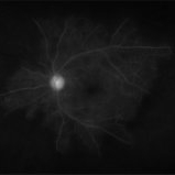
 Branches Starved of Flow, Yet Nature Strives to Grow
Apr 1 2025 by rohan jain
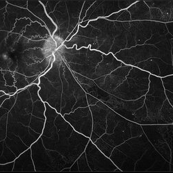
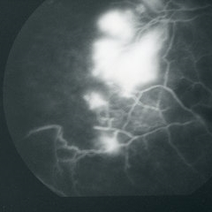
Tufts of NVE's in a case of Branch Retinal Vein Occlusion
Photographer: Dr. ROHAN JAIN
Condition/keywords: branch retinal vein occlusion (BRVO), capillary nonperfusion, non-perfused branch retinal vein occlusion (BRVO), non-perfusion, NVE, OCT Angiography, ST BRVO
-
 Chronic CRVO
Dec 12 2024 by Korey Starkey
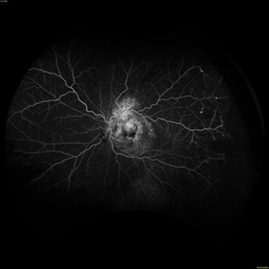
Fluorescein Angiography of a 62 year-old man with chronic central retinal vein occlusion. Vision is 20/200.
Photographer: Korey Starkey
Imaging device: Optos
Condition/keywords: capillary nonperfusion, central retinal vein occlusion (CRVO), FLUORESCEIN ANGIOGRAPHY, ischemia, microaneurysms, Optos
-
 Diabetic Retinopathy
Nov 20 2024 by Korey Starkey
64 year old female being monitored for moderate-severe diabetic retinopathy.
Photographer: Korey Starkey
Condition/keywords: capillary nonperfusion, FA, FLUORESCEIN ANGIOGRAPHY, microaneurysms, nonproliferative diabetic retinopathy, Optos, OPTOS CALIFORNIA, tortuous vessels
-
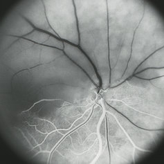
 Central Retinal Vein Occlusion
Sep 27 2024 by Jeffrey Barker
65 year old male with a Central Retinal Vein Occlusion and Macular Edema and Capillary Nonperfusion.
Photographer: Jeffrey P. Barker
Condition/keywords: central retinal vein occlusion (CRVO), macular edema
-
 Old BRVO
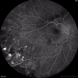
Oct 18 2023 by Anand Temkar
LE widefield FA montage of a 68 years old male with history of Old BRVO, showing peripheral capillary non perfusion and some temporal laser marks (staining ).
Photographer: Dr.Anand Temkar- Retina Foundation, Ahmedabad
Imaging device: Mirante
Condition/keywords: branch retinal vein occlusion (BRVO), capillary nonperfusion
-
 Coats Disease Fluorescein Angiography
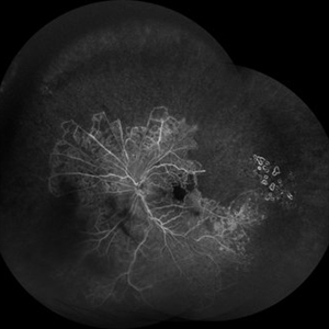
Sep 2 2022 by FLOR ANGELICA JACOME GUTIERREZ
Fluorescein angiography of a patient with Coats disease where we found telangiectatic vessels, aneurysms, peripheral capillary nonperfusion and perivascular leak.
Photographer: Dr.Guillermo Salcedo Villanueva
Imaging device: Zeiss CLARUS 700 (FA)
Condition/keywords: Coats' disease, epiretinal membrane (ERM)
-
 Proliferative Diabetic Retinopathy and SC Disease
Aug 27 2021 by Caesar K. Luo, MD, FASRS
53 year-old male with SC disease complicated by proliferative diabetic retinopathy with severe peripheral non perfusion and small, central retained island.
Photographer: Fred Hanamoto, Bay Area Retina Associates
Imaging device: Optos California
Condition/keywords: capillary nonperfusion, peripheral ischemia, proliferative diabetic retinopathy (PDR), retinal ischemia, sickle cell retinopathy
-
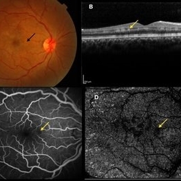 Paracentral Acute Middle Maculopathy
Oct 25 2019 by Gayathri Mohan
Multimodal images of a case of a 29-year-old female with paracentral acute middle maculopathy. A-color fundus photograph showing multiple confluent white retinal patches. B- On OCT the acute lesions of PAMM characteristically appear as placoid, hyperreflective bands at the level of the INL C-Fundus fluorescein angiography showing a capillary nonperfusion area D-flow void areas in deep capillary plexus
Photographer: Akshar Soni
Imaging device: Heidelberg, Nidek
Condition/keywords: fundus albipunctatus, optical coherence tomography (OCT), paracentral acute middle maculopathy
-
 Branch Retinal Vein Occlusion with Acute on Chronic Subhyaloid Hemorrhage
Oct 24 2019 by Nichole Lewis
60-year-old male with a branch retinal vein occlusion and subhyaloid hemorrhage and retinal neovascularization. VA HM.
Photographer: Nichole
Condition/keywords: branch retinal vein occlusion (BRVO), capillary nonperfusion, retinal neovascularization, subhyaloid hemorrhage
-
 OCTA in Vasculitis
Sep 3 2019 by Manish Nagpal, MD, FRCS (UK), FASRS
OCTA in a case of vasculitis highlighting the capillary non perfusion areas closing in on macula.
Photographer: Gayathri Mohan
Condition/keywords: capillary nonperfusion, vasculitis
-
 Capillary Non-Perfusion
Aug 26 2019 by Narciso F. Atienza, MD, MBA, FASRS, FPCS, FPAO.
FA at 1 min 44 sec showing capillary non-perfusion and blocked fluoresence of the infero-temporal area.
Photographer: Narciso F Atienza, Jr. MD, MBA
Imaging device: Topcon TRC
Condition/keywords: capillary nonperfusion
-
 Capillary Non-Perfusion
Aug 26 2019 by Narciso F. Atienza, MD, MBA, FASRS, FPCS, FPAO.
FA at 1 min showing capillary non-perfusion and blocked fluoresence of the inferior macula to infero-temporal area.
Photographer: Narciso F Atienza, Jr. MD, MBA
Imaging device: Topcon TRC
Condition/keywords: capillary nonperfusion
-
 Capillary Non-Perfusion
Aug 26 2019 by Narciso F. Atienza, MD, MBA, FASRS, FPCS, FPAO.
FA at 51 sec showing capillary non-perfusion and blocked fluoresence of the inferior macula and infero-temporal area with transit of dye on previously noted infero-temporal branch vein.
Photographer: Narciso F Atienza, Jr. MD, MBA
Imaging device: Topcon TRC
Condition/keywords: capillary nonperfusion
-
 Capillary Non-Perfusion
Aug 26 2019 by Narciso F. Atienza, MD, MBA, FASRS, FPCS, FPAO.
FA at 17 sec showing capillary non-perfusion and blocked fluoresence of the infero-temporal area.
Photographer: Narciso F Atienza, Jr. MD, MBA
Imaging device: Topcon TRC
Condition/keywords: capillary nonperfusion
-
 Capillary Non-Perfusion
Aug 26 2019 by Narciso F. Atienza, MD, MBA, FASRS, FPCS, FPAO.
FA at 13 sec showing capillary non-perfusion and blocked fluoresence of the infero-temporal area.
Photographer: Narciso F Atienza, Jr. MD, MBA
Imaging device: Topcon TRC
Condition/keywords: capillary nonperfusion
-
 Early Venous Phase
Aug 26 2019 by Narciso F. Atienza, MD, MBA, FASRS, FPCS, FPAO.
Early venous phase shows asymmetrical transit of dye perfusion of the infero-temporal arcade. Infero-temporal arcade shows beginning perfusion. Areas of non perfusion are also more prominent.
Photographer: Narciso F Atienza, Jr. MD, MBA
Imaging device: Topcon TRC
Condition/keywords: capillary nonperfusion, inferotemporal arcade
-
 Laser Induced BRAO in IRVAN Syndrome
May 3 2019 by Deependra Vikram Singh, MD FASRS
Fundus photograph of a 26-year-old man with IRVAN syndrome referred for vitreous surgery in OS for secondary rhegmatogenous retinal detachment. OD has received laser photocoagulation for capillary nonperfusion areas and retinal artery macroaneurysm associated with retinal vasculitis. Fundus photograph of OD shows laser induced nasal BRAO. Case re-emphasizes why laser for macroaneurysm should be avoided in cases with IRVAN.
Photographer: Deependra V Singh, Eye-Q Superspecialty Eye Hospitals. Gurugram, India
Imaging device: Zeiss Visucam 500
Condition/keywords: arteriolar macroaneurysm, branch retinal artery occlusion (BRAO), laser photocoagulation
-
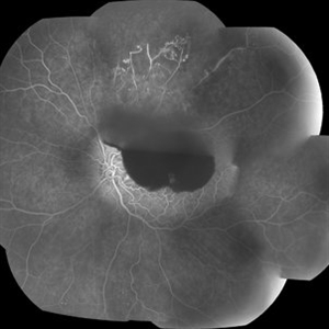
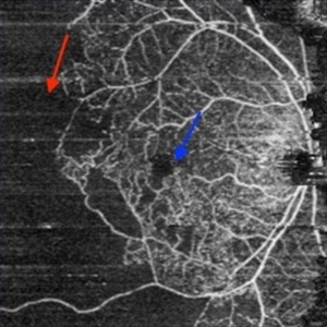
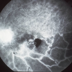 Nonperfused BRVO with Collateral Vessels
Apr 8 2019 by Gary R. Cook, MD, FACS
Late-phase fluorescein angiogram image of the left eye of a 73-year-old African-American female with a nonperfused BRVO showing flow through the collateral vessels, marked loss of the capillary bed, disc leakage from some NVD, and ischemic staining of the retinal veins; V.A. = 20/70-1
Imaging device: Topcon VT-50
Condition/keywords: branch retinal vein occlusion (BRVO), capillary nonperfusion, collaterals, disc leakage, FA late phase, fluorescein angiogram (FA)
-
 Eales Disease
Apr 1 2019 by Gary R. Cook, MD, FACS
Late-phase fluorescein angiogram image of the retinal periphery of a 23-year-old Vietnamese female with Eales disease showing peripheral capillary nonperfusion, vaso-occlusion and peripheral retinal neovascularization; V.A.= 20/80.
Imaging device: Topcon VT-50
Condition/keywords: Eales disease, FA late phase, fluorescein angiogram (FA), peripheral retinal neovascularization, vaso-occlusive disease
-
 Hemi-CRAO
Mar 26 2019 by Gary R. Cook, MD, FACS
Mid-phase (laminar venous return) fluorescein angiogram image of an embolic superior hemi-CRAO showing marked delay in filling of the superior retinal arteriolar and venous vasculature and total loss of the retinal capillary bed in the superior hemisphere OD.
Condition/keywords: capillary closure, capillary nonperfusion, central retinal artery occlusion (CRAO), FA mid phase, fluorescein angiogram (FA)
-
 Retinal Ischemia Secondary to Diabetic Retinopathy
Aug 29 2018 by Olivia Rainey
Fluorescein angiogram series of a 57-year-old male patient with proliferative diabetic retinopathy of the right eye. Patient has delayed AV transit with significant retinal ischemia and retinal capillary nonperfusion. The ischemia is extensive resulting in neovascularization of the iris and consequently neovascular glaucoma.
Photographer: Olivia Rainey
Imaging device: Optos
Condition/keywords: diabetes, disc hyperfluorescene, fluorescein angiogram (FA), non-perfusion, Optos, proliferative diabetic retinopathy (PDR), retinal ischemia, ultra-wide field imaging, vitreous hemorrhage
-
 Proliferative Diabetic Retinopathy
Jul 12 2018 by Nichole Lewis
68-year-old male with proliferative diabetic retinopathy and capillary nonperfusion. Returning for pan retinal photocoagulation. VA 20/25
Photographer: Nichole Lewis
Condition/keywords: capillary nonperfusion, diabetes, non-perfusion, pan-retinal photocoagulation (PRP), proliferative diabetic retinopathy (PDR)
-
 Proliferative Diabetic Retinopathy with Neovascular Glaucoma
Jul 12 2018 by Nichole Lewis
68-year-old male with proliferative diabetic retinopathy, severe capillary nonperfusion and neovascular glaucoma. Treated with Avastin intra-ocular injection and future pan-retinal photocoagulation. VA 20/300.
Photographer: Nichole Lewis
Condition/keywords: capillary nonperfusion, diabetes, neovascular glaucoma, non-perfusion, proliferative diabetic retinopathy (PDR)
-
 Wyburn-Mason Fluorescein Angiography
Apr 29 2018 by Sarina M Amin, MD
Wide-field fluorescein angiography of a 32-year-old woman with Wyburn-Mason syndrome showing temporal periphery capillary nonperfusion.
Photographer: Sarah Ellano, Retinal Consultants of Arizona, Phoenix, Arizona
Imaging device: Optos
Condition/keywords: Wyburn-Mason
-
 Capillary Nonperfusion
Apr 12 2018 by SUSHIL BHATT
OPTOS ultra wide field angiogram of an 45 years old diabetic male patient shows capillary nonperfusion areas with inadequate laser.
Photographer: Bhatt Sushil PGIMER chandigarh INDIA
Imaging device: OPTOS Ultra wide Field
Condition/keywords: capillary nonperfusion
 Loading…