Search results (19 results)
-
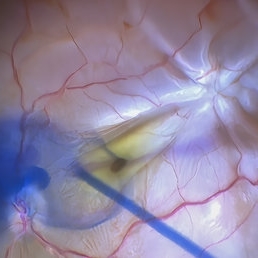 Star Folds in a Chronic Retinal Detachment
Jul 3 2024 by Anjana Mirajkar, MS Ophthalmology
Intra-operative still RE showing a star fold at the parafoveal area causing traction at the macula. Brilliant blue dye being injected to the stain the ILM.
Photographer: Dr. Anjana Mirajkar -Retina Foundation, Ahmedabad
Condition/keywords: brilliant blue staining, proliferative vitreoretinopathy (PVR), star folds
-
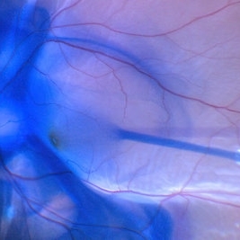
 BBG Dye injection to Stain ILM during Vitrectomy Surgery | Intra-Operative Still
Apr 28 2023 by Veer Singh, MS, FVRS, FMRF, FICO (Retina)
BBG Dye injection to Stain ILM during Vitrectomy Surgery | Intra-Operative Still
Photographer: Dr. Veer Singh
Condition/keywords: brilliant blue staining, ERM, ILM staining
-
Vitrectomy for Macular Hole
Jan 13 2023 by Manish Nagpal, MD, FRCS (UK), FASRS
This is a case of Macular hole for which vitrectomy is being done. After doing core vitrectomy triamcinolone dye is injected to stain the hyaloid. High aspiration is used on cutter to engage the hyaloid and gradually pull it anteriorly. PVD induction is carried out. After this brilliant blue dye is injected to stain the internal limiting membrane. ILM is peeled using a 25 gauge forceps in a tangential manner. After this i use a instrument called the massager which we have developed to gently and atraumatically massage concentrically the edges of the hole. This releases the subtle contaction on the edges of the hole and relaxes the margins. After this air fluid exchange is carried out followed by low vacuum aspiration over the hole. The hole approximates itself gradually as the aspiration dries up the edges.
Condition/keywords: forceps, hyaloid, ILM, macular hole, peeling, staining, video, vitrectomy
-
Vitrectomy for Sub ILM blood over macula
Jan 2 2023 by Manish Nagpal, MD, FRCS (UK), FASRS
This is a case of non resolving ILM hemorrhage over macula. Vitrectomy is carried out and hyaloid is removed after traimcinolone staining. After this brilliant blue dye is injected to stain the ILM. Internal limiting membrane is then removed with a forceps. Once the sub ilm blood is exposed , it easily aspirates with the cutter. The origin is probably from a macroaneurysm and there is a small component of subretinal residual blood noted at the end of the surgery.
Condition/keywords: brilliant blue, hyaloid, internal limiting membrane, macula, microaneurysm, retina, sub ILM blood, sub ILM hemorrhage, triamcinolone, video, vitrectomy
-
Macular hole in myopic patient
Nov 4 2022 by Manish Nagpal, MD, FRCS (UK), FASRS
This is a case earlier operated three times for myopic retinal detachment. Buckling was done followed by vilion oil injection and then silicon oil removal, now the patient developed a macular hole. Staining with brilliant blue followed by ILM peeling with a longer length forceps was carried out. Air fluid exchange with drainage over the hole and gas injection was done
Condition/keywords: brilliant blue staining, ILM peeling, myopia, myopic macular hole, video, vitrectomy
-
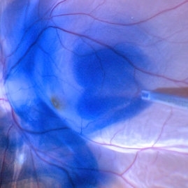
 Brilliant Blue Dye Injection to Stain ILM in a Macular Hole with Retinal Detachment
Feb 4 2022 by Manish Nagpal, MD, FRCS (UK), FASRS
Intraoperative still of a Brilliant blue dye injection being done to stain the ILM.
Photographer: Manish Nagpal, Director, Retina Foundation, Ahmedabad
Imaging device: Sony PMW -10 MD surgical camera
Condition/keywords: full thickness macular hole, macula, retina
-
 Brilliant blue dye injection to stain ILM in a macular hole with retinal detachment
Feb 4 2022 by Manish Nagpal, MD, FRCS (UK), FASRS
Intraoperative still of brilliant blue dye injection in process to initiate ILM peel in a patient who has a retinal detachment with a macular hole
Photographer: Manish Nagpal, Director, Retina Foundation, Ahmedabad
Imaging device: Sony PMW -10 MD surgical camera
Condition/keywords: brilliant blue, ILM flap, macular hole
-
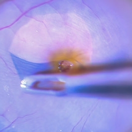
 Brilliant Blue Dye Injected in a Case of Macular Hole to Stain the ILM
Feb 4 2022 by Manish Nagpal, MD, FRCS (UK), FASRS
Intraoperative still of a brilliant blue dye being injected to stain the ILM.
Photographer: Manish Nagpal, Director, Retina Foundation, Ahmedabad
Imaging device: Sony PMW -10 MD surgical camera
Condition/keywords: brilliant blue, ILM flap, ILM staining, macular hole, retina, retina surgery
-
 Internal Limiting Membrane Peeling
Feb 2 2022 by Manish Nagpal, MD, FRCS (UK), FASRS
Intraoperative photo of an ILM peeling being done after brilliant blue staining with 25 gauge forceps.
Photographer: Manish Nagpal, Retina Foundation, Ahmedabad, India
Imaging device: Sony PMW -10 MD surgical camera
Condition/keywords: ILM flap, ILM staining, internal limiting membrane (ILM) peeling
-
 Stained ILM with a Flap
Feb 2 2022 by Manish Nagpal, MD, FRCS (UK), FASRS
Intraoperative photo of an ILM peeling. A flap initiation has been achieved with a pinch and peel technique using forceps and after this the ILM is peeled.
Photographer: Manish Nagpal, Retina Foundation, Ahmedabad, india
Imaging device: Sony PMW -10 MD surgical camera
Condition/keywords: brilliant blue, ILM flap, ILM staining, internal limiting membrane (ILM) peeling
-
 Internal Limiting Membrane Peeling
Jan 10 2022 by Manish Nagpal, MD, FRCS (UK), FASRS
Intraoperative image of internal limiting membrane being peeled using a 25 gauge ILM forceps. Brilliant blue dye has been used to stain the ILM.
Photographer: Manish Nagpal, Director, Retina Foundation, Ahmedabad
Imaging device: Sony PMW -10 MD surgical camera
Condition/keywords: internal limiting membrane (ILM) peeling
-
Epimacular Membrane
Oct 14 2021 by Islam bechakh
A vitrectomy is performed in our 25 G transconjunctival patient after careful decontamination of the cul-de-sacs by washing with povidone-iodine (Betadine®) 5% for 2 minutes. The panoramic system associated with the operating microscope makes it possible to control the traction on the retinal periphery and to facilitate the manipulation of the dye (Brilliant Blue G) during the surgery. The peeling of the membrane is extended to the whole macular area by trying, by a superficial grip begun in the sub-foveolar, to peel only the membrane. The internal limiting is then stained a second time and the total or partial decision is discussed on a case-by-case basis depending on the severity of the retraction and the type of diffuse or cystoid edema.
Photographer: Islam Bechakh
Condition/keywords: Epimacular membrane, vitrectomy
-
 ILM Removal
Apr 5 2018 by Mohamed Tawfik, MD
Steps Of ILM peel stained with brilliant blue under PFO.
Photographer: Mohamed A,Tawfik MD,FRSCed
Imaging device: intra opeative Photography Screen shoot
Condition/keywords: internal limiting membrane (ILM) peeling
-
 Video ILM Peeling With 25-Gauge Diamond Dusted Brush and Brilliant Blue Dye
Jun 5 2016 by Thomas A. Ciulla, MD, MBA, FASRS
Video ILM peeling with 25-gauge diamond dusted brush and brilliant blue dye.
Condition/keywords: brilliant blue, internal limiting membrane (ILM) peeling, macular hole, vitrectomy
-
ILM Peeling With 25 Gauge Diamond Dusted Membrane Brush and Brilliant Blue Dye
Jun 5 2016 by Thomas A. Ciulla, MD, MBA, FASRS
ILM peeling with 25-gauge diamond dusted membrane brush and brilliant blue dye.
Condition/keywords: brilliant blue, internal limiting membrane (ILM) peeling, macular hole, pars plana vitrectomy (PPV)
-
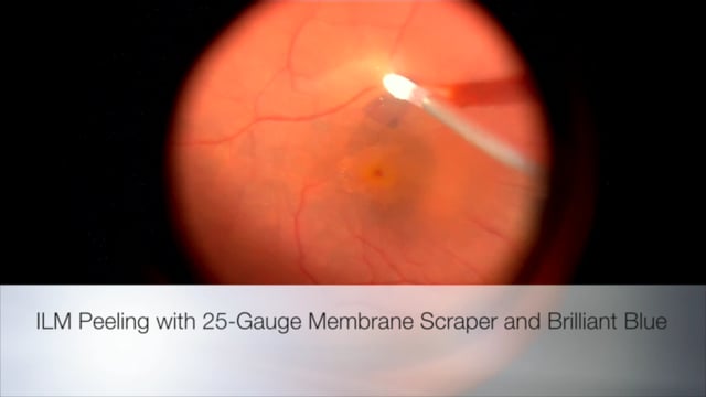
Jun 5 2016 by Thomas A. Ciulla, MD, MBA, FASRS
ILM peeling with 25-gauge membrane scraper and brilliant blue.
Condition/keywords: brilliant blue, internal limiting membrane (ILM) peeling, macular hole, vitrectomy
-
 Lutein: A New Dye for Chromovitrectomy
May 16 2014 by Mauricio Maia, MD, PhD
This video shows a new dye for vitreoretinal surgery comprised of soluble lutein/zeaxanthin 1% and brilliant blue 0.025 %. The green dye was deposited on the posterior pole; vigorous dye flushing into the vitreous cavity was unnecessary. The dye indirectly shows the posterior hyaloid by deposition of the golden lutein crystals. The ILM stained greenish-blue; No evidence of toxicity was observed.
Photographer: Mauricio Maia, Federal University of São Paulo
Condition/keywords: chromovitrectomy, internal limiting membrane (ILM) peeling, lutein
-
 ILM peeling
Apr 11 2014 by Subhendu Kumar Boral, MBBS, MD(AIIMS), DNB, FASRS (USA)
Brilliant blue stained ILM peeling in a case of idiopathic full thickness macular hole in a 61-year-old lady.
Photographer: Subhendu Kumar Boral
Condition/keywords: internal limiting membrane (ILM) peeling
-
 Vitreomacular Traction
Sep 27 2012 by Virgilio Morales-Canton, MD
Intraoperative image of a patient with a macular traction syndrome. Brilliant blue was used. DORC 27 Ga system.
Photographer: Virgilio Morales-Canton
Imaging device: optronics surgical camera
Condition/keywords: brilliant blue, macular traction, vitreomacular interface disorders
 Loading…