Search results (162 results)
-
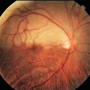
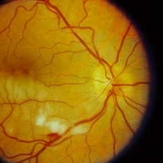
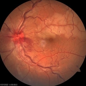
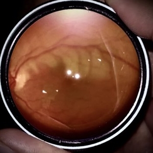 Branch Retinal Artery Occlusion
Oct 1 2024 by Angel Enrique Flores Pineda
Fundus photograph of a 78-year-old woman with poorly controlled systemic arterial hypertension and dyslipidemia. Hollenhorst plaque can be observed.
Photographer: Angel Enrique Flores Pineda, Hospital General de Zona #20
Imaging device: Smartphone (IPhone 15 plus)
Condition/keywords: branch retinal artery occlusion (BRAO)
-
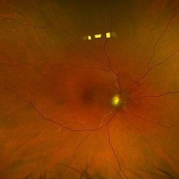
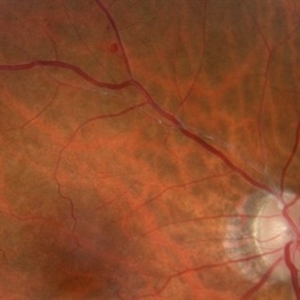 Hollenhorst Plaque
Jun 25 2024 by Virginia Gebhart
75 year female with complaint of shadow in the bottom of her vision for many years. Hollenhorst plaque on superior pole of the disc and sclerotic superotemporal arteriole. Also DBHs superiorly most likely due to combined BRAO/BRVO.
Photographer: Virginia Gebhart
Imaging device: Topcon 50DX
Condition/keywords: branch retinal artery occlusion (BRAO), branch retinal vein occlusion (BRVO), hollenhorst plaque, sclerotic arteriole
-
 Combined Central Retinal Vein Occlusion with Branch Retinal Artery Occlusion
Apr 29 2024 by KANWALJEET HARJOT MADAN, M.S. (Ophthalmology), FAICO (Vitreous - Retina)
This is fundus photograph of a 46-year male patient who presented with sudden diminution of vision in his right eye (RE) for 3 days. He was hypertensive but non diabetic. On examination, his best corrected vision in RE was 6/12. His left eye (LE) was normal. His fundus examination in RE revealed multiple intra retinal hemorrhages in all quadrants with tortuosity of veins suggestive of central retinal vein occlusion (CRVO) with mild disc edema. An ischemic area was seen superior to fovea suggestive of branch retinal artery occlusion. OCT depicted thickening of inner retinal layers with little evidence of macular edema. Hematological and cardio vascular investigations were done. He had bilateral thickening of intimal and medial walls of carotid arteries. He was under cardiology treatment. His vision improved to 6/6.
Photographer: Dr. Kanwaljeet Harjot Madan, M.S. (Ophthalmologist) Fellow in Vitrous & Retina. Thind Eye Hospital, Jalandhar City. Punjab. India
Condition/keywords: branch retinal artery occlusion (BRAO), central retinal vein occlusion (CRVO)
-
 Combined Central Retinal Vein Occlusion with Branch Retinal Artery Occlusion
Apr 28 2024 by KANWALJEET HARJOT MADAN, M.S. (Ophthalmology) Fellow in Vitreous & Retina
This is fundus photograph of a 46-year male patient who presented with sudden diminution of vision in his right eye (RE) for 3 days. He was hypertensive but non diabetic. On examination, his best corrected vision in RE was 6/12. His left eye (LE) was normal. His fundus examination in RE revealed multiple intra retinal hemorrhages in all quadrants with tortuosity of veins suggestive of central retinal vein occlusion (CRVO) with mild disc edema. An ischemic area was seen superior to fovea suggestive of branch retinal artery occlusion. OCT depicted thickening of inner retinal layers with little evidence of macular edema. Hematological and cardio vascular investigations were done. He had bilateral thickening of intimal and medial walls of carotid arteries. He was under cardiology treatment. His vision improved to 6/6.
Photographer: Dr Kanwaljeet Harjot Madan
Condition/keywords: branch retinal artery occlusion (BRAO), central retinal vein occlusion
-
 Superior Hemi-Central Retinal Artery Occlusion
Apr 24 2024 by Mosab Salah
Fundus photograph -inverted view- taken by smartphone fundus photography, of a young man with sudden onset altitudinal field defect, a Superior Hemi-Central Retinal Artery Occlusion noted.
Photographer: Dr Mosab Salah, The Islamic Hospital, Amman, Jordan
Imaging device: smartphone fundus photography and 30 D Lens
Condition/keywords: arterial occlusion, branch retinal artery occlusion (BRAO), BRAO, CRAO, Hemi-Central Retinal Artery Occlusion (CRAO), occlusive vasculitis, smartphone fundus photography
-
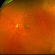
 Branch Retinal Artery Oclussion
Mar 17 2024 by César Adrián Gomez Valdivia, MD
Decreased arterial blood flow to the retina leading to ischemic damage.
Photographer: Erika Paulina Ornelas Cazares
Imaging device: Topcon TRC-50DX
Condition/keywords: branch retinal artery occlusion (BRAO), oclussion
-
 Neovascularization at the Disc
Nov 10 2023 by Philip Conkling, MD
Fluorescein angiogram of a patient with a history of a branch retinal artery occlusion who developed neovascularization at the disc.
Condition/keywords: branch retinal artery occlusion (BRAO), Neovascularisation at the Disc (NVD)
-
 Neovascularization at the Disc
Nov 10 2023 by Philip Conkling, MD
Fluorescein angiogram of a patient with a history of a branch retinal artery occlusion who developed neovascularization at the disc.
Condition/keywords: branch retinal artery occlusion (BRAO), Neovascularisation at the Disc (NVD)
-
 Branch Retinal Artery Occlusion (BRAO)
Sep 26 2023 by Ben Serar
Fundus photograph of LE showing retinal edema and opacification along the superotemporal arcade, with cherry red spot at the macula, in a case of Branch Retinal Artery Occlusion (BRAO).
Condition/keywords: branch retinal artery occlusion (BRAO), cherry red spot
-
 Branch Retinal Artery Occlusion (BRAO)
Sep 21 2023 by Ben Serar
Fundus photograph of RE showing retinal edema and opacification along the inferotemporal vessel arcade, with cotton wool spots and flame shaped haemorrhage, in a case of Branch Retinal Artery Occlusion (BRAO).
Condition/keywords: branch retinal artery occlusion (BRAO)
-
 Branch Retinal Artery Occlusion (BRAO)
Sep 12 2023 by Ben Serar
Fundus photograph of the LE showing arterial occlusion along the inferotemporal vessel arcade with surrounding retinal edema and cotton-wool spots, in a case of Branch Retinal Artery Occlusion (BRAO).
Condition/keywords: branch retinal artery occlusion (BRAO), cotton wool spots, retinal edema
-
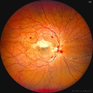
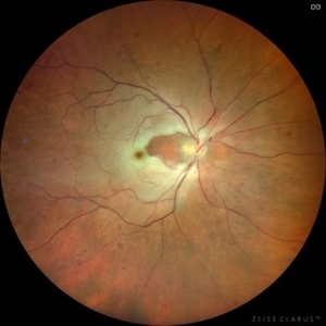 BRAO (Branch Retinal Artery Occlusion)
Jun 24 2023 by Mauricio Galvan Chavez, MD
Fundus photograph of a BRAO (Branch Retinal Artery Occlusion)
Photographer: Mauricio Galvan, Clinica de Retina, Guadalajara Jalisco.
Imaging device: Zeiss Clarus 700
Condition/keywords: arterial occlusion, branch retinal artery occlusion (BRAO), BRAO
-
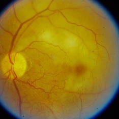
 Branch retinal artery occlusion
Jan 24 2023 by Rayna Marshall
Widefield fundus autofluorescence image of a 54-year-old female with an asymptomatic chronic branch retinal artery occlusion in the left eye. Hyper-autofluorescent embolus present at proximal inferior arcade, hypo-autoflorescence temporally corresponding to hyper-pigmentation. Vision was 20/20.
Photographer: Drew H. Scoles, MD, PhD, University of Pennsylvania
Condition/keywords: branch retinal artery occlusion (BRAO), BRAO, embolus
-
 Branch retinal artery occlusion
Jan 24 2023 by Rayna Marshall
Widefield fundus image of a 54-year-old female with an asymptomatic chronic branch retinal artery occlusion in the left eye. Peripheral schisis-like changes with pigmentation and temporal dot-blot hemorrhages. Vision was 20/20.
Photographer: Drew H. Scoles, MD, PhD, University of Pennsylvania
Condition/keywords: branch retinal artery occlusion (BRAO), BRAO, embolus
-
 Branch retinal artery occlusion
Jan 24 2023 by Rayna Marshall
OCT image of a 54-year-old female with an asymptomatic chronic branch retinal artery occlusion in the left eye showing inner retinal atrophy in the inferior macula corresponding to the region of chronic ischemia. Vision was 20/20.
Photographer: Drew H. Scoles, MD, PhD, University of Pennsylvania
Condition/keywords: branch retinal artery occlusion (BRAO), BRAO, embolus
-
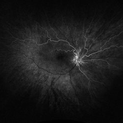
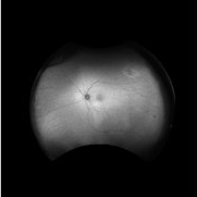
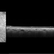
 Branch Retinal Artery Occlusion
Dec 15 2022 by Christopher R. Adam, M.D.
OCT
Condition/keywords: branch retinal artery occlusion (BRAO), BRAO
-
 Branch Retinal Artery Occlusion
Dec 15 2022 by Christopher R. Adam, M.D.
FA 32 seconds
Condition/keywords: branch retinal artery occlusion (BRAO), BRAO
-
 Branch Retinal Artery Occlusion
Dec 15 2022 by Christopher R. Adam, M.D.
AF
Condition/keywords: branch retinal artery occlusion (BRAO), BRAO
-
 Branch Retinal Artery Occlusion
Dec 15 2022 by Christopher R. Adam, M.D.
Color
Condition/keywords: branch retinal artery occlusion (BRAO)
-
 Combined central retinal vein occlusion and branch retinal arteriolar occlusion
Sep 13 2022 by Ruchir Mehta, DO, DNB, FRCS
Fundus photograph of left eye of a 63 years old female with known type 2 DM and HTN showing combined central retinal venous occlusion and superior branch retinal arteriolar occlusion
Photographer: Ruchir Mehta, Mehta Superspeciality Eye Hospital, Jamnagar, Gujarat, India
Imaging device: Zeiss Visucam 500
Condition/keywords: branch retinal artery occlusion (BRAO), central retinal vein occlusion (CRVO), COMBINED
-
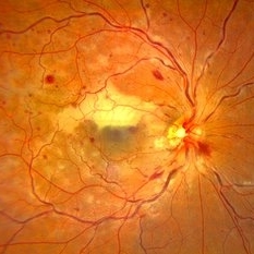
 Branch Retinal Artery occlusion
Sep 3 2022 by Tandava Krishnan
Branch Retinal artery occlusion with a visible y shaped embolus at the point of division of arteriole
Photographer: Tandava Krishnan
Condition/keywords: branch retinal artery occlusion (BRAO), embolic, embolus
-
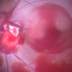
 Intraocular Foreign Body with BRAO
Jan 12 2022 by Manish Nagpal, MD, FRCS (UK), FASRS
Intraoperative photo of a foreign body which was impacted on the edge of the disc leading to a BRAO. On table, the IOFB was loosened from the impact site and this photo was taken just prior to removal of the same using a magnet.
Photographer: Manish Nagpal, Director, Retina Foundation, Ahmedabad
Imaging device: Sony PMW -10 MD surgical camera
Condition/keywords: branch retinal artery occlusion (BRAO), BRAO, intraocular foreign body
-
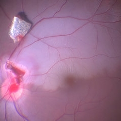
 Impacted IOFB over the Disc with Blood Stained Hyaloid
Jan 11 2022 by Manish Nagpal, MD, FRCS (UK), FASRS
Intraoperative photo of a foreign body impacted on the edge of disc leading to a BRAO and blood staining of the hyaloid is noted.
Photographer: Manish Nagpal, Retina Foundation, Ahmedabad, India
Imaging device: Sony PMW -10 MD surgical camera
Condition/keywords: branch retinal artery occlusion (BRAO), intraocular foreign body
-
 Branch Retinal Artery Occlusion after COVID-19 Vaccine
Aug 24 2021 by Narciso F. Atienza, MD, MBA, FASRS, FPCS, FPAO.
77 year old female who had loss of vision in her right eye 6 hours after her vaccination for COVID-19. The patient has hypertension with poor control, maintained on oral medications on an irregular basis. She has no prior hospitalizations related to COVID-19.
Photographer: Narciso F Atienza, Jr. MD MBA, FASRS, FPCS, FPAO. Legazpi Eye Center
Imaging device: Topcpn TRC
Condition/keywords: branch retinal artery occlusion (BRAO), COVID-19
 Loading…