Search results (59 results)
-
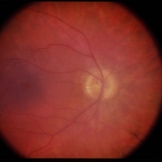
 Retinitis Pigmentosa
Apr 1 2025 by Jordyn Beckman
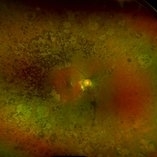
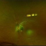
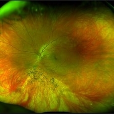
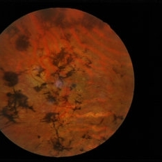
63 year old woman with Retinitis Pigmentosa observed over time with peripheral loss. Over the span of 5 years BCVA changed from 20/25 to 20/50.
Photographer: Jordyn Beckman, Retina Consultants of Carolina, P.A.
Imaging device: Optos California
Condition/keywords: atrophy, bone spicules, retinitis pigmentosa
-
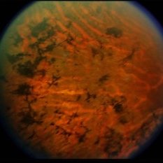
 Asteroid Hyalosis in Retinitis Pigmentosa
Dec 9 2024 by Mauricio Bayram-Suverza, MD
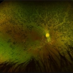
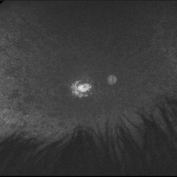
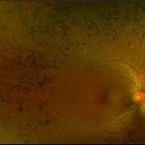
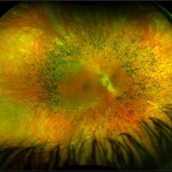
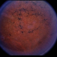
A 54 year-old male patient presented with asteroid hyalosis. Retinal examination revealed the presence of bone spicules, primarily located in the mid-periphery. Genetic testing identified a pathogenic variant in the RHO gene.
Photographer: Mauricio Bayram-Suverza, Casey Eye Institute, OHSU.
Imaging device: Optos California
Condition/keywords: Asteroid hyalosis, retinal dystrophy, Retinitis Pigmentosa, vitreous
-
 Magnification of Enucleated Eye: Phthisis Bulbi
May 18 2020 by McGill University Health Centre
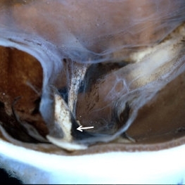
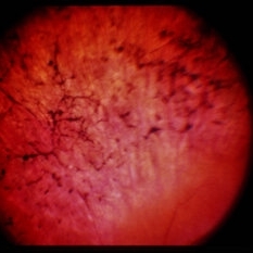
Higher magnification of enucleated eye with phthisis bulbi revealing bone spicules (arrow) corresponding to ossification due to metaplastic changes of the retinal pigmented epithelium (RPE) cells toward osteoblasts.
Condition/keywords: phthisis bulbi
-
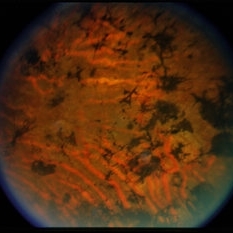
 Pigmentary Retinal Dystrophy
Mar 29 2019 by Jessica Norkus
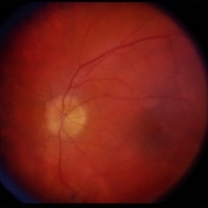
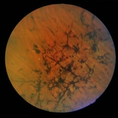
Optos ultra wide field image of 41-year-old male patient with pigmentary retinal dystrophy. Atypical findings due to unilateral presentation. Patient has been experiencing symptoms for 15 years, notes significant nyctalopia.
Photographer: Jessica Norkus
Imaging device: Optos Ultra Wide Field Camera
Condition/keywords: abnormal fundus, bone spicule, color fundus photograph, color photo, fundus photograph, Optos, peripheral bone spicules, pigment changes, ultra-wide field imaging, unilateral blindness
-
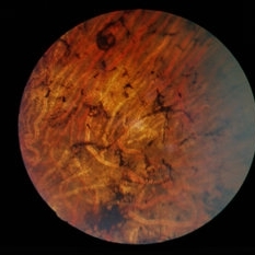
 Pigmentary Retinal Dystrophy
Mar 29 2019 by Jessica Norkus
Optos ultra wide field image of 41-year-old male patient with pigmentary retinal dystrophy. Atypical findings due to unilateral presentation. Patient has been experiencing symptoms for 15 years, notes significant nyctalopia.
Photographer: Jessica Norkus
Imaging device: Optos Ultra Wide Field Camera
Condition/keywords: abnormal fundus, bone spicule, color fundus photograph, color photo, fundus autofluorescence (FAF), fundus photograph, Optos, peripheral bone spicules, pigment changes, ultra-wide field imaging, unilateral blindness
-
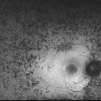
 Retinitis Pigmentosa - Fluorescein Angiogram OS
Jun 18 2018 by Hosam Attia, MD
38-year-old African American female with unilateral retinitis pigmentosa.
Imaging device: Optos California
Condition/keywords: bone spicule, peripheral bone spicules, retinitis pigmentosa
-
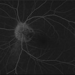
 Retinitis Pigmentosa - Fluorescein Angiogram OD
Jun 18 2018 by Hosam Attia, MD
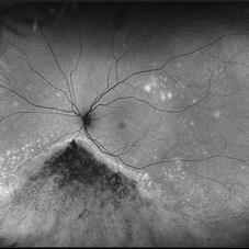
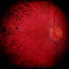
Ultra-wide fluorescein angiogram of a 38-year-old African, American female with degenerative myopia, Unilateral RP variant, depicting abnormal fluorescence pattern with extensive mid-peripheral bone spicules hypofluorescence, extending further into the periphery w/ relative sparing of the macula OD. VF 30-V showed severe peripheral constriction OD, enlarged BS OS & OCT showed severe ellipsoid zone degeneration with saucerization and cystoid macular degeneration w/ No obvious late macular leakage on FA (Both, not shown)
Imaging device: Optos California
Condition/keywords: bone spicule, peripheral bone spicules, retinitis pigmentosa
-
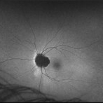 Retinitis Pigmentosa - Autofluorescence OS
Jun 18 2018 by Hosam Attia, MD
Retinitis Pigmentosa
Imaging device: Optos California
Condition/keywords: bone spicule, peripheral bone spicules, retinitis pigmentosa
-
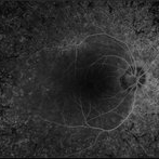
 Retinitis Pigmentosa - Autofluorescence OD
Jun 18 2018 by Hosam Attia, MD
Ultra-wide fundus auto-fluorescence photograph of a 38-year-old African, American female with degenerative myopia, unilateral RP variant, depicting extensive mid-peripheral bone spicules hypo-autofluorescence, extending further into the periphery w/ relative sparing of the macula OD VF 30-V showed severe peripheral constriction OD, enlarged BS OS and OCT showed severe ellipsoid zone degeneration with saucerization and cystoid macular degeneration with no obvious late macular leakage on FA (Both, not shown)
Imaging device: Optos California
Condition/keywords: bone spicule, peripheral bone spicules, retinitis pigmentosa
-
 Retinitis Pigmentosa - Color OS
Jun 18 2018 by Hosam Attia, MD
38-year-old female with unilateral retinitis pigmentosa.
Imaging device: Optos California
Condition/keywords: bone spicule, peripheral bone spicules, retinitis pigmentosa
-
 Retinitis Pigmentosa - Color OD
Jun 18 2018 by Hosam Attia, MD
Pseudo-color ultra-wide fundus photograph of a 38-year-old African American female with degenerative myopia and Unilateral RP variant, depicting extensive mid-peripheral bone spicules, extending further into the periphery, with relative sparing of the macula OD. VF 30-V showed severe peripheral constriction OD, enlarged BS OS and OCT showed severe ellipsoid zone degeneration with saucerization and cystoid macular degeneration with no obvious late macular leakage on FA (Both, not shown)
Imaging device: Optos California
Condition/keywords: bone spicule, peripheral bone spicules, retinitis pigmentosa
-
 Retinitis Pigmentosa
May 26 2017 by Olivia Rainey
Ultra-wide-field pseudocolor image of the left eye of an 39-year-old female with Retinitis Pigmentosa. She had slightly atypical appearance due to asymmetry: sectoral atrophy in left eye, compared to 360 degree bone spicule formation in right eye. Ddx: Pigmentary degeneration vs infection vs X-linked RP carrier due to asymmetry. Recommended genetic testing through My Retina Tracker, as well as visual field and ERG testing. Patient's vision was sc20/100 PH 20/70 in the right eye and sc20/80 PH 20/40 in the left eye.
Photographer: Olivia Rainey
Imaging device: Optos California
Condition/keywords: autofluorescence imaging, bone spicule, hyperautofluorescent ring, hypoautofluorescence, Optos, peripheral bone spicules, retinitis pigmentosa, ultra-wide field imaging
-
 Retinitis Pigmentosa
May 26 2017 by Olivia Rainey
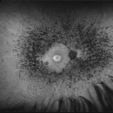
Ultra-wide-field fundus autofluorescence image of the left eye of an 39-year-old female with Retinitis Pigmentosa. She had slightly atypical appearance due to asymmetry: sectoral atrophy in left eye, compared to 360 degree bone spicule formation in right eye. Ddx: Pigmentary degeneration vs infection vs X-linked RP carrier due to asymmetry. Recommended genetic testing through My Retina Tracker, as well as visual field and ERG testing. Patient's vision was sc20/100 PH 20/70 in the right eye and sc20/80 PH 20/40 in the left eye.
Photographer: Olivia Rainey
Imaging device: Optos
Condition/keywords: autofluorescence imaging, hyperautofluorescence, hypoautofluorescence, left eye, Optos, peripheral bone spicules, retinitis pigmentosa, ultra-wide field imaging
-
 Retinitis Pigmentosa
May 26 2017 by Olivia Rainey
Ultra-wide-field pseudocolor image of the left eye of an 39-year-old female with Retinitis Pigmentosa. She had slightly atypical appearance due to asymmetry: sectoral atrophy in left eye, compared to 360 degree bone spicule formation in right eye. Ddx: Pigmentary degeneration vs infection vs X-linked RP carrier due to asymmetry. Recommended genetic testing through My Retina Tracker, as well as visual field and ERG testing. Patient's vision was sc20/100 PH 20/70 in the right eye and sc20/80 PH 20/40 in the left eye.
Photographer: Olivia Rainey
Imaging device: Optos California
Condition/keywords: bone spicule, fundus photograph, left eye, Optos, peripheral bone spicules, pseudocolor, retinitis pigmentosa, ultra-wide field imaging
-
 Retinitis Pigmentosa
May 26 2017 by Olivia Rainey
Ultra-wide-field pseudocolor image of the right eye of an 39-year-old female with Retinitis Pigmentosa. She had slightly atypical appearance due to asymmetry: sectoral atrophy in left eye, compared to 360 degree bone spicule formation in right eye. Ddx: Pigmentary degeneration vs infection vs X-linked RP carrier due to asymmetry. Recommended genetic testing through My Retina Tracker, as well as visual field and ERG testing. Patient's vision was sc20/100 PH 20/70 in the right eye and sc20/80 PH 20/40 in the left.
Photographer: Olivia Rainey
Imaging device: Optos California
Condition/keywords: bone spicule, fundus photograph, Optos, peripheral bone spicules, pseudocolor, retinitis pigmentosa, ultra-wide field imaging
-
 Retinitis Pigmentosa
Dec 22 2014 by H. Michael Lambert, MD
Fundus photo with bone spicules.
Condition/keywords: retinitis pigmentosa
-
 Retinitis Pigmentosa
Dec 22 2014 by H. Michael Lambert, MD
Fundus photo with bone spicules.
Condition/keywords: retinitis pigmentosa
-
 Retinitis Pigmentosa
Dec 22 2014 by H. Michael Lambert, MD
Fundus photo with bone spicules.
Condition/keywords: retinitis pigmentosa
-
 Retinitis Pigmentosa
Dec 22 2014 by H. Michael Lambert, MD
Fundus photo with bone spicules.
Condition/keywords: retinitis pigmentosa
-
 Retinitis Pigmentosa
Dec 22 2014 by H. Michael Lambert, MD
Fundus photo with bone spicules.
Condition/keywords: retinitis pigmentosa
-
 Retinitis Pigmentosa
Dec 22 2014 by H. Michael Lambert, MD
Fundus photo with bone spicules.
Condition/keywords: retinitis pigmentosa
-
 Retinitis Pigmentosa
Dec 22 2014 by H. Michael Lambert, MD
Fundus photo with bone spicules.
Condition/keywords: retinitis pigmentosa
-
 Retinitis Pigmentosa
Dec 22 2014 by H. Michael Lambert, MD
Fundus photo with bone spicules.
Condition/keywords: retinitis pigmentosa
-
 Retinitis Pigmentosa
Dec 22 2014 by H. Michael Lambert, MD
Fundus photo with bone spicules
Condition/keywords: retinitis pigmentosa
-
 Retinitis Pigmentosa
Dec 22 2014 by H. Michael Lambert, MD
Fundus photo with bone spicules.
Condition/keywords: retinitis pigmentosa
 Loading…